

Color.Atlas.of.Endodontics-ublog.tk
.pdfChapter Eleven Traumatic Injuries to the Permanent Dentition |
1 73 |
C, D |
E |
FIGURE 11-6 A and B, Clinical photographs of a maxillary right lateral incisor exhibiting a crown root fracture. C, Radiographic examination reveals normal structures, and the fracture is not apparent. D and E, Removal of the fractured segment permits evaluation of the tooth for restorability.
conjunction with required endodontic and restorative procedures; extraction also may be cons idered. 25,26 Because most of these fractures involve anterior teeth, esthetics is a major concern. Extrusion may result in a tooth that exhibits a smaller mesial-distal dimension and decreased crown-root ratio. Crown lengthening results in elongation of the tooth with diminished bone support.
An alternative treatment technique is surgical extrusion. 27 This technique is only appropriate for teeth with complete apical development. After administering local anesthesia, the clinician gently luxates the apical segment, moves it in a coronal direction, and stabilizes it with sutures or a splint. Root canal treatment can be performed after initial healing takes place, and restorative treatment can begin within 6 to 8 weeks after periodontal healing.
ROOT FRACTURES
Horizontal root fractures frequently occur as the result of facial trauma to the anterior teeth (Figure 11-7). They are frequently oblique and therefore often difficult to detect.
Clinical features that suggest root fracture are tenderness to percussion and palpation, tooth mobility, displacement, and bleeding from the gingival sulcus. Teeth with root fractures often do not respond to pulp testing. Recent evidence suggests that a negative initial response is significantly related to later pulp necrosis.28 Several radiographic projections may be required to identify the fracture line ( see Figure 11-1). 29 If the initial parallel film does not demonstrate a fracture, occlusal films may be required. Variation of the vertical angle may also be useful.
The pulp of the majority of teeth with intact root structure remains vital. Treatment procedures are designed to enhance the healing of osseous and supporting periodontal structures. Four types of healing responses have been described for root fractures30:
1.Calcific healing with callus formation between the closely approximated segments (Figure 11-8)
2.Connective tissue healing whereby the segments are separated by a fibrous attachment (see Figure 11-7)
3.A combination of bone and connective tissue healing in which bone grows between the segments

1 74 |
Color Atlas of Endodontics |
FIGURE 11-7 A, Clinical photograph of a patient who fell while playing tennis. Note the hemorrhage from the gingival sulcus of the maxillary left central incisor. B, A parallel periapical radiograph demonstrates a horizontal root fracture C, The fracture is less apparent on a maxillary occlusal film. D, A postoperative photograph of a rigid wire and composite splint. E, Postoperative film taken after splint placement. F, A photograph of the dentition after splint removal. G, On recall examination the tooth was responsive and exhibited normal mobility. Note that during the recall interval, tooth #8 required endodontic treatment because of pulp necrosis.
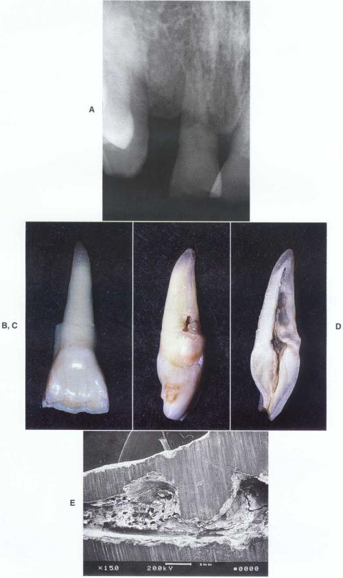
FIGURE 11-8 A, A radiograph of a patient who related having his maxillary right central incisor fractured in World War II. B, Healing of the fracture site was noted on extraction. C, Note the resorptive lesion on the external surface of the root below the fracture site. D, A vertical section reveals the resorptive lesion to be internal and demonstrates destruction of the facial canal wall. As the resorption progressed, external perforation occurred. E, Scanning electron microscopy of the resorptive defect.

176 |
Color Atlas of Endodontics |
and periodontal ligament formation occurs between the osseous tissues and the root segments (Figure 11-9)
4.Nonunion, with granulation tissue formation at the fracture site resulting from necrosis of the coronal pulp tissue, which initiates an inflammatory reaction at the fracture line (Figure 11-10); the inflammation then spreads laterally into the
alveolar bone, inducing resorption and formation of a "butterfly" radiolucent area
Treatment procedures for teeth exhibiting root fracture should enhance the potential for union of the fractured segments. In cases of root fracture in which no displacement has occurred and the tooth exhibits normal mobility, treatment may not be required. In
FIGURE 11-9 Fracture healing with connective tissue and bone.
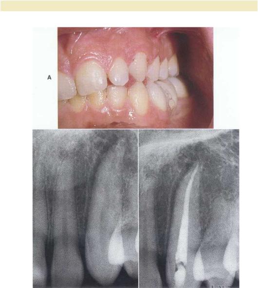
FIGURE 11-10 A, A periapical radiograph depicting horizontal root fractures of the maxillary central incisors. The left maxillary central incisor became necrotic and was opened for drainage. The maxillary right central incisor is responsive to pulp testing. B, The coronal segment was treated and the apical segment, which exhibited resorption, was surgically removed. C, A recall radiograph exhibits osseous regeneration. The maxillary right central incisor remains responsive to testing.

Chapter Eleven Traumatic Injuries to the Permanent Dentition |
1 77 |
cases where the tooth exhibits displacement or mobility, the clinician should reduce the fracture immediately and place a rigid splint to immobilize the segments. Radiographic verification of the tooth position after splinting should be obtained.
Fractures that have a vertical component or extend near the gingival sulcus have a poor prognosis because of bacterial contamination from the oral environment and subsequent periodontal breakdown. Options for treatment of these teeth include removal of the coronal segment and crown lengthening, orthodontic extrusion, and extraction.
The splint should remain in place for 8 to 12 weeks to permit the calcified tissues to heal. The clinical recall evaluations should assess the pulpal status, tooth mobility, and periodontal probing depths. A lack of response to pulp testing does not indicate that necrosis has occurred as long as no other signs or symptoms are associated with the tooth. The pulp remains vital in the ma- j ority of cases.31 Signs of necrosis include discoloration, sensitivity to percussion and palpation, and radiographic evidence of resorption or a radiolucent lesion. When necrosis occurs it generally affects only the coronal segment, with the apical segment remaining vital.32 Mobility after 8 to 12 weeks of splinting may require continued splinting.
If pulp necrosis develops, the coronal segment can be treated with nonsurgical endodontic techniques as long as no radiolucent area is associated with the apical segment, the apical segment exhibits hemorrhage, and an adequate constricted area is available in the coronal segment for instrumentation and obturation with gutta-per- cha (see Figure 11-10). If the apical segment exhibits signs of necrosis, both segments can be treated nonsurgically or the coronal segment can be treated and the apical segment removed surgically (see Figure 11-10). If a constriction cannot be detected or created, apexification of the coronal segment is indicated before obturation.
A common finding on recall examination of teeth with root fractures is canal obliteration and discoloration resulting from calcification. The majority of teeth with canal obliteration do not become necrotic.33 Endodontic treatment of teeth exhibiting calcification is not warranted unless evidence of necrosis exists.
Treatment is generally palliative. Recall examination is essential to assess pulpal status. If a tooth that is initially responsive becomes unresponsive over time, necrosis has most likely occurred. A tooth that is initially unresponsive can become responsive after the injury, indicating a positive pulpal response . 34 Of the luxation injuries, concussion has the best pulpal prognosis, with only 3 % becoming necrotic. 35
Subluxation
Subluxation is defined as a tooth that is slightly mobile after a traumatic episode but not displaced from the socket (Figure 11-12). On clinical examination, bleeding from the gingival sulcus may be observed and the tooth may be unresponsive to pulp testing. Radiography may reveal widening of the periodontal ligament.
Treatment is palliative, although the mobility may require splinting. Because the injury is primarily to the attachment apparatus, a physiologic splint of monofila ment fishing line and composite resin should be placed for 7 to 14 days when needed.
Recall examination is performed to assess pulpal status. Pulpal necrosis occurs in approximately 6% of all cases. 35
Lateral and Extrusive Luxation
Lateral and extrusive luxation is a traumatic injury that results in a tooth being displaced from its socket (Figure 11-13). Because the tooth is moved from the socket, frac- ture of the alveolar process is a common finding. Often numerous teeth are involved. Pulp testing is unreliable. Displacement disrupts the vascular and neural elements, frequently resulting in pulp necrosis. Occlusal radiographs are often valuable in demonstrating tooth displacement. 9
Teeth that are displaced without damage to the socket can be repositioned and stabilized with a physiologic splint consisting of monofilament fishing line or orthodontic wire and composite for a period of 7 to 14 days. If a fracture of the alveolar process has occurred, the tooth should be repositioned and a rigid splint placed for 4 to 6 weeks. Because the incidence of pulp necrosis is high and often remains undetected,
root canal treatment should be initiated after initial stabilization.35,36
LUXATION INJURIES
Concussion
Concussion is defined as traumatic injury to a tooth that does not result in mobility or displacement from the socket (Figure 11-11). The primary clinical finding is tenderness to biting pressure and percussion resulting from injury to the supporting structures. An additional clinical finding is discoloration, although this is not common. Pulp testing may reveal responsiveness, but the pulp also may not be responsive to initial testing. Radiographic findings reveal normal structures.
Intrusive Luxation
The intruded tooth is displaced centrally into bone (Figure 11-14). Clinical examination reveals a short clinical crown with gingival hemorrhage. Because the tooth is displaced into the bone, mobility is absent. Radiographic examination demonstrates the tooth position. Because of the crushing injury, the periodontal ligament space may not be evident. Treatment of teeth that are intruded depends on the extent of apical development. In cases where root formation is incomplete, the teeth may reerupt. If the roots are completely formed, the clinician
178 |
Color Atlas o f Endodontics |
FIGURE 11-11 A, A photograph of a patient who sustained a concussion-type injury. Note the hemorrhage into the dentin. B, A periapical radiograph reveals horizontal bone loss consistent with previous periodontal disease and normal apical structures. C, Clinical photograph taken after nonsurgical root canal treatment and internal bleaching. D, A postoperative radiograph.

Chapter Eleven Traumatic Injuries to the Permanent Dentition |
179 |
|
B
A
C
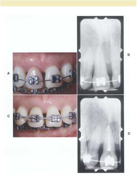
FIGURE 11-13 A, A preoperative radiograph of a maxillary right lateral incisor after a lateral ex-
trusive luxation injury. B, A recall radiograph taken after endodontic treatment and orthodontic repositioning. Note the calcification present in the maxillary central incisors. C, Clinical examination re-
veals normal tissues and tooth coloration.
18 0 |
Color Atlas of Endodontics |
B C
FIGURE 11-14 A, A clinical photograph of a lateral intrusive traumatic injury to the maxillary left canine. B, A preoperative radiograph demonstrates a widened mesial periodontal ligament space. C, A postoperative radiograph taken after endodontic treatment.
must reposition the tooth surgically with forceps or through orthodontic movement. If surgical repositioning is performed, the tooth should be splinted. Orthodontic treatment is recommended because it produces decreased resorption and preserves the crestal bone. 38 Orthodontic treatment should be initiated promptly because the development of replacement resorption or ankylosis may prevent tooth movement.
Root canal treatment should also be initiated because of the poor pulpal prognosis with this type of injury. 15,17 Although the placement of calcium hydroxide is recommended to prevent root resorption, it should take place only after initial healing is complete because calcium hydroxide has been shown to increase resorption when placed immediately after injury. 39
AVULSION
Avulsion is defined as complete displacement of a tooth from the socket (Figure 11-15). Although time is of the essence in treating an avulsed tooth, obtaining a complete medical and dental history and performing a clinical and radiographic examination are essential. Treatment procedures are directed toward management of the pulp and the more significant periodontal reactions to injury.
Periodontal Reactions to Avulsion
After replantation a coagulum forms between the root and alveolar bone. After 1 week the epithelial attachment is reestablished at the cementoenamel junction and the gingival collagen fibers become spliced. 40 This limits bacterial invasion and permits healing to continue. Four

Chapter Eleven Tramnatic Injuries to the Permanent Dentition |
1 8 1 |
FIGURE 11-15 A, A photograph of a patient who avulsed his maxillary right lateral and both central incisors while playing baseball. B, A preoperative radiograph demonstrating complete avulsion
with intact socket walls. C, A photograph of the teeth after replantation. D, An orthodontic appliance and wire splint in place. E, Splint removal after the initiation of nonsurgical root canal treatment at 2 weeks. F, Calcium hydroxide placement.

182 |
Color Atlas o f Endodontics |
FIGURE 11-16 A, A clinical photograph of a maxillary left central incisor that was previously avulsed. B, A radiograph of the involved tooth demonstrating replacement resorption.
FIGURE 11-17 A, A radiograph of a mandibular left central incisor with a history of lateral luxation and a lateral incisor that was avulsed and demonstrates inflammatory resorption. B, Clinical view of a sinus tract that developed as a result of pulp necrosis.