- •Foreword
- •Preface
- •Contents
- •About the Editors
- •Contributors
- •1: Tracheobronchial Anatomy
- •Trachea
- •Introduction
- •External Morphology
- •Internal Morphology
- •Mucous Layer
- •Blood Supply
- •Anatomo-Clinical Relationships
- •Bronchi
- •Main Bronchi
- •Bronchial Division
- •Left Main Bronchus (LMB)
- •Right Main Bronchus (RMB)
- •Blood Supply
- •References
- •2: Flexible Bronchoscopy
- •Introduction
- •History
- •Description
- •Indications and Contraindications
- •Absolute Contraindications
- •Procedure Preparation
- •Technique of FB Procedure
- •Complications of FB Procedure
- •Basic Diagnostic Procedures
- •Bronchoalveolar Lavage (BAL)
- •Transbronchial Lung Biopsy (TBLB)
- •Transbronchial Needle Aspiration (TBNA)
- •Bronchial Brushings
- •Advanced Diagnostic Bronchoscopy
- •EBUS-TBNA
- •Ultrathin Bronchoscopy
- •Transbronchial Lung Cryobiobsy (TBLC)
- •Therapeutic Procedures Via FB
- •LASER Bronchoscopy
- •Electrocautery
- •Argon Plasma Coagulation (APC)
- •Cryotherapy
- •Photodynamic Therapy
- •Airway Stent Placement
- •Endobronchial Valve Placement
- •Conclusion
- •References
- •History and Historical Perspective
- •Indications and Contraindications
- •Procedure Description
- •Procedure Planning
- •Target Approximation
- •Sampling
- •Complications
- •Future Directions
- •Summary and Recommendations
- •References
- •4: Rigid Broncoscopy
- •Innovations
- •Ancillary Equipment
- •Rigid Bronchoscopy Applications
- •Laser Bronchoscopy
- •Tracheobronchial Prosthesis
- •Transbronchial Needle Aspiration (TBNA)
- •Rigid Bronchoscope in Other Treatments for Bronchial Obstruction
- •Mechanical Debridement
- •Pediatric Rigid Bronchoscopy
- •Tracheobronchial Dilatation
- •Foreign Bodies Removal
- •Other Indications
- •Complications
- •The Procedure
- •Some Conclusions
- •References
- •History and Historical Perspective
- •Indications and Contraindications
- •Preprocedural Evaluation and Preparation
- •Physical Examination
- •Procedure-Related Indications
- •Application of the Technique
- •Topical Anesthesia
- •Anesthesia of the Nasal Mucosa and Nasopharynx
- •Anesthesia of the Mouth and Oropharynx
- •Superior Laryngeal Nerve Block
- •Recurrent Laryngeal Nerve Block (RLN)
- •Conscious Sedation
- •Monitored Anesthesia Care (MAC)
- •General Anesthesia
- •Monitoring the Depth of Anesthesia
- •Interventional Bronchoscopy Suites
- •Airway Devices
- •Laryngeal Mask Airway (LMA)
- •Endotracheal Tube (ETT)
- •Rigid Bronchoscope
- •Modes of Ventilation
- •Spontaneous Ventilation
- •Assisted Ventilation
- •Noninvasive Positive Pressure Ventilation (NIV)
- •Positive Pressure Controlled Mechanical Ventilation
- •Jet Ventilation
- •Electronic Mechanical Jet Ventilation
- •Postprocedure Care
- •Special Consideration
- •Anesthesia for Peripheral Diagnostic and Therapeutic Bronchoscopy
- •Anesthesia for Interventional Bronchoscopic Procedures During the COVID-19 Pandemic
- •Summary and Recommendations
- •Conclusion
- •References
- •Background
- •Curricular Structure and Delivery
- •What Is a Bronchoscopy Curriculum?
- •Tradition, Teaching Styles, and Beliefs
- •Using Assessment Tools to Guide the Educational Process
- •The Ethics of Teaching
- •When Learners Teach: The Journey from Novice to Mastery and Back Again
- •The Future Is Now
- •References
- •Interventional Procedure
- •Assessment of Flow–Volume Curve
- •Dyspnea
- •Analysis of Pressure–Pressure Curve
- •Conclusions
- •References
- •Introduction
- •Adaptations of the IP Department
- •Environmental Control
- •Personal Protective Equipment
- •Procedure Performance
- •Bronchoscopy in Intubated Patients
- •Other Procedures in IP Unit
- •References
- •Introduction
- •Safety
- •Patient Safety
- •Provider Safety
- •Patient Selection and Screening
- •Lung Cancer Diagnosis and Staging
- •Inpatients
- •COVID-19 Clearance
- •COVID Clearance: A Role for Bronchoscopy
- •Long COVID: A Role for Bronchoscopy
- •Preparing for the Next Pandemic
- •References
- •Historical Perspective
- •Indications and Contraindications
- •Evidence-Based Review
- •Summary and Recommendations
- •References
- •Introduction
- •Clinical Presentation
- •Diagnosis
- •Treatment
- •History and Historical Perspectives
- •Indications and Contraindications
- •Benign and Malignant Tumors
- •Tumors with Uncertain Prognosis
- •Application of the Technique
- •Evidence Based Review
- •Summary and Recommendations
- •References
- •12: Cryotherapy and Cryospray
- •Introduction
- •Historical Perspective
- •Equipment
- •Cryoadhesion
- •Indications
- •Cryorecanalization
- •Cryoadhesion and Foreign Body Removal
- •Cryoadhesion and Mucus Plugs/Blood Clot Retrieval
- •Endobronchial Cryobiopsy
- •Transbronchial Cryobiopsy for Lung Cancer
- •Safety Concerns and Contraindications
- •Cryoablation
- •Indications
- •Evidence
- •Safety Concerns and Contraindications
- •Cryospray
- •Indications
- •Evidence
- •Safety Concerns and Contraindications
- •Advantages of Cryotherapy
- •Limitations
- •Future Research Directions
- •References
- •13: Brachytherapy
- •History and Historical Perspective
- •Indications and Contraindications
- •Application of the Technique
- •Evidence-Based Review
- •Adjuvant Treatment
- •Palliative Treatment
- •Complications
- •Summary and Recommendations
- •References
- •14: Photodynamic Therapy
- •Introduction
- •Photosensitizers
- •First-Generation Photosensitizers
- •M-Tetrahidroxofenil Cloro (mTHPC) (Foscan®)
- •PDT Reaction
- •Tumor Damage Process
- •Procedure
- •Indications
- •Curative PDT Indications
- •Palliative PDT Indications
- •Contraindications
- •Rationale for Use in Early-Stage Lung Cancer
- •Rationale
- •PDT in Combination with Other Techniques for Advanced-Stage Non-small Cell Lung Cancer
- •Commentary
- •Complementary Endoscopic Methods for PDT Applications
- •New Perspectives
- •Other PDT Applications
- •Conclusions
- •References
- •15: Benign Airways Stenosis
- •Etiology
- •Congenital Tracheal Stenosis
- •Iatrogenic
- •Infectious
- •Idiopathic Tracheal Stenosis
- •Distal Bronchial Stenosis
- •Diagnosis Methods
- •Patient History
- •Imaging Techniques
- •Bronchoscopy
- •Pulmonary Function Test
- •Treatment
- •Endoscopic Treatment
- •Dilatation
- •Laser Therapy
- •Stents
- •How to Proceed
- •Stent Placement
- •Placing a Montgomery T Tube
- •The Rule of Twos for Benign Tracheal Stenosis (Fig. 15.23)
- •Surgery
- •Summary and Recommendations
- •References
- •16: Endobronchial Prostheses
- •Introduction
- •Indications
- •Extrinsic Compression
- •Intraluminal Obstruction
- •Stump Fistulas
- •Esophago-respiratory Fistulas (ERF)
- •Expiratory Central Airway Collapse
- •Physiologic Rationale for Airway Stent Insertion
- •Stent Selection Criteria
- •Stent-Related Complications
- •Granulation Tissue
- •Stent Fracture
- •Migration
- •Contraindications
- •Follow-Up and Patient Education
- •References
- •Introduction
- •Overdiagnosis
- •False Positives
- •Radiation
- •Risk of Complications
- •Lung Cancer Screening Around the World
- •Incidental Lung Nodules
- •Management of Lung Nodules
- •References
- •Introduction
- •Minimally Invasive Procedures
- •Mediastinoscopy
- •CT-Guided Transthoracic Biopsy
- •Fluoroscopy-Guided Transthoracic Biopsies
- •US-Guided Transthoracic Biopsy
- •Thoracentesis and Pleural Biopsy
- •Thoracentesis
- •Pleural Biopsy
- •Surgical or Medical Thoracoscopy
- •Image-Guided Pleural Biopsy
- •Closed Pleural Biopsy
- •Image-Guided Biopsies for Extrathoracic Metastases
- •Tissue Acquisition, Handling and Processing
- •Implications of Tissue Acquisition
- •Guideline Recommendations for Tissue Acquisition in Mediastinal Staging
- •Methods to Overcome Challenges in Tissue Acquisition and Genotyping
- •Rapid on-Site Evaluation (ROSE)
- •Sensitive Genotyping Assays
- •Liquid Biopsy
- •Summary, Recommendations and Highlights
- •References
- •History
- •Data Source and Methodology
- •Tumor Size
- •Involvement of the Main Bronchus
- •Atelectasis/Pneumonitis
- •Nodal Staging
- •Proposal for the Revision of Stage Groupings
- •Small Cell Lung Cancer (SCLC)
- •Discussion
- •Methodology
- •T Descriptors
- •N Descriptors
- •M Descriptors
- •Summary
- •References
- •Introduction
- •Historical Perspective
- •Fluoroscopy
- •Radial EBUS Mini Probe (rEBUS)
- •Ultrasound Bronchoscope (EBUS)
- •Virtual Bronchoscopy
- •Trans-Parenchymal Access
- •Cone Beam CT (CBCT)
- •Lung Vision
- •Sampling Instruments
- •Conclusions
- •References
- •History and Historical Perspective
- •Narrow Band Imaging (NBI)
- •Dual Red Imaging (DRI)
- •Endobronchial Ultrasound (EBUS)
- •Optical Coherence Tomography (OCT)
- •Indications and Contraindications
- •Confocal Laser Endomicroscopy and Endocytoscopy
- •Raman Spectrophotometry
- •Application of the Technique
- •Supplemental Technology for Diagnostic Bronchoscopy
- •Evidence-Based Review
- •Summary and Recommendations, Highlight of the Developments During the Last Three Years (2013 on)
- •References
- •Introduction
- •History and Historical Perspective
- •Endoscopic AF-OCT System
- •Preclinical Studies
- •Clinical Studies
- •Lung Cancer
- •Asthma
- •Airway and Lumen Calibration
- •Obstructive Sleep Apnea
- •Future Applications
- •Summary
- •References
- •23: Endobronchial Ultrasound
- •History and Historical Perspective
- •Equipment
- •Technique
- •Indication, Application, and Evidence
- •Convex Probe Ultrasound
- •Equipment
- •Technique
- •Indication, Application, and Evidence
- •CP-EBUS for Malignant Mediastinal or Hilar Adenopathy
- •CP-EBUS for the Staging of Non-small Cell Lung Cancer
- •CP-EBUS for Restaging NSCLC After Neoadjuvant Chemotherapy
- •Complications
- •Summary
- •References
- •Introduction
- •What Is Electromagnetic Navigation?
- •SuperDimension Navigation System (EMN-SD)
- •Computerized Tomography
- •Computer Interphase
- •The Edge Catheter: Extended Working Channel (EWC)
- •Procedural Steps
- •Planning
- •Detecting Anatomical Landmarks
- •Pathway Planning
- •Saving the Plan and Exiting
- •Registration
- •Real-Time Navigation
- •SPiN System Veran Medical Technologies (EMN-VM)
- •Procedure
- •Planning
- •Navigation
- •Biopsy
- •Complications
- •Limitations
- •Summary
- •References
- •Introduction
- •Image Acquisition
- •Hardware
- •Practical Considerations
- •Radiation Dose
- •Mobile CT Studies
- •Future Directions
- •Conclusion
- •References
- •26: Robotic Assisted Bronchoscopy
- •Historical Perspective
- •Evidence-Based Review
- •Diagnostic Yield
- •Monarch RAB
- •Ion Endoluminal Robotic System
- •Summary
- •References
- •History and Historical Perspective
- •Indications and Contraindications
- •General
- •Application of the Technique
- •Preoperative Care
- •Patient’s Position and Operative Field
- •Incision and Initial Dissection
- •Palpation
- •Biopsy
- •Control of Haemostasis and Closure
- •Postoperative Care
- •Complications
- •Technical Variants
- •Extended Cervical Mediastinoscopy
- •Mediastinoscopic Biopsy of Scalene Lymph Nodes
- •Inferior Mediastinoscopy
- •Mediastino-Thoracoscopy
- •Video-Assisted Mediastinoscopic Lymphadenectomy
- •Transcervical Extended Mediastinal Lymphadenectomy
- •Evidence-Based Review
- •Summary and Recommendations
- •References
- •Introduction
- •Case 1
- •Adrenal and Hepatic Metastases
- •Brain
- •Bone
- •Case 1 Continued
- •Biomarkers
- •Case 1 Concluded
- •Case 2
- •Chest X-Ray
- •Computerized Tomography
- •Positive Emission Tomography
- •Magnetic Resonance Imaging
- •Endobronchial Ultrasound with Transbronchial Needle Aspiration
- •Transthoracic Needle Aspiration
- •Transbronchial Needle Aspiration
- •Endoscopic Ultrasound with Needle Aspiration
- •Combined EUS-FNA and EBUS-TBNA
- •Case 2 Concluded
- •Case 3
- •Standard Cervical Mediastinoscopy
- •Extended Cervical Mediastinoscopy
- •Anterior Mediastinoscopy
- •Video-Assisted Thoracic Surgery
- •Case 3 Concluded
- •Case 4
- •Summary
- •References
- •29: Pleural Anatomy
- •Pleural Embryonic Development
- •Pleural Histology
- •Cytological Characteristics
- •Mesothelial Cells Functions
- •Pleural Space Defense Mechanism
- •Pleura Macroscopic Anatomy
- •Visceral Pleura (Pleura Visceralis or Pulmonalis)
- •Parietal Pleura (Pleura Parietalis)
- •Costal Parietal Pleura (Costalis)
- •Pleural Cavity (Cavitas Thoracis)
- •Pleural Apex or Superior Pleural Sinus [12–15]
- •Anterior Costal-Phrenic Sinus or Cardio-Phrenic Sinus
- •Posterior Costal-Phrenic Sinus
- •Cost-Diaphragmatic Sinus or Lateral Cost-Phrenic Sinus
- •Fissures18
- •Pleural Vascularization
- •Parietal Pleura Lymphatic Drainage
- •Visceral Pleura Lymphatic Drainage
- •Pleural Innervation
- •References
- •30: Chest Ultrasound
- •Introduction
- •The Technique
- •The Normal Thorax
- •Chest Wall Pathology
- •Pleural Pathology
- •Pleural Thickening
- •Pneumothorax
- •Pulmonary Pathology
- •Extrathoracic Lymph Nodes
- •COVID and Chest Ultrasound
- •Conclusions
- •References
- •Introduction
- •History of Chest Tubes
- •Overview of Chest Tubes
- •Contraindications for Chest Tube Placement
- •Chest Tube Procedural Technique
- •Special Considerations
- •Pneumothorax
- •Empyema
- •Hemothorax
- •Chest Tube Size Considerations
- •Pleural Drainage Systems
- •History of and Introduction to Indwelling Pleural Catheters
- •Indications and Contraindications for IPC Placement
- •Special Considerations
- •Non-expandable Lung
- •Chylothorax
- •Pleurodesis
- •Follow-Up and IPC Removal
- •IPC-Related Complications and Management
- •Competency and Training
- •Summary
- •References
- •32: Empyema Thoracis
- •Historical Perspectives
- •Incidence
- •Epidemiology
- •Pathogenesis
- •Clinical Presentation
- •Radiologic Evaluation
- •Biochemical Analysis
- •Microbiology
- •Non-operative Management
- •Prognostication
- •Surgical Management
- •Survivorship
- •Summary and Recommendations
- •References
- •Evaluation
- •Initial Intervention
- •Pleural Interventions for Recurrent Symptomatic MPE
- •Especial Circumstances
- •References
- •34: Medical Thoracoscopy
- •Introduction
- •Diagnostic Indications for Medical Thoracoscopy
- •Lung Cancer
- •Mesothelioma
- •Other Tumors
- •Tuberculosis
- •Therapeutic Indications
- •Pleurodesis of Pneumothorax
- •Thoracoscopic Drainage
- •Drug Delivery
- •Procedural Safety and Contraindications
- •Equipment
- •Procedure
- •Pre-procedural Preparations and Considerations
- •Procedural Technique [32]
- •Medical Thoracoscopy Versus VATS
- •Conclusion
- •References
- •Historical Perspective
- •Indications and Contraindications
- •Evidence-Based Review
- •Endobronchial Valves
- •Airway Bypass Tracts
- •Coils
- •Other Methods of ELVR
- •Summary and Recommendations
- •References
- •36: Bronchial Thermoplasty
- •Introduction
- •Mechanism of Action
- •Trials
- •Long Term: Ten-Year Study
- •Patient Selection
- •Bronchial Thermoplasty Procedure
- •Equipment
- •Pre-procedure
- •Bronchoscopy
- •Post-procedure
- •Conclusion
- •References
- •Introduction
- •Bronchoalveolar Lavage (BAL)
- •Technical Aspects of BAL Procedure
- •ILD Cell Patterns and Diagnosis from BAL
- •Technical Advises for Conventional TLB and TLB-C in ILD
- •Future Directions
- •References
- •Introduction
- •The Pediatric Airway
- •Advanced Diagnostic Procedures
- •Endobronchial Ultrasound
- •Virtual Navigational Bronchoscopy
- •Cryobiopsy
- •Therapeutic Procedures
- •Dilation Procedures
- •Thermal Techniques
- •Mechanical Debridement
- •Endobronchial Airway Stents
- •Metallic Stents
- •Silastic Stents
- •Novel Stents
- •Endobronchial Valves
- •Bronchial Thermoplasty
- •Discussion
- •References
- •Introduction
- •Etiology
- •Congenital ADF
- •Malignant ADF
- •Cancer Treatment-Related ADF
- •Benign ADF
- •Iatrogenic ADF
- •Diagnosis
- •Treatment Options
- •Endoscopic Techniques
- •Stents
- •Clinical Results
- •Stent Complications
- •Other Available Stents
- •Other Endoscopic Methods
- •References
- •Introduction
- •Anatomy and Physiology of Swallowing
- •Functional Physiology of Swallowing
- •Epidemiology and Risk Factors
- •Types of Foreign Bodies
- •Organic
- •Inorganic
- •Mineral
- •Miscellaneous
- •Clinical Presentation
- •Acute FB
- •Retained FB
- •Radiologic Findings
- •Bronchoscopy
- •Airway Management
- •Rigid Vs. Flexible Bronchoscopy
- •Retrieval Procedure
- •Instruments
- •Grasping Forceps
- •Baskets
- •Balloons
- •Suction Instruments
- •Ablative Therapies
- •Cryotherapy
- •Laser Therapy
- •Electrocautery and APC
- •Surgical Management
- •Complications
- •Bleeding and Hemoptysis
- •Distal Airway Impaction
- •Iron Pill Aspiration
- •Follow-Up and Sequelae
- •Conclusion
- •References
- •Vascular Origin of Hemoptysis
- •History and Historical Perspective
- •Diagnostic Bronchoscopy
- •Therapeutic Bronchoscopy
- •General Measures
- •Therapeutic Bronchoscopy
- •Evidence-Based Review
- •Summary
- •Recommendations
- •References
- •History
- •“The Glottiscope” (1807)
- •“The Esophagoscope” (1895)
- •The Rigid Bronchoscope (1897–)
- •The Flexible Bronchoscope (1968–)
- •Transbronchial Lung Biopsy (1972) (Fig. 42.7)
- •Laser Therapy (1981–)
- •Endobronchial Stents (1990–)
- •Electromagnetic Navigation (2003–)
- •Bronchial Thermoplasty (2006–)
- •Endobronchial Microwave Therapy (2004–)
- •American Association for Bronchology and Interventional Pulmonology (AABIP) and Journal of Bronchology and Interventional Pulmonology (JOBIP) (1992–)
- •References
- •Index
4 Rigid Broncoscopy |
57 |
|
|
Applications andContraindications
RB’s most important applications are therapeutic, and include: laser application, electrocautery, argon plasma coagulation or cryotherapy, dilatation of tracheobronchial stenosis using balloon dilatation or directly with the rigid tube, airway stent placement, and foreign bodies’ removal, particularly in children. Massive hemoptysis is also another therapeutic indication. Diagnostic applications are: hemoptysis and the need for deep biopsies, better obtained with the rigid biopsy forceps (Table 4.1, [6]).
There are not many absolute contraindications for the use of the rigid bronchoscope: unstable cardiovascular state, signifcant cardiac arrhythmias, severe hypoxemia that will not improve with the procedure, and cervical spine instability. The most important contraindication is lack of appropriately trained personnel [7].
Some clinic situations, however, must be considered as relative contraindications for RB. An unstable neck that makes unsafe the excessive mobilization during the bronchoscopy, microstomy, maxillofacial trauma or other oral lesions that prevent an appropriate mouth opening to introduce the rigid tube, and technical diffculties related with cervical ankylosis and severe kyphoscoliosis, among the most important ones.
Table 4.1 Indications for rigid bronchoscopya
Foreign body removal
Hemoptysis
Tracheobronchial stenosis
Tracheobronchomalacia
Central airway obstruction
Extrinsic compression
Therapeutic procedures:
Stents
Laser
Electrocautery
Cryotherapy
Argon plasma coagulation
Dilatational balloons
a Modifed from Lamb and Beamis [6]
Rigid Bronchoscopy Applications
Laser Bronchoscopy
Laser bronchoscopy application has diminished in the last few years. Reasons include high cost of the equipment, lack of adequate training, need for RB in most of the cases, long procedure time, the absence of improvement in mortality when applied to malignant conditions (even though quality of life and survival defnitely get better), and the insuffcient number of patients in some centers. In addition to this, other therapeutic modalities such as electrocautery and argon plasma coagulation have become more popular given their availability, low cost, and similar good results.
However, the application of laser therapy through the RB has not been replaced in some indications and it is still the technique offering the best results. RB laser resection is an important tool in treating central airway obstructions (benign or malignant) and provides an immediate reopening of the trachea or bronchus when stenotic lesions are found. For most of the treatments, Neodymium-Yttrium-Aluminum-Garnet (Nd-YAG) or Neodymium-Yttrium-Aluminum- Phosphate (Nd-YAP) is used. Diodos laser is also equally useful and has become more popular given its lower cost.
In a published series about laser applications in malignant lesions, 1585 patients were treated with 2253 therapy sessions of Nd-YAG laser during a period of 11 years. More than 93% showed immediate good results. Complications included 18 hemorrhages, 6 pneumothorax, and
10deaths [8].
Similar results have been published on low-
grade malignant tumors that are unresectable or present in nonsurgical candidates with advanced age or severe cardiorespiratory insuffciency. In a prospective study on 19 patients who presented with carcinoid tumor and cylindroma with inoperability criteria, the use of laser was associated with an immediate symptomatic improvement
58 |
J. P. Díaz-Jiménez and A. N. Rodríguez |
|
|
following the treatment in 100% of the cases. Fifteen patients were free from disease during a follow-up time of average 20 months (from 6 to 50 months) and 2 patients died of unrelated causes at 21 and 6 months of treatment. Although low-grade malignant tumor recurrence is hard to predict, the use of laser is an excellent way to keep inoperable patients free from symptoms [9].
In a retrospective review on laser bronchoscopy application, laser resection was offered to 17 patients with inoperable lung carcinoma requiring mechanical ventilation secondary to acute respiratory failure. All of them received Nd-YAG laser treatment through an RB, with respiratory assistance (jet ventilation) at the operating room. A subgroup of seven patients could be weaned from mechanical ventilation and were able to receive other therapies showing an improved survival. The rest of the patients had tumoral extrinsic compression of the airway or submucosal growing of the tumor and had almost no beneft from laser application. They died on mechanical ventilation or after being extubated when the order “comfort measures only” was established. Survival improvement seen in the frst group of patients (p = 0.0038) was associated with the presence of obstructive endobronchial tumor as the cause of respiratory insuffciency [10]. These results show that even those patients with acute respiratory failure due to obstructive lesions can be treated with laser bronchoscopy with good results.
Tracheobronchial Prosthesis
In the last years, tracheobronchial stenosis has received much interest from bronchoscopists due to the several available techniques to treat this problem. Endoscopic treatment of tracheobronchial stenosis can be achieved through balloon dilatation, stent placements, laser resection, and even with dilatation with the rigid bronchoscope.
Balloon dilatation can be done through an RB or through a fber bronchoscope with a wide working channel. The balloons are designed for esophagus dilatation but are also used in the airway; angioplasty balloons can be used as well.
RB dilatation is performed by applying a smooth rotation to the rigid tube, simultaneously advancing, and passing through the stenotic area several times until a safe airway diameter is achieved. Laser resection can be applied before this dilatation if needed. All fbrous stenoses treated by mechanical dilatation have the tendency to recur and repeated procedures are needed to keep the airway open. In addition, sometimes forceful maneuvers cause mucosal damage with more scar formation, and in the long term they can worsen the stenosis. Thus, mechanical dilatation is only recommended to solve an acute situation and as a bridge to a more defnitive treatment. Benign airway stenosis is discussed in detail in a dedicated chapter of this book.
Tracheobronchial prostheses can be indicated in benign or malignant airway stenosis [11].
Several types of prosthesis are available to use with both the RB and the exible bronchoscope. Many of the auto-expandable metallic prostheses have been designed specially to allow placement with the exible bronchoscope under uoroscopic control. Airway prosthesis is discussed in detail elsewhere in this book. However, we have to say that the RB is the only instrument suited for silicone prosthesis placement. We recommend the use of silicone prosthesis to treat most of the airway lesions, particularly benign conditions since metallic stents are associated with signifcant complications that have been recognized by many experts and made clear by the U.S. Food and Drug Administration (FDA) during 2005, when it recommended against metallic stent application to airway benign conditions. (Available at: www.fda.gov/cdrh/safety/072905- tracheal.html.)
Results on the application of the RB are presented in a study where this instrument was used under general anesthesia to insert silicone prostheses (Dumon) in 31 adult patients with more than 50% malignant airway obstruction. After laser resection, a stent was placed and all patients presented immediate improvement in respiratory symptoms. All patients but three tolerated the prostheses well. Stents were placed in the trachea in 14 cases, right main bronchus 13,
Данная книга находится в списке для перевода на русский язык сайта https://meduniver.com/
4 Rigid Broncoscopy |
59 |
|
|
left main bronchus 8, and intermedius bronchus 3. Complications included: migration 5, mucous obstruction 2, and hemoptysis in 1 patient [12].
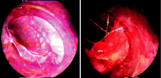
We consider training in RB use crucial to any interventional pulmonologist. Regardless of the type of stent selected for a given treatment, expertise working with the RB will be needed at some point during the course of therapy. For instance, when a complication arises (i.e., migration, stent disruption) and the prosthesis needs to be removed or replaced, the best instrument to retrieve it is the RB. In addition, most of the prostheses placed via FB are very diffcult to remove with fbrobronchoscope, requiring the application of the RB to extract or adjust them. When metallic uncovered stents stay for a given period of time within the airway, they become embedded in the mucosa. In order to remove them, the beveled end of the RB should be placed between the metallic stent wall and the tracheal mucosa, and with soft rotating movements the RB is advanced distally “dissecting” the stent from the airway wall until it is totally detached. Then it can be removed with forceps (Fig. 4.11).
Likewise, the growth of tumor tissue through uncovered metallic stents requires RB and laser to relieve the obstruction, remove the prosthesis, and replace it in case of need. Training in RB is one of the most important skills that an interven-
tionist has to learn and be profcient at, and is a requisite when placing silicone (Dumon) prosthesis [12, 13]. Such training also involves the staff assisting and collaborating during the procedure: assisting nurse or scrub nurse, anesthesiologist, circulating assistant, etc.
Transbronchial Needle Aspiration (TBNA)
TBNA of subcarinal and paratracheal nodules was described in 1950. Wang, in 1978, reported a diagnostic sensibility of 90% for this technique when applied with the RB [14]. After the introduction of the FB during the 1960s, most of the bronchoscopists have been using this instrument to perform TBNA in lymph nodes located subcarinal and parahilar. Diagnostic sensibility for TBNA when performed with the FB has been reported as 80–89%, especially when the 19-gauge needle is used [15, 16].
The appearance of endobronchial ultrasound (EBUS) has completely changed the approach to lymph node sampling, and this technique has virtually replaced all blind procedures given the high diagnostic yield, particularly in mediastinal sampling [17]. However, in spite of EBUS generalized use, it can still be a place for blind TBNA
Fig. 4.11 Metallic prosthesis removal with the rigid bronchoscope
60 |
J. P. Díaz-Jiménez and A. N. Rodríguez |
|
|
applied both with the RB and the FB, particularly where EBUS is not available given its high cost.
A study published in 1996 described results on needle aspiration through the RB. Twenty- four procedures were performed in 24 patients using RB and a 2-cm long rigid needle, under general anesthesia and guided with computerized tomography. Samples taken were: tracheal wall (n = 11), main carina (n = 3), right secondary carina (n = 3), left principal bronchus (n = 2), and right principal bronchus (n = 3). The average amount of samples was 6 (from 1 to 19). An in situ cytopathologist immediately reviewed the samples to determine the number of samples needed. Diagnostic sensibility and specifcity was 88% and 100%, respectively. TBNA was diagnostic in 18 patients. Findings helped in therapeutic decisions in 21 patients. There were no false-positives during a follow-up period of six months. Three false-negatives were present, and follow-up showed that these three patients ultimately had malignant lesions. There were no complications [18]. Those fndings suggest that even though the technique has been improved by using EBUS or blind TBNA with the FB, the RB can have a role in the diagnosis of intrathoracic lymphadenopathies if no other method is available.
Rigid Bronchoscope in Other Treatments for Bronchial Obstruction
Laser treatments in tracheobronchial obstructions are effective, but expensive. As a result, other therapeutic options have been developed and applied with good results. Electrocautery is broadly available and results in airway resections comparable to laser. Also, cryotherapy and argon plasma coagulation can be applied with RB.
Results on electrocautery application with the RB are depicted in a study that performed this procedure under general anesthesia in 29 patients with tracheobronchial obstruction, 24 of which had malignant conditions. In nine patients, stents were placed immediately after electrocoagulation. All patients but one presented immediate improvement in the symptoms, and an objective
improvement in the pulmonary function was also observed in eight patients who had been tested with spirometry before surgery. There were neither intraoperative deaths nor complications [19]. Electrocautery can be also applied through the FB, but, similar to laser applications, procedures are more time consuming since the RB allows better vision, optimal suction, and the possibility to remove large tumoral pieces. Cryotherapy has been presented as an alternative therapy for obstructions. However, it is called a “slow” opening method since it lacks immediate effects. Initially, all treatments with cryotherapy were performed with RB but more recently, the cryotherapy probes have been designed for application with the FB and new modalities of cryotherapy are available, such as cryoextraction or cryoresection and also cryospray, which makes this technique more versatile and can be applied as a fast method to open the airway.
Balloon dilatation can be applied both with the RB and FB.
Mechanical Debridement
Even though laser, electrocautery, cryotherapy, and argon plasma coagulation are useful for coagulating during debridement of airway lesions, most of the obstructive tumors are generally extracted in a mechanical mode. In fact, all opening procedures involve the use of forceps. When performed with an FB, this procedure is invariably long and tedious, especially if large tumors are involved. The removal of big tumor pieces through the narrow channel of FB is very complicated, since the biggest pieces that can be extracted ft in small biopsy forceps. It is obvious that a bigger channel such as the one of the RB will accomplish the same task in a shorter period of time.
Most of the experienced bronchoscopists use laser only to coagulate the tumor and when that is accomplished, dissect large tumoral pieces with the beveled rigid tube (Figs. 4.12 and 4.13), obtaining a much effcient procedure [20]. Grillo et al. [21] affrm that the use of auxiliary methods like laser is not necessary to reopen the airway
Данная книга находится в списке для перевода на русский язык сайта https://meduniver.com/