

- •Foreword
- •Preface
- •Contents
- •About the Editors
- •Contributors
- •1: Tracheobronchial Anatomy
- •Trachea
- •Introduction
- •External Morphology
- •Internal Morphology
- •Mucous Layer
- •Blood Supply
- •Anatomo-Clinical Relationships
- •Bronchi
- •Main Bronchi
- •Bronchial Division
- •Left Main Bronchus (LMB)
- •Right Main Bronchus (RMB)
- •Blood Supply
- •References
- •2: Flexible Bronchoscopy
- •Introduction
- •History
- •Description
- •Indications and Contraindications
- •Absolute Contraindications
- •Procedure Preparation
- •Technique of FB Procedure
- •Complications of FB Procedure
- •Basic Diagnostic Procedures
- •Bronchoalveolar Lavage (BAL)
- •Transbronchial Lung Biopsy (TBLB)
- •Transbronchial Needle Aspiration (TBNA)
- •Bronchial Brushings
- •Advanced Diagnostic Bronchoscopy
- •EBUS-TBNA
- •Ultrathin Bronchoscopy
- •Transbronchial Lung Cryobiobsy (TBLC)
- •Therapeutic Procedures Via FB
- •LASER Bronchoscopy
- •Electrocautery
- •Argon Plasma Coagulation (APC)
- •Cryotherapy
- •Photodynamic Therapy
- •Airway Stent Placement
- •Endobronchial Valve Placement
- •Conclusion
- •References
- •History and Historical Perspective
- •Indications and Contraindications
- •Procedure Description
- •Procedure Planning
- •Target Approximation
- •Sampling
- •Complications
- •Future Directions
- •Summary and Recommendations
- •References
- •4: Rigid Broncoscopy
- •Innovations
- •Ancillary Equipment
- •Rigid Bronchoscopy Applications
- •Laser Bronchoscopy
- •Tracheobronchial Prosthesis
- •Transbronchial Needle Aspiration (TBNA)
- •Rigid Bronchoscope in Other Treatments for Bronchial Obstruction
- •Mechanical Debridement
- •Pediatric Rigid Bronchoscopy
- •Tracheobronchial Dilatation
- •Foreign Bodies Removal
- •Other Indications
- •Complications
- •The Procedure
- •Some Conclusions
- •References
- •History and Historical Perspective
- •Indications and Contraindications
- •Preprocedural Evaluation and Preparation
- •Physical Examination
- •Procedure-Related Indications
- •Application of the Technique
- •Topical Anesthesia
- •Anesthesia of the Nasal Mucosa and Nasopharynx
- •Anesthesia of the Mouth and Oropharynx
- •Superior Laryngeal Nerve Block
- •Recurrent Laryngeal Nerve Block (RLN)
- •Conscious Sedation
- •Monitored Anesthesia Care (MAC)
- •General Anesthesia
- •Monitoring the Depth of Anesthesia
- •Interventional Bronchoscopy Suites
- •Airway Devices
- •Laryngeal Mask Airway (LMA)
- •Endotracheal Tube (ETT)
- •Rigid Bronchoscope
- •Modes of Ventilation
- •Spontaneous Ventilation
- •Assisted Ventilation
- •Noninvasive Positive Pressure Ventilation (NIV)
- •Positive Pressure Controlled Mechanical Ventilation
- •Jet Ventilation
- •Electronic Mechanical Jet Ventilation
- •Postprocedure Care
- •Special Consideration
- •Anesthesia for Peripheral Diagnostic and Therapeutic Bronchoscopy
- •Anesthesia for Interventional Bronchoscopic Procedures During the COVID-19 Pandemic
- •Summary and Recommendations
- •Conclusion
- •References
- •Background
- •Curricular Structure and Delivery
- •What Is a Bronchoscopy Curriculum?
- •Tradition, Teaching Styles, and Beliefs
- •Using Assessment Tools to Guide the Educational Process
- •The Ethics of Teaching
- •When Learners Teach: The Journey from Novice to Mastery and Back Again
- •The Future Is Now
- •References
- •Interventional Procedure
- •Assessment of Flow–Volume Curve
- •Dyspnea
- •Analysis of Pressure–Pressure Curve
- •Conclusions
- •References
- •Introduction
- •Adaptations of the IP Department
- •Environmental Control
- •Personal Protective Equipment
- •Procedure Performance
- •Bronchoscopy in Intubated Patients
- •Other Procedures in IP Unit
- •References
- •Introduction
- •Safety
- •Patient Safety
- •Provider Safety
- •Patient Selection and Screening
- •Lung Cancer Diagnosis and Staging
- •Inpatients
- •COVID-19 Clearance
- •COVID Clearance: A Role for Bronchoscopy
- •Long COVID: A Role for Bronchoscopy
- •Preparing for the Next Pandemic
- •References
- •Historical Perspective
- •Indications and Contraindications
- •Evidence-Based Review
- •Summary and Recommendations
- •References
- •Introduction
- •Clinical Presentation
- •Diagnosis
- •Treatment
- •History and Historical Perspectives
- •Indications and Contraindications
- •Benign and Malignant Tumors
- •Tumors with Uncertain Prognosis
- •Application of the Technique
- •Evidence Based Review
- •Summary and Recommendations
- •References
- •12: Cryotherapy and Cryospray
- •Introduction
- •Historical Perspective
- •Equipment
- •Cryoadhesion
- •Indications
- •Cryorecanalization
- •Cryoadhesion and Foreign Body Removal
- •Cryoadhesion and Mucus Plugs/Blood Clot Retrieval
- •Endobronchial Cryobiopsy
- •Transbronchial Cryobiopsy for Lung Cancer
- •Safety Concerns and Contraindications
- •Cryoablation
- •Indications
- •Evidence
- •Safety Concerns and Contraindications
- •Cryospray
- •Indications
- •Evidence
- •Safety Concerns and Contraindications
- •Advantages of Cryotherapy
- •Limitations
- •Future Research Directions
- •References
- •13: Brachytherapy
- •History and Historical Perspective
- •Indications and Contraindications
- •Application of the Technique
- •Evidence-Based Review
- •Adjuvant Treatment
- •Palliative Treatment
- •Complications
- •Summary and Recommendations
- •References
- •14: Photodynamic Therapy
- •Introduction
- •Photosensitizers
- •First-Generation Photosensitizers
- •M-Tetrahidroxofenil Cloro (mTHPC) (Foscan®)
- •PDT Reaction
- •Tumor Damage Process
- •Procedure
- •Indications
- •Curative PDT Indications
- •Palliative PDT Indications
- •Contraindications
- •Rationale for Use in Early-Stage Lung Cancer
- •Rationale
- •PDT in Combination with Other Techniques for Advanced-Stage Non-small Cell Lung Cancer
- •Commentary
- •Complementary Endoscopic Methods for PDT Applications
- •New Perspectives
- •Other PDT Applications
- •Conclusions
- •References
- •15: Benign Airways Stenosis
- •Etiology
- •Congenital Tracheal Stenosis
- •Iatrogenic
- •Infectious
- •Idiopathic Tracheal Stenosis
- •Distal Bronchial Stenosis
- •Diagnosis Methods
- •Patient History
- •Imaging Techniques
- •Bronchoscopy
- •Pulmonary Function Test
- •Treatment
- •Endoscopic Treatment
- •Dilatation
- •Laser Therapy
- •Stents
- •How to Proceed
- •Stent Placement
- •Placing a Montgomery T Tube
- •The Rule of Twos for Benign Tracheal Stenosis (Fig. 15.23)
- •Surgery
- •Summary and Recommendations
- •References
- •16: Endobronchial Prostheses
- •Introduction
- •Indications
- •Extrinsic Compression
- •Intraluminal Obstruction
- •Stump Fistulas
- •Esophago-respiratory Fistulas (ERF)
- •Expiratory Central Airway Collapse
- •Physiologic Rationale for Airway Stent Insertion
- •Stent Selection Criteria
- •Stent-Related Complications
- •Granulation Tissue
- •Stent Fracture
- •Migration
- •Contraindications
- •Follow-Up and Patient Education
- •References
- •Introduction
- •Overdiagnosis
- •False Positives
- •Radiation
- •Risk of Complications
- •Lung Cancer Screening Around the World
- •Incidental Lung Nodules
- •Management of Lung Nodules
- •References
- •Introduction
- •Minimally Invasive Procedures
- •Mediastinoscopy
- •CT-Guided Transthoracic Biopsy
- •Fluoroscopy-Guided Transthoracic Biopsies
- •US-Guided Transthoracic Biopsy
- •Thoracentesis and Pleural Biopsy
- •Thoracentesis
- •Pleural Biopsy
- •Surgical or Medical Thoracoscopy
- •Image-Guided Pleural Biopsy
- •Closed Pleural Biopsy
- •Image-Guided Biopsies for Extrathoracic Metastases
- •Tissue Acquisition, Handling and Processing
- •Implications of Tissue Acquisition
- •Guideline Recommendations for Tissue Acquisition in Mediastinal Staging
- •Methods to Overcome Challenges in Tissue Acquisition and Genotyping
- •Rapid on-Site Evaluation (ROSE)
- •Sensitive Genotyping Assays
- •Liquid Biopsy
- •Summary, Recommendations and Highlights
- •References
- •History
- •Data Source and Methodology
- •Tumor Size
- •Involvement of the Main Bronchus
- •Atelectasis/Pneumonitis
- •Nodal Staging
- •Proposal for the Revision of Stage Groupings
- •Small Cell Lung Cancer (SCLC)
- •Discussion
- •Methodology
- •T Descriptors
- •N Descriptors
- •M Descriptors
- •Summary
- •References
- •Introduction
- •Historical Perspective
- •Fluoroscopy
- •Radial EBUS Mini Probe (rEBUS)
- •Ultrasound Bronchoscope (EBUS)
- •Virtual Bronchoscopy
- •Trans-Parenchymal Access
- •Cone Beam CT (CBCT)
- •Lung Vision
- •Sampling Instruments
- •Conclusions
- •References
- •History and Historical Perspective
- •Narrow Band Imaging (NBI)
- •Dual Red Imaging (DRI)
- •Endobronchial Ultrasound (EBUS)
- •Optical Coherence Tomography (OCT)
- •Indications and Contraindications
- •Confocal Laser Endomicroscopy and Endocytoscopy
- •Raman Spectrophotometry
- •Application of the Technique
- •Supplemental Technology for Diagnostic Bronchoscopy
- •Evidence-Based Review
- •Summary and Recommendations, Highlight of the Developments During the Last Three Years (2013 on)
- •References
- •Introduction
- •History and Historical Perspective
- •Endoscopic AF-OCT System
- •Preclinical Studies
- •Clinical Studies
- •Lung Cancer
- •Asthma
- •Airway and Lumen Calibration
- •Obstructive Sleep Apnea
- •Future Applications
- •Summary
- •References
- •23: Endobronchial Ultrasound
- •History and Historical Perspective
- •Equipment
- •Technique
- •Indication, Application, and Evidence
- •Convex Probe Ultrasound
- •Equipment
- •Technique
- •Indication, Application, and Evidence
- •CP-EBUS for Malignant Mediastinal or Hilar Adenopathy
- •CP-EBUS for the Staging of Non-small Cell Lung Cancer
- •CP-EBUS for Restaging NSCLC After Neoadjuvant Chemotherapy
- •Complications
- •Summary
- •References
- •Introduction
- •What Is Electromagnetic Navigation?
- •SuperDimension Navigation System (EMN-SD)
- •Computerized Tomography
- •Computer Interphase
- •The Edge Catheter: Extended Working Channel (EWC)
- •Procedural Steps
- •Planning
- •Detecting Anatomical Landmarks
- •Pathway Planning
- •Saving the Plan and Exiting
- •Registration
- •Real-Time Navigation
- •SPiN System Veran Medical Technologies (EMN-VM)
- •Procedure
- •Planning
- •Navigation
- •Biopsy
- •Complications
- •Limitations
- •Summary
- •References
- •Introduction
- •Image Acquisition
- •Hardware
- •Practical Considerations
- •Radiation Dose
- •Mobile CT Studies
- •Future Directions
- •Conclusion
- •References
- •26: Robotic Assisted Bronchoscopy
- •Historical Perspective
- •Evidence-Based Review
- •Diagnostic Yield
- •Monarch RAB
- •Ion Endoluminal Robotic System
- •Summary
- •References
- •History and Historical Perspective
- •Indications and Contraindications
- •General
- •Application of the Technique
- •Preoperative Care
- •Patient’s Position and Operative Field
- •Incision and Initial Dissection
- •Palpation
- •Biopsy
- •Control of Haemostasis and Closure
- •Postoperative Care
- •Complications
- •Technical Variants
- •Extended Cervical Mediastinoscopy
- •Mediastinoscopic Biopsy of Scalene Lymph Nodes
- •Inferior Mediastinoscopy
- •Mediastino-Thoracoscopy
- •Video-Assisted Mediastinoscopic Lymphadenectomy
- •Transcervical Extended Mediastinal Lymphadenectomy
- •Evidence-Based Review
- •Summary and Recommendations
- •References
- •Introduction
- •Case 1
- •Adrenal and Hepatic Metastases
- •Brain
- •Bone
- •Case 1 Continued
- •Biomarkers
- •Case 1 Concluded
- •Case 2
- •Chest X-Ray
- •Computerized Tomography
- •Positive Emission Tomography
- •Magnetic Resonance Imaging
- •Endobronchial Ultrasound with Transbronchial Needle Aspiration
- •Transthoracic Needle Aspiration
- •Transbronchial Needle Aspiration
- •Endoscopic Ultrasound with Needle Aspiration
- •Combined EUS-FNA and EBUS-TBNA
- •Case 2 Concluded
- •Case 3
- •Standard Cervical Mediastinoscopy
- •Extended Cervical Mediastinoscopy
- •Anterior Mediastinoscopy
- •Video-Assisted Thoracic Surgery
- •Case 3 Concluded
- •Case 4
- •Summary
- •References
- •29: Pleural Anatomy
- •Pleural Embryonic Development
- •Pleural Histology
- •Cytological Characteristics
- •Mesothelial Cells Functions
- •Pleural Space Defense Mechanism
- •Pleura Macroscopic Anatomy
- •Visceral Pleura (Pleura Visceralis or Pulmonalis)
- •Parietal Pleura (Pleura Parietalis)
- •Costal Parietal Pleura (Costalis)
- •Pleural Cavity (Cavitas Thoracis)
- •Pleural Apex or Superior Pleural Sinus [12–15]
- •Anterior Costal-Phrenic Sinus or Cardio-Phrenic Sinus
- •Posterior Costal-Phrenic Sinus
- •Cost-Diaphragmatic Sinus or Lateral Cost-Phrenic Sinus
- •Fissures18
- •Pleural Vascularization
- •Parietal Pleura Lymphatic Drainage
- •Visceral Pleura Lymphatic Drainage
- •Pleural Innervation
- •References
- •30: Chest Ultrasound
- •Introduction
- •The Technique
- •The Normal Thorax
- •Chest Wall Pathology
- •Pleural Pathology
- •Pleural Thickening
- •Pneumothorax
- •Pulmonary Pathology
- •Extrathoracic Lymph Nodes
- •COVID and Chest Ultrasound
- •Conclusions
- •References
- •Introduction
- •History of Chest Tubes
- •Overview of Chest Tubes
- •Contraindications for Chest Tube Placement
- •Chest Tube Procedural Technique
- •Special Considerations
- •Pneumothorax
- •Empyema
- •Hemothorax
- •Chest Tube Size Considerations
- •Pleural Drainage Systems
- •History of and Introduction to Indwelling Pleural Catheters
- •Indications and Contraindications for IPC Placement
- •Special Considerations
- •Non-expandable Lung
- •Chylothorax
- •Pleurodesis
- •Follow-Up and IPC Removal
- •IPC-Related Complications and Management
- •Competency and Training
- •Summary
- •References
- •32: Empyema Thoracis
- •Historical Perspectives
- •Incidence
- •Epidemiology
- •Pathogenesis
- •Clinical Presentation
- •Radiologic Evaluation
- •Biochemical Analysis
- •Microbiology
- •Non-operative Management
- •Prognostication
- •Surgical Management
- •Survivorship
- •Summary and Recommendations
- •References
- •Evaluation
- •Initial Intervention
- •Pleural Interventions for Recurrent Symptomatic MPE
- •Especial Circumstances
- •References
- •34: Medical Thoracoscopy
- •Introduction
- •Diagnostic Indications for Medical Thoracoscopy
- •Lung Cancer
- •Mesothelioma
- •Other Tumors
- •Tuberculosis
- •Therapeutic Indications
- •Pleurodesis of Pneumothorax
- •Thoracoscopic Drainage
- •Drug Delivery
- •Procedural Safety and Contraindications
- •Equipment
- •Procedure
- •Pre-procedural Preparations and Considerations
- •Procedural Technique [32]
- •Medical Thoracoscopy Versus VATS
- •Conclusion
- •References
- •Historical Perspective
- •Indications and Contraindications
- •Evidence-Based Review
- •Endobronchial Valves
- •Airway Bypass Tracts
- •Coils
- •Other Methods of ELVR
- •Summary and Recommendations
- •References
- •36: Bronchial Thermoplasty
- •Introduction
- •Mechanism of Action
- •Trials
- •Long Term: Ten-Year Study
- •Patient Selection
- •Bronchial Thermoplasty Procedure
- •Equipment
- •Pre-procedure
- •Bronchoscopy
- •Post-procedure
- •Conclusion
- •References
- •Introduction
- •Bronchoalveolar Lavage (BAL)
- •Technical Aspects of BAL Procedure
- •ILD Cell Patterns and Diagnosis from BAL
- •Technical Advises for Conventional TLB and TLB-C in ILD
- •Future Directions
- •References
- •Introduction
- •The Pediatric Airway
- •Advanced Diagnostic Procedures
- •Endobronchial Ultrasound
- •Virtual Navigational Bronchoscopy
- •Cryobiopsy
- •Therapeutic Procedures
- •Dilation Procedures
- •Thermal Techniques
- •Mechanical Debridement
- •Endobronchial Airway Stents
- •Metallic Stents
- •Silastic Stents
- •Novel Stents
- •Endobronchial Valves
- •Bronchial Thermoplasty
- •Discussion
- •References
- •Introduction
- •Etiology
- •Congenital ADF
- •Malignant ADF
- •Cancer Treatment-Related ADF
- •Benign ADF
- •Iatrogenic ADF
- •Diagnosis
- •Treatment Options
- •Endoscopic Techniques
- •Stents
- •Clinical Results
- •Stent Complications
- •Other Available Stents
- •Other Endoscopic Methods
- •References
- •Introduction
- •Anatomy and Physiology of Swallowing
- •Functional Physiology of Swallowing
- •Epidemiology and Risk Factors
- •Types of Foreign Bodies
- •Organic
- •Inorganic
- •Mineral
- •Miscellaneous
- •Clinical Presentation
- •Acute FB
- •Retained FB
- •Radiologic Findings
- •Bronchoscopy
- •Airway Management
- •Rigid Vs. Flexible Bronchoscopy
- •Retrieval Procedure
- •Instruments
- •Grasping Forceps
- •Baskets
- •Balloons
- •Suction Instruments
- •Ablative Therapies
- •Cryotherapy
- •Laser Therapy
- •Electrocautery and APC
- •Surgical Management
- •Complications
- •Bleeding and Hemoptysis
- •Distal Airway Impaction
- •Iron Pill Aspiration
- •Follow-Up and Sequelae
- •Conclusion
- •References
- •Vascular Origin of Hemoptysis
- •History and Historical Perspective
- •Diagnostic Bronchoscopy
- •Therapeutic Bronchoscopy
- •General Measures
- •Therapeutic Bronchoscopy
- •Evidence-Based Review
- •Summary
- •Recommendations
- •References
- •History
- •“The Glottiscope” (1807)
- •“The Esophagoscope” (1895)
- •The Rigid Bronchoscope (1897–)
- •The Flexible Bronchoscope (1968–)
- •Transbronchial Lung Biopsy (1972) (Fig. 42.7)
- •Laser Therapy (1981–)
- •Endobronchial Stents (1990–)
- •Electromagnetic Navigation (2003–)
- •Bronchial Thermoplasty (2006–)
- •Endobronchial Microwave Therapy (2004–)
- •American Association for Bronchology and Interventional Pulmonology (AABIP) and Journal of Bronchology and Interventional Pulmonology (JOBIP) (1992–)
- •References
- •Index

Pleural Anatomy |
29 |
|
|
Juan Antonio Moya Amorós |
|
Pleural Embryonic Development
Once the embryonic disc has been formed in the trilaminar phase, which contains three germ layers, namely, ectoderm, mesoderm, and endoderm, the embryo will experience morphological changes following a basic body structuring plan, based on:
\(a)\ Craniocaudal differentiation gradient with the formation of the notochord, and the neural plate.
\(b)\ Body regular and transversal segmentation pattern, which evokes the evolutionary phases of the phylogenetic past ( sh, reptiles, etc.).
\(c)\ Body lateral folding, approaching the midline, converting its fat, trilaminar shape into a cylindrical structure with the ectoderm on the outside, the endoderm on the inside, and the mesoderm between the two.
It is precisely from the mesoderm that the pleura derives, among other embryo intermediate structures, forming a serous membrane that along with the peritoneum, internally covers the future coelomic cavity (parietal serosa), as well as
J. A. Moya Amorós (*)
Thoracic Surgery Department, Bellvitge University Hospital, Barcelona, Spain
e-mail: juan.moya@bellvitgehospital.cat
externally covers the organs and viscera. contained inside that cavity (visceral serosa).
The rst pleural vestige appears in the 22–23- day embryo, when it has already acquired three blastoderm sheets: ectoderm, mesoderm, and endoderm [1]. From this moment on, in the fat embryo thickness, the mesoderm (hereinafter mesenchyme) in turn undergoes fragmentation changes in three territories:
\1.\ Medial territory or chordamesoderm, from which the notochord will derive.
\2.\ Two lateral masses or lateral mesoderms, on each side of the embryo, from which they derive:
\(a)\ The somites, precursors of muscles, and bones.
\(b)\ The gononephrotome, precursor of the kidney, and the gonads.
\(c)\ The somatic mesoderm or somatopleura (future parietal pleura), and the splanchnic mesoderm or splanchnopleura (visceral pleura precursor) (Fig. 29.1).
The somatopleura is con gured by the somatic mesoderm fusion with the ectoderm internal part, so that the “coalescence” of both structures will end up forming the parietal pleura, which will internally cover the ribs and the intercostal space elements (costal pleura), with which it is kept separated by the endothoracic fascia.
© The Author(s), under exclusive license to Springer Nature Switzerland AG 2023 |
507 |
J. P. Díaz-Jiménez, A. N. Rodríguez (eds.), Interventions in Pulmonary Medicine, https://doi.org/10.1007/978-3-031-22610-6_29
508 |
J. A. Moya Amorós |
|
|
The splanchnopleura, however, is produced by the fusion of the splanchnic mesoderm with the endoderm external part, so that the serosa resulting from this fusion will be the visceral pleura that will intimately surround the lung mesenchyme (lung parenchyma).
Approximately at the fourth week [2], the embryo has already evolved from having a fat shape to progressively acquiring a tubular-
|
|
|
2 |
1 |
|
|
4 |
|
|
|
|
|
|
|
|
5 |
|
3 |
|
|
|
|
|
|
6 |
7 |
|
8 |
|
|
|
|
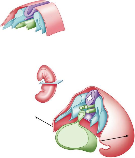
Fig. 29.1 Diagram of a 23-day embryo with 8 somites, at the neural canal stage in the lateral and ventral folding phas. (1) Ectoderm, (2) Neural canal, (3) Notochord, (4) Somite, (5) Gononephrotome, (6) Somatopleura, (7) Splacnopleura, (8) Endoderm
cylindrical shape due to a folding process of its lateral walls, both in the cranial-caudal and lateral-medial directions. The cause still does not have a coherent explanation, although as a consequence of it, there is an approximation and fusion in all the embryonic leaves midline, except at the point where the omphalo-mesenteric duct or future umbilical cord emerges.
From this development moment, the embryo will have a closed body or trunk with a single cavity inside or coelomic cavity, which contains the future viscera (Fig. 29.2).
In parallel, embryonic changes have also been taking place at the level of the endoderm foregut anterior face. Immediately below the III to IV pharyngeal pouch, an endodermal cells outgrowth appears in the form of a laryngo-tracheo-bronchial bud [3] that progressively and after 24 divisions comes to constitute both adult lungs tracheobronchial tree. In this sense, the lung can be considered as an enormous gland that, from the endodermal duct, invaginates towards the mesenchyme depth, maintaining communication with the outside through the embryo primitive mouth (stomodeum).
Fig. 29.2 Embryo of 25 days with 8–10 somites in the initial stage of tubular closure.
(1)Closed neural tube,
(2)Notochord, (3) Dorsal aorta, (4) Endoderm connected toon (5) Extraembryonic coelom (yolk vesicle),
(6)Somite, (7) Somatopleura, (8) Splacnopleura
1
6
3 |
2 |
|
|
4 |
|
7
8
5
Данная книга находится в списке для перевода на русский язык сайта https://meduniver.com/
29 Pleural Anatomy |
|
|
|
|
|
|
|
509 |
||
|
|
|
|
|
|
|
|
|
|
|
Fig. 29.3 A 28-day |
|
|
|
|
|
|
|
|
|
|
embryo with 12 somites, |
|
|
|
|
|
|
|
|
|
|
in the single cavitation |
|
|
|
|
|
|
|
|
|
|
stage of the coelom. (1) |
1 |
4 |
2 |
2 |
1 |
|||||
Ectoderm, (2) Neural |
||||||||||
|
|
|
|
|
|
|
|
|
||
tube, (3) Notochord, (4) |
|
|
|
|
|
|
|
|
|
|
Aorta, (5) Somite, (6) |
|
|
|
|
|
|
|
|
|
|
Somatopleura, (7) |
|
|
|
|
|
|
|
|
|
|
Splacnopleura, (8) |
|
|
|
|
|
|
|
|
|
|
Endoderm, (9) |
|
|
|
|
|
|
|
|
|
|
Omphalo-mesenteric |
|
|
|
|
|
|
|
|
|
|
duct, (10) Caudal |
|
|
|
5 |
|
|
|
|
|
|
Tubercle, (11) Left |
|
|
|
|
|
|
|
|
||
|
|
|
|
|
|
|
|
|
||
caudal appendage |
|
|
|
|
|
|
|
|
|
|
|
|
|
|
|
|
|
|
|
||
(outline of the lower |
|
|
|
|
|
|
|
|
|
|
limb) |
|
|
|
|
8 |
|
|
|
|
|
|
6 |
|
|
|
|
|
|
|
|
|
|
|
|
|
|
|
|
|
|
||
|
7 |
|
|
|
|
|
|
|
|
|
11
9
10
Between the fourth and ninth weeks, inside the coelomic cavity, a new transverse septation process is outlined at the expense of a mesoderm septum internal growth (transverse septum), which will form the future diaphragm. As a consequence of this septation the coelomic cavity, (until now the only one) it is divided into two interconnected cavities, one cranial or thoracic and caudal or abdominal.
Parallel to these embryonic movements, in the paraxial regions inside the thoracic cavity, some cardinal folds emerge that protrude towards the cavity interior, pushing the somatopleura [4] in a dorso-ventral and cranio-caudal direction, in
relation to vascular structures formation contained within. As a consequence of these folds progressive growth, the de nitive septation of the thoracic cavity results in three cavities: a central one or pericardial cavity and two lateral ones or pleural cavities [4, 5] (Fig. 29.3).
Pleural Molecular Biology During
Embryogenesis
Since 1990, molecular biology techniques application has made it possible to understand the correlation between the fundamental embryologic
510 |
J. A. Moya Amorós |
|
|
morphological changes, and the molecular aspects involved in genes maintenance and conservation that direct or guide the body’s normal development.
Currently, molecular families that direct embryonic development are already recognized, and in this sense, genetic sequencing studies have shown a phylogenetic conservatism from the rudimentary species of worms to humans, so that there are very few changes in the developmental regulatory genes nucleotide bases. It is also known that the same gene can express different functions in the different ontogenesis phases, and can even act in different organs.
There is the possibility that the same speci c gene acts even differently both in the embryonic phase and after birth. To all this molecular complexity is added the fact that mutated genes presence can induce changes even to the point of converting a normal cell into tumoral cells (proto oncogenes). The fundamental molecular processes during this body structuring period are grouped into categories that act as:
\1.\ Transcription factors, which are proteins with domains that bind speci c genes DNA, or even act on RNA polymerase II and, consequently, regulate the amount of RNA- messenger that the gene produces. Speci cally in humans the 38 homologous genes are called Hox genes, POU genes, and Pax genes.
They belong to this group that are collectively called Homeobox:
•\ Basic protein helix-loop-helix. •\ Zinc nger proteins.
•\ Homeodomain proteins.
\2.\ Activation or signal factors, most of which are proteins that act as peptide growth factors. The rst one obtained in the 1950s was neural growth factor, later on TGF-β (transforming growth factor β), and FGF ( broblast growth factor), involved in mesenchymal cells broblastic proliferation capacity. Another important family of activation molecules are the
hedgehog proteins, of which the sonic hedgehog is the most peculiar in that it undergoes autoproteolysis that allows obtaining a peptide capable of stimulating the target cell to directly or indirectly produce new differentiation pathways.
Speci cally, the pleura, as a mesoderm derivative, is constituted as a morphogenetic eld that remains at the mercy of molecular signals infuenced from the ectoderm, neural tube, and notochord (Speman’s induction theory, 1938) [5]. These molecular signals cause very curious but necessary events such as the transformation of mesenchymal cells into epithelial cells, and vice versa.
Once the epithelial somites have formed, their ventromedial cells undergo the inductive stimulus of activation by sonic hedgehog [6] (which originates in the notochord and neural tube). The response to this induction is Pax-1 and Pax-91 expression inside the somite, and as a nal consequence, there is a cell adhesion molecule loss, speci cally N-cadherin6, which again favors the “transformation of epithelial cells into mesenchymal cells,” thus acquiring the ability to migrate towards the embryo midline after forming the secondary mesoderm.
Contrary to these events, the somite is exposed to the infuence of products secreted by neural tube Wnt gene [6], which establish a sonic hedgehog inhibitory action, and the somatic cells remain under Pax-3, Pax-7 and paraxis expression, obtaining dermatome and myotome morphogenesis.
On the other hand, under BMP-4 infuence from the ectoderm [6], lateral mesoderm cells also begin to produce it. This molecule has the ability to infuence the paraxial or lateral mesoderm to assume lateral mesoderm properties. All these biological facts together allow to establish a balance between the medializing forces coming from the neural tube and the notochord, against the lateralizing forces coming from the ectoderm (Fig. 29.4).
Данная книга находится в списке для перевода на русский язык сайта https://meduniver.com/