


54 |
Chapter 6 |
C.Course of the Lateral Corticospinal Tract
The lateral corticospinal tract runs in the posterior limb of the internal capsule to enter the middle three-fifths of the crus cerebri of the midbrain, through the basilar pons and into the medullary pyramids. Between 85% and 90% of the corticospinal fibers cross in the pyramidal decussation as the lateral corticospinal tract, which is found in the posterior aspect of the lateral funiculus. The remaining 10% to 15% of the fibers continue as the anterior corticospinal tract. The fibers of the anterior corticospinal tract decussate in the spinal cord at the level of innervation of the lower motor neurons of the anterior horn that they innervate.
D.Transection of the Lateral Corticospinal Tract
1.Superior to the motor decussation, transection results in contralateral spastic paresis and Babinski sign (upward fanning of the toes).
2.In the spinal cord, transection results in ipsilateral spastic paresis and Babinski sign.
CASE 6-2
A 17-year-old man complained of pain on the left side of his chest and progressive weakness of his left lower limb for 2 months before coming to the clinic. What is the most likely diagnosis?
Relevant Physical Exam Findings
●Neurologic evaluation revealed weakness in the left lower limb; spasticity and hyperreflexia at the knee and ankle were also observed.
●On the left side, a loss of two-point discrimination, vibratory sense, and conscious proprioception below the hip was observed. A loss of pain and temperature sensation below the T7 dermatome was observed on the right side.
Diagnosis
●Brown-Séquard syndrome, resulting from an upper motor neuron lesion at T5-T6 spinal cord levels, represents an incomplete spinal cord lesion characterized by symptoms indicative of hemisection of the spinal cord. It involves ipsilateral hemiplegia with contralateral pain and temperature deficits.
IDiseases of the Motor Neurons and Corticospinal
Tracts (Figures 6-9 and 6-10)
A.Upper Motor Neuron (UMN) Lesions are caused by lesions of the corticospinal tract or destruction of the cortical cells of origin. UMN lesions result in spastic paresis with pyramidal signs (Babinski sign) and hyperreflexia.
B.Lower Motor Neuron (LMN) Lesions are caused by damage to the motor neurons. They result in flaccid paralysis, hyporeflexia, atrophy, fasciculations, and fibrillations. Poliomyelitis or Werdnig–Hoffmann disease (Figure 6-10A) results from damage to motor neurons.
C.An Example of a Combined UMN and LMN Disease is Amyotrophic Lateral Sclerosis (ALS, or Lou Gehrig Disease) (Figure 6-10D). ALS is caused by damage to the corticospinal tracts, with pyramidal signs, and by damage to the LMNs, with LMN symptoms. Patients with ALS have no sensory deficits.

|
|
Spinal Cord |
55 |
|
Fasciculus gracilis |
Ipsilateral loss of fine touch, conscious |
|
|
proprioception, and vibratory sense |
|
|
|
|
from lower limb |
|
Fasciculus cuneatus |
Loss of of fine touch, conscious |
|
|
|
|
proprioception, and vibratory sense |
|
Posterior |
|
from upper limb |
|
|
Ipsilateral segmental anesthesia |
|
|
spinocerebellar |
|
||
and areflexia |
|
||
tract |
|
|
|
|
Ipsilateral lower limb |
|
|
|
|
|
|
Lat. |
Posterior |
dystaxia |
|
corticospinal |
horn |
Ipsilateral spastic |
|
tract |
|
paresis with |
|
|
|
pyramidal signs |
|
Lat. |
|
Contralateral lower |
|
spinothalamic |
|
|
|
Anterior |
limb dystaxia |
|
|
tract |
|
||
horn |
|
|
|
|
Contralateral loss of |
|
|
Anterior |
|
|
|
|
pain and temperature |
|
|
spinocerebellar |
|
sensation one segment |
|
tract |
|
below lesion |
|
|
|
Ipsilateral flaccid paralysis |
|
|
|
in affected myotomes |
|
Anterior white commissure |
Bilateral loss of pain and temperature |
|
|
|
|
sensation within dermatomes |
|
|
|
of involved segments |
|
Anterior corticospinal tract |
Mild contralateral muscle weakness |
|
|
in proximal muscles |
|
||
Figure 6-9 Transverse section of the cervical spinal cord. The clinically important ascending and descending pathways are shown on the left. Clinical deficits that result from the interruption of these pathways are shown on the right.
II |
Sensory Pathway Lesions. An example of a condition caused by these |
|
lesions is posterior column disease (tabes dorsalis) (Figure 6-10C). This disease is seen in |
|
patients with neurosyphilis. It is characterized by a loss of fine touch, conscious proprioception, |
|
and vibratory sense. Irritative involvement of the posterior roots results in pain and paresthesias. |
|
Patients have a Romberg sign. (Subject stands with feet together and, when their eyes are closed, |
|
loses balance. This is a sign of posterior column ataxia.) |
IIICombined Motor and Sensory Lesions
A.Spinal Cord Hemisection (Brown-Séquard Syndrome) (Figure 6-10E) is caused by damage to the following structures:
1.Posterior columns (gracile [leg] and cuneate [arm] fasciculi). Damage results in ipsilateral loss of fine touch, conscious proprioception, and vibratory sense.
2.Lateral corticospinal tract. Damage results in ipsilateral spastic paresis with pyramidal signs inferior to the lesion.
3.Lateral spinothalamic tract. Damage results in contralateral loss of pain and temperature sensation one to two segments inferior to the lesion.
4.Hypothalamospinal tract. Damage results in ipsilateral Horner syndrome (i.e., miosis, ptosis, hemianhidrosis, and apparent enophthalmos).
5.Anterior horn. Damage results in ipsilateral flaccid paralysis.
B.Anterior Spinal Artery Occlusion (Figure 6-10F) causes infarction of the anterior twothirds of the spinal cord but spares the posterior columns and horns. It results in damage to the following structures:
56 |
Chapter 6 |
A B
C D
E F
G H
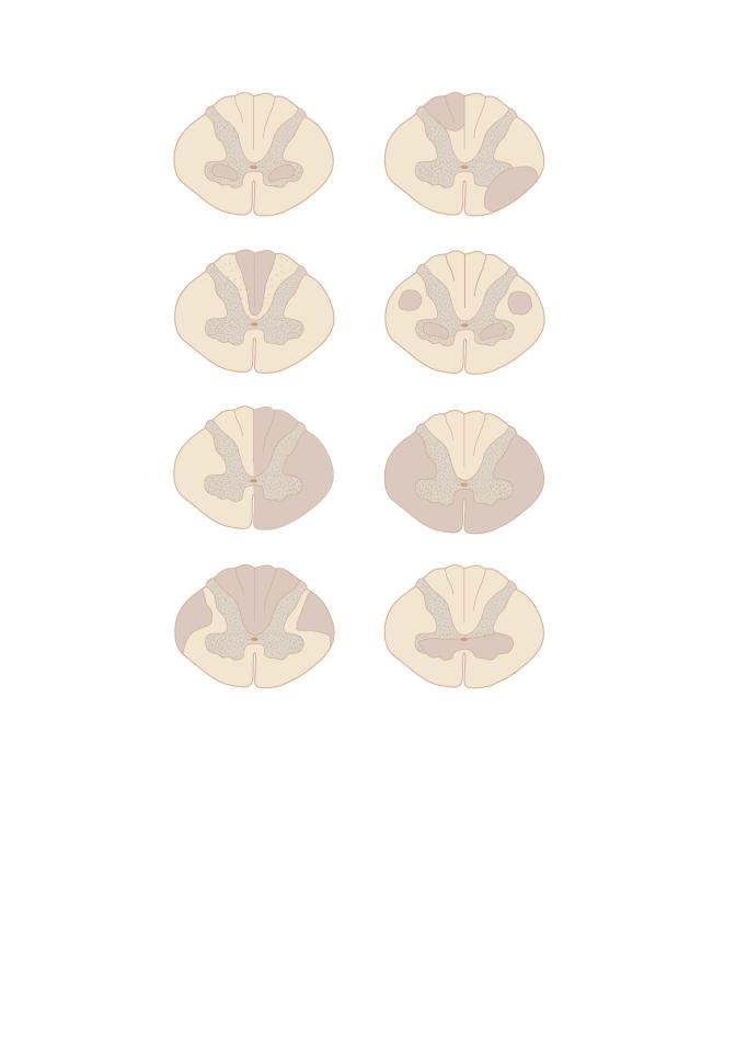
Figure 6-10 Classic lesions of the spinal cord. A. Poliomyelitis and progressive infantile muscular atrophy (Werdnig– Hoffmann disease). B. Multiple sclerosis. C. Posterior column disease (tabes dorsalis). D. Amyotrophic lateral sclerosis. E. Hemisection of the spinal cord (Brown-Séquard syndrome). F. Complete anterior spinal artery occlusion. G. Subacute combined degeneration (vitamin B12 neuropathy). H. Syringomyelia.
1.Lateral corticospinal tracts. Damage results in bilateral spastic paresis with pyramidal signs inferior to the lesion.
2.Lateral spinothalamic tracts. Damage results in bilateral loss of pain and temperature sensation inferior to the lesion.
3.Hypothalamospinal tract. Damage results in bilateral Horner syndrome.
4.Anterior horns. Damage results in bilateral flaccid paralysis.
5.Corticospinal tracts to the sacral parasympathetic centers at S2-S4. Damage results in loss of voluntary bladder and bowel control.
C.Subacute Combined Degeneration (Vitamin B12 Neuropathy) (Figure 6-10G) is caused by pernicious (megaloblastic) anemia. It results from damage to the following structures:
1.Posterior columns (gracile and cuneate fasciculi). Damage results in bilateral loss of fine touch, conscious proprioception, and vibratory sense.
Spinal Cord |
57 |
2.Lateral corticospinal tracts. Damage results in bilateral spastic paresis with pyramidal signs.
3.Spinocerebellar tracts. Damage results in bilateral arm and leg dystaxia.
D.Syringomyelia (Figure 6-10H) is a central cavitation of the spinal cord, resulting from a congenital defect, trauma, hemorrhage, or infection. Expansion of the syrinx may result in damage to nearby structures, including:
1.Anterior white commissure. Damage to decussating lateral spinothalamic axons causes bilateral loss of pain and temperature sensation.
2.Anterior horns. LMN lesions result in flaccid paralysis, hyperreflexia and wasting of the affected musculature.
E.Friedreich Ataxia—has the same spinal cord pathology and symptoms as subacute combined degeneration.
F.Multiple Sclerosis (Figure 6-10B), demyelination primarily involves the white matter of the spinal cord. Damage is random and asymmetric.
IV Peripheral Nervous System (PNS) Lesions. An example of
a PNS lesion is Guillain–Barré syndrome (acute idiopathic polyneuritis, or postinfectious polyneuritis). It primarily affects the motor fibers of the anterior roots and peripheral nerves, and it produces LMN symptoms (i.e., muscle weakness, flaccid paralysis, and hyporeflexia). Guillain–Barré syndrome has the following features:
A.Demyelination and edema.
B.Upper cervical root (C4) involvement and respiratory paralysis are common.
C.Caudal cranial nerve involvement with facial diplegia is present in 50% of cases.
D.Elevated protein levels may cause papilledema.
E.Sensory fibers may be affected, resulting in paresthesias.
F.The protein level in the cerebrospinal fluid is elevated but without pleocytosis (albuminocytologic dissociation).
VIntervertebral Disk Herniation is seen at the L4 to L5 or L5 to S1
interspace in 90% of cases. It appears at the C5 to C6 or C6 to C7 interspace in 10% of cases.
A.Intervertebral disk herniation consists of prolapse, or herniation, of the nucleus pulposus through the defective anulus fibrosus and into the vertebral canal.
B.The nucleus pulposus impinges on the spinal roots, resulting in spinal root symptoms (i.e., paresthesias, pain, sensory loss, hyporeflexia, and muscle weakness).
VI Cauda Equina Syndrome (Spinal Roots L3 to C0) may
result from a nerve root tumor, an ependymoma, a dermoid tumor, or from a lipoma of the terminal spinal cord. It is characterized by:
A.Severe radicular unilateral pain.
B.Sensory distribution in a unilateral saddle-shaped area.