


Blood Supply |
29 |
VI Venous Dural Sinuses
A.Superior Sagittal Sinus receives blood from the bridging veins and emissary veins (a potential route for transmission of extracranial infection into the brain). The superior sagittal sinus also receives cerebrospinal fluid (CSF) through the arachnoid villi.
B.Cavernous Sinus contains CNs III, IV, V1 and V2, and VI and the postganglionic sympathetic fibers. It also contains the siphon of the internal carotid artery (Figure 4-4).
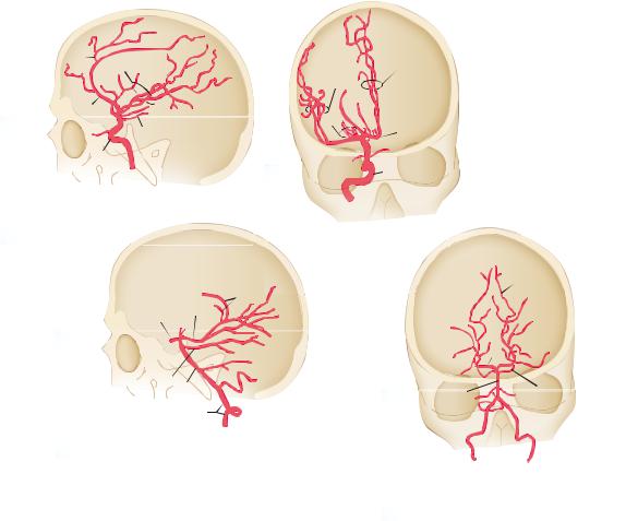
VII Angiography
A.Carotid Angiography. Figure 4-5A,B shows the internal carotid artery, anterior cerebral artery, and middle cerebral artery.
B.Vertebral Angiography. Figure 4-5C,D shows the vertebral artery, PICA and AICA, basilar artery, superior cerebellar artery, and posterior cerebral artery (Figures 4-6 and 4-7).
C.Veins and Dural Sinuses. Figure 4-7 shows the internal cerebral vein, superior cerebral veins, great cerebral vein, superior ophthalmic vein, and major dural sinuses.
D.Digital Subtraction Angiography. See Figures 4-8 to 4-11.
VIII The Middle Meningeal Artery, a branch of the maxillary artery,
enters the cranium through the foramen spinosum. It supplies most of the dura, including its calvarial portion. Laceration results in epidural hemorrhage (hematoma) (Figures 4-12 and
4-13). A classic “lucid interval” is seen in 50% of cases.
30 |
Chapter 4 |
|
|
|
A |
|
12 |
B |
|
|
6 |
|
|
|
|
10 |
|
|
7 |
|
|
|
10 2 |
|
|
1 |
|
15 |
|
|
2 |
|
|
|
|
|
9 |
1 |
|
|
11 |
15 |
|
|
|
|
|
|
|
|
8 |
|
11 |
|
|
|
|
8 |
|
|
|
|
|
C
14
13
15
5
|
3 |
17 |
16 |
|
4 |
|
18 |
List of structures:
1.Anterior cerebral artery
2.Anterior choroidal artery
3.Anterior inferior cerebellar artery
4.Basilar artery
5.Calcarine artery
6Callosomarginal artery
7.Callosmarginal and pericallosal arteries (of anterior cerebral artery)
8.Internal carotid artery
9.Lateral striate arteries
D 
5
|
13 |
|
4 |
3 |
17 |
|
16
18
10.Middle cerebral artery
11.Ophthalmic artery
12.Pericallosal artery
13.Posterior cerebral artery
14.Posterior choridal arteries
15.Posterior communicating artery
16.Posterior inferior cerebellar artery
17.Superior cerebellar artery
18.Vertebral artery
Figure 4-5 A. Carotid angiogram, lateral projection. B. Carotid angiogram, anteroposterior projection. C. Vertebral angiogram, lateral projection. D. Vertebral angiogram, anteroposterior projection.

Blood Supply |
31 |
B
A C
Figure 4-6 Arterial anatomy of a magnetic resonance section; axial (A), sagittal (B, C). ACAcm, anterior cerebral artery, callosal marginal branch; A2 and A1, branches of the anterior cerebral artery; ACApc, pericallosal branch of the anterior cerebral artery; ACoA, anterior communicating artery; AICA, anterior inferior cerebellar artery; M1 and M2, segments of the middle cerebral artery (MCA); MCAb, bifurcation; ICAs, internal carotid artery siphon; ICAc, internal carotid artery cavernous; PCA, posterior cerebral artery; PCoA, posterior communicating artery; BA, basilar artery; SCA, superior cerebellar artery. (Reprinted from Grossman CB, Magnetic Resonance Imaging and Computed Tomography of the Head and Spine. 2nd ed. Philadelphia, PA: Williams & Wilkins; 1996:124, with permission.)
Inferior sagittal sinus
Superior ophthalmic vein
Cavernous sinus 
Internal cerebral vein
Superior sagittal sinus
Superior cerebral veins (bridging veins)
Great cerebral vein (of Galen)
 Straight sinus
 Confluence of sinuses
 Transverse sinus
 Sigmoid sinus
Figure 4-7 Carotid angiogram, venous phase, showing the cerebral veins and venous sinuses.

32 |
Chapter 4 |
Callosomarginal artery of ACA
Pericallosal artery of ACA
Frontopolar branch |
|
MCA M1 segment |
of ACA |
|
|
|
||
|
|
Ophthalmic artery |
|
|
|
PCoM |
|
|
|
||
|
|
|
|
Cavernous ICA
Petrous ICA
Cervical ICA
Figure 4-8 Carotid angiogram, lateral projection. Identify the cortical branches of the anterior cerebral artery (ACA) and middle cerebral artery (MCA). Follow the course of the internal carotid artery (ICA). Remember that aneurysms of the posterior communicating artery (PCoM) may result in third-nerve palsy. The paracentral lobule is irrigated by the callosomarginal artery.
ACA |
|
|
Cortical branches of MCA |
|
|||
A1 segment of ACA |
|
|
Lateral striate branches |
|
|
|
|
|
|
|
of MCA |
ACoM |
|
|
M1 segment of MCA |
|
|
||
|
|
|
|
Cavernous part of ICA |
|
|
Supraclinoid part of ICA |
|
|
||
|
|
|
|
|
|
|
Petrous part of ICA |
|
|
|
Cervical part of ICA |
Figure 4-9 Carotid angiogram, anteroposterior projection. Identify the anterior cerebral artery (ACA), middle cerebral artery (MCA), and internal carotid artery (ICA). The horizontal branches of the MCA perfuse the basal nuclei and internal capsule. ACoM, anterior communicating artery.

Blood Supply |
33 |
Posterior choroidal arteries
Parieto-occipital branches of PCA
PCA, P1 segment
 Calcarine branches
of PCA
Thalamoperforating
arteries
Hemispheric branches
PCoM |
|
of SCA |
|
Superior cerebellar artery
PICA
Basilar artery
Vertebral artery
Vertebral artery
Figure 4-10 Vertebral angiogram, lateral projection. Two structures are found between the posterior cerebral artery (PCA) and the superior cerebellar artery (SCA): the tentorium and the third cranial nerve. PCoM, posterior communicating artery; PICA, posterior inferior cerebellar artery.
Calcarine artery of PCA
PCA
Temporal branches
of PCA
Superior cerebellar artery
Basilar artery
PICA 
Vertebral artery
Figure 4-11 Vertebral angiogram, anteroposterior projection. Which artery supplies the visual cortex? The calcarine artery, a branch of the posterior cerebral artery (PCA). Occlusion of the PCA (calcarine artery) results in a contralateral homonymous hemianopia, with macular sparing. PICA, posterior inferior cerebellar artery.

34 |
Chapter 4 |
Outer table
Diploë
Dura mater

 Periosteal dura
Figure 4-12 An epidural hematoma results from laceration of the middle meningeal artery. Note the biconvex clot. (Reprinted from Osburn AG, Tong KA. Handbook of Neuroradiology: Brain and Skull. St. Louis, MO: Mosby; 1996:191, with permission.)
Dura mater
Arachnoid
Figure 4-13 A subdural hematoma (SDH) results from lacerated bridging veins. SDHs are frequently accompanied by traumatic subarachnoid hemorrhages and cortical contusions. (Reprinted from Osburn AG, Tong KA. Handbook of Neuroradiology: Brain and Skull. St. Louis, MO: Mosby; 1996:192, with permission.)

Blood Supply |
35 |
CASE 4-1
A 62-year-old man comes to the clinic complaining of problems with his vision and a horrible headache that began earlier in the day. He reports bumping into objects and not being able to read half the printed page of the newspaper. He has a history of hypertension and diabetes mellitus. What is the most likely diagnosis?
Relevant Physical Exam Findings
●Complete hemianopia
●Contralateral face and limb sensory loss
Relevant Lab Findings
●Computed tomography scan of the brain determined the presence of ischemic infarction with hemorrhagic change.
Diagnosis
● Posterior cerebral artery infarct