


118 Chapter 15
D.The cingulate gyrus (Brodmann areas 23 and 24)
E.The entorhinal area (Brodmann area 28)
F.Back to the hippocampal formation
IV Clinical Correlations
A.Klüver–Bucy Syndrome results from bilateral ablation of the anterior temporal lobes, to include the amygdaloid nuclei. It causes psychic blindness (visual agnosia), hyperphagia, docility (placidity), and hypersexuality.
B.Amnestic (Confabulatory) Syndrome results from bilateral infarction of the hippocampal formation (i.e., hippocampal branches of the posterior cerebral arteries and anterior choroidal
arteries of the internal carotid arteries). It causes anterograde amnesia (i.e., inability to learn and retain new information). Memory loss suggests hippocampal pathology.
C.Foster Kennedy Syndrome results from meningioma of the olfactory groove. The meningioma compresses the olfactory tract and optic nerve. Ipsilateral anosmia and optic atrophy and contralateral papilledema occur as a result of increased intracranial pressure.
D.The hippocampus is the most epileptogenic part of the cerebrum. Lesions may cause psychomotor attacks. Sommer sector is very sensitive to ischemia.
E.Bilateral transection of the fornix may cause the acute amnestic syndrome (i.e., inability to consolidate short-term memory into long-term memory).
F.Wernicke Encephalopathy results from a thiamine (vitamin B1) deficiency. The clinical triad includes ocular disturbances and nystagmus, gait ataxia, and mental dysfunction. Pathologic features include mammillary nuclei (bodies), dorsomedial nuclei of the thalamus, periaqueductal gray, and the pontine tegmentum.
G.Strachan Syndrome results from high-dose thiamine (vitamin B1) therapy. The clinical triad includes spinal ataxia, optic atrophy, and nerve deafness.
H.Bilateral destruction or removal of the cingulate gyri causes loss of initiative and inhibition and dulling of the emotions. Memory is unaffected. Lesions of the anterior cingulate gyri cause placidity. Cingulectomy is used to treat severe anxiety and depression (Figure 15-3).
Limbic System |
119 |
Fimbria of fornix
Schaffer collateral
To mamillary nucleus 
To septal area 
Dentate gyrus |
|
CA3 |
|
|
|
|
|
|
|
Granule cell |
CA4 |
CA2 |
|
|
Mossy fiber |
|
|
||
|
|
|
|
|
Hippocampal sulcus |
|
|
Polymorphic layer |
|
|
|
|
|
|
|
|
|
Pyramidal layer |
Hippocampus |
|
|
|
Molecular layer |
proper |
Perforant path |
|
CA1 |
|
|
|
|
|
||
Parahippocampal |
|
|
Alveus |
|
gyrus |
|
|
|
|
Alveolar path |
|
Subiculum |
|
|
|
|
|
|
|
Entorhinal cortex |
|
From cingulate gyrus |
|
|
(Area 28) |
|
|
||
|
From association cortices |
|
||
|
|
|
||
Parahippocampal gyrus 
Collateral sulcus
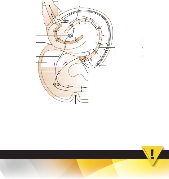
Figure 15-3 Major connections of the hippocampal formation. The hippocampal formation (HF) consists of three parts: hippocampus, dentate gyrus, and subiculum. The two major hypothalamic output pathways are (1) granule cell via mossy fiber to pyramidal cell via precommissural fornix to septal nuclei and (2) subicular neuron via postcommissural fornix to the medial mammillary nucleus. The HF plays an important role in learning and memory, and lesions of the HF result in short-term memory defects. In Alzheimer disease, loss of cells in the HF and entorhinal cortex leads to loss of memory and cognitive function. CA, cornu ammonis. The sector CA1 is very sensitive to hypoxia (cardiac arrest or stroke). (Reprinted from Fix JD. BRS Neuroanatomy. 3rd ed. Baltimore, MD: Williams & Wilkins; 1996:332, with permission.)
CASE 15-1
A 15-year-old boy was knocked out after several rounds of boxing with friends. Computed tomography scanning showed acute subdural hematoma associated with the right cerebral hemisphere. After regaining consciousness, the boy no longer experienced normal fear and anger and demonstrated aberrant sexual behaviors and excessive oral tendencies. He also complained of being very hungry all the time. What is the most likely diagnosis?
Relevant Lab Findings
●Computed tomography and magnetic resonance imaging revealed lesions of the right temporal lobe and right-dominant orbitofrontal regions, including bilateral rectal and medial orbital gyri, and an intact left temporal lobe.
Diagnosis
●Klüver-Bucy syndrome occurs when both the right and left medial temporal lobes malfunction, with frequent involvement of the amygdala. The cardinal symptom is excessive oral tendencies where the patient puts all types of objects into the mouth. Such patients also have an irresistible impulse to touch objects and demonstrate placidity (lack of emotional response) and a marked increase in sexual activity, without concern for social appropriateness.
C H A P T E R 1 6
Basal Nuclei and
Extrapyramidal Motor
System
Objectives
1.List the components of the basal nuclei.
2.Define striatum, corpus striatum, and claustrum.
3.Describe the anatomical structures that make up the extrapyramidal system and outline their functions.
4.Describe the anatomy, clinical signs, and treatment of Parkinson Disease, Huntington Disease, hemiballism, Wilson Disease, and tardive dyskinesia.
IBasal Nuclei (Ganglia) (Figure 16-1)
A.Components
1.Caudate nucleus
2.Putamen
3.Globus pallidus
B.Grouping of the Basal Nuclei
1.The striatum consists of the caudate nucleus and putamen.
2.The lentiform nucleus consists of the globus pallidus and putamen.
3.The corpus striatum consists of the lentiform nucleus and caudate nucleus.
4.The claustrum lies between the lentiform nucleus and the insular cortex. It has reciprocal connections between the sensory cortices (i.e., visual cortex) (Figures 16-2 to 16-4).
II The Extrapyramidal (Striatal) Motor System (see Figure 16-1)
plays a role in the initiation and execution of somatic motor activity, especially willed movement. It is also involved in automatic stereotyped postural and reflex motor activity (e.g., normal subjects swing their arms when they walk).
A.Structure. The striatal motor system includes the following structures:
1.Neocortex
2.Striatum (caudatoputamen or neostriatum)
120

Corpus callosum
Lateral ventricle
Caudate nucleus
Thalamus
Internal capsule
Third ventricle
Optic tract
Substantia nigra
Basal Nuclei and Extrapyramidal Motor System |
121 |
Fornix
|
|
|
|
|
Putamen |
|
|
|
|
|
|
|
Globus |
Lentiform |
|
|
|
|
|
|
pallidus |
nucleus |
|
|
VA |
|
VA |
|
|
Insula |
|
|
|
|
|
|
|||
|
|
|
|
|
|||
|
VLCM |
|
CMVL |
|
|
|
|
|
|
|
|
|
|
||
|
|
|
|
|
Subthalamic |
||
|
|
|
|
|
nucleus |
|
|
|
|
|
|
|
Lateral ventricle |
|
|
|
Mamillary |
|
|
|
|
||
Amygdala |
body |
Hippocampus |
|
||||
|
|
|
|
||||
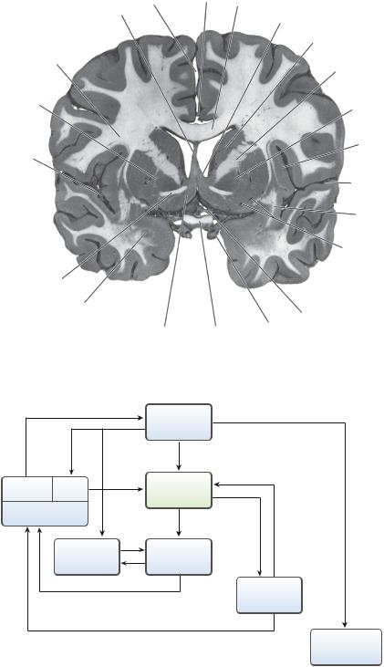
Figure 16-1 Coronal section through the midthalamus at the level of the mammillary bodies. The basal nuclei (ganglia) are all prominent at this level and include the striatum and lentiform nucleus. The subthalamic nucleus and substantia nigra are important components of the striatal motor system. CM, centromedian nucleus; VA, ventral anterior nucleus; VL, ventral lateral nucleus.
3.Globus pallidus
4.Subthalamic nucleus
5.Substantia nigra (i.e., pars compacta and pars reticularis)
6.Thalamus (ventral anterior, ventral lateral, and centromedian nuclei)
B.Figure 16-5 shows the major afferent and efferent connections of the striatal system.
C.Neurotransmitters are seen in Figure 16-6.
IIIClinical Correlation
A.Parkinson Disease. This is a degenerative disease that affects the substantia nigra and its projections to the striatum.
1.Results of Parkinson disease are a depletion of dopamine in the substantia nigra and striatum as well as a loss of melanin-containing dopaminergic neurons in the substantia nigra.
2.Clinical signs are bradykinesia, stooped posture, shuffling gait, cogwheel rigidity, pill-rolling tremor, and masked facies. Lewy bodies are found in the melanin-containing neurons of the substantia nigra. Progressive supranuclear palsy is associated with Parkinson disease.
3.Treatment has been successful with L-dopa. Surgical intervention includes pallidotomy (rigidity) and ventral thalamotomy (tremor).
122 |
Chapter 16 |
|
|
|
Thalamus, |
|
|
|
internal |
Thalamic |
|
|
|
|
|
|
medullary fasciculus |
Thalamus, DM |
|
|
Thalamus, lamina |
|
Fornix, crus |
|
Thalamus, VPM |
|
|
|
|
|
|
|
anterior |
|
Thalamus, pulvinar |
|
Cingulate sulcus |
|
|
|
Mamillothalamic |
|
Pretectal area |
|
tract |
|
|
Thalamic |
|
|
|
|
fasciculus |
|
|
|
|
Caudate |
|
|
Calcarine |
|
|
|
fissure |
||
nucleus, |
|
|
|
|
head |
|
|
|
|
Zona |
|
|
|
|
incerta |
|
|
Lateral |
|
|
|
|
||
|
|
|
lemniscus |
|
Lenticular |
|
|
Fastigial |
|
fasciculus |
|
|
||
|
|
nucleus |
||
|
|
|
||
Ansa |
|
|
Superior |
|
|
|
cerebellar |
||
lenticularis |
|
|
||
|
|
peduncle |
||
|
|
|
||
Optic nerve |
|
|
Medullary striae |
|
|
|
|
||
|
|
|
of fourth ventricle |
|
Posterior |
|
|
Medial lemniscus |
|
|
|
Corticospinal |
||
cerebral |
Red |
Substantia |
||
atery |
nucleus |
tract |
||
nigra |
||||
|
|
|
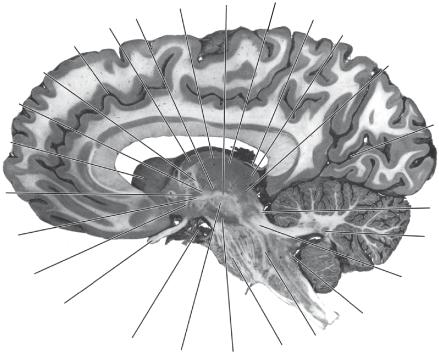
Figure 16-2 A parasagittal section through the caudate nucleus and the substantia nigra.
B.Methylphenyltetrahydropyridine (MPTP)-induced Parkinsonism. MPTP is an analog of meperidine (Demerol). It destroys dopaminergic neurons in the substantia nigra.
C.Huntington Disease (chorea). This is an inherited autosomal dominant movement disorder that is traced to a single gene defect on chromosome 4.
1.It is associated with degeneration of the cholinergic and g-aminobutyric acid (GABA)- ergic neurons of the striatum. It is accompanied by gyral atrophy in the frontal and temporal lobes.
2.Glutamate (GLU) excitotoxicity results when GLU is released in the striatum and binds to its receptors on striatal neurons, culminating in an action potential. GLU is removed from the extracellular space by astrocytes. In Huntington disease, GLU is bound to the N-methyl-D-aspartate receptor, resulting in an influx of calcium ions and subsequent cell death. This cascade of events with neuronal death most likely occurs in cerebrovascular accidents (e.g., stroke).
3.Clinical signs include choreiform movements, hypotonia, and progressive dementia.
D.Other Choreiform Dyskinesias
1.Sydenham chorea (St. Vitus dance) is the most common cause of chorea overall. It occurs primarily in girls, typically after a bout of rheumatic fever.
2.Chorea gravidarum usually occurs during the second trimester of pregnancy. Many patients have a history of Sydenham chorea.
E.Hemiballism is a movement disorder that usually results from a vascular lesion of the subtha-
lamic nucleus. Clinical signs include violent contralateral flinging (ballistic) movements of one or both extremities.
Hypothalamus, lateral preoptic nucleus
Internal capsule, anterior limb
Globus pallidus, medial medullary lamina
Globus pallidus, GPe
Insula, short gyri
Globus pallidus, GPi
Insula, long gyrus
Internal capsule, posterior limb
Middle cerebral artery, branch
Internal capsule, retrolenticular part
Basal Nuclei and Extrapyramidal Motor System |
123 |
Fornix, Cingulate column gyrus
Anterior commissure
Caudate nucleus, head
Putamen
Lateral sulcus
Hypothalamus, paraventricular nucleus
Globus pallidus, lateral medullary lamina
Temporal operculum
Interthalamic
adhesion
Thalamus, VL
Thalamus, DM
Choroid plexus of |
|
Optic radiation |
|
lateral ventricle |
|
|
|
Thalamus, pulvinar |
|
Hippocampus, tail |
|
|
|
||
Great |
Longitudinal |
Stria medullaris |
|
cerebral |
of thalamus |
||
cerebral |
|||
vein |
|
||
fissure |
|
||
|
|
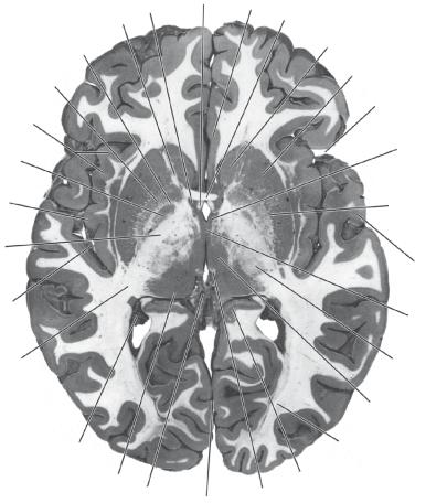
Figure 16-3 An axial (horizontal) section through the anterior commissure and the massa intermedia.
F. Hepatolenticular Degeneration (Wilson Disease) is an autosomal recessive disorder that is caused by a defect in the metabolism of copper. The gene locus is on chromosome 13.
1.Clinical signs include choreiform or athetotic movements, rigidity, and wing-beating tremor. Tremor is the most common neurologic sign.
2.Lesions are found in the lentiform nucleus. Copper deposition in the limbus of the cornea gives rise to the corneal Kayser–Fleischer ring, which is a pathognomonic sign. Deposition of copper in the liver leads to multilobular cirrhosis.
3.Psychiatric symptoms include psychosis, personality disorders, and dementia.
4.The diagnosis is based on low serum ceruloplasmin, elevated urinary excretion of copper, and increased copper concentration in a liver biopsy specimen.
5.Treatment includes penicillamine, a chelator.
G.Tardive Dyskinesia is a syndrome of repetitive choreic movement that affects the face and trunk. It results from treatment with phenothiazines, butyrophenones, or metoclopramide.
|
|
Anterior |
|
|
|
Longitudinal |
cerebral artery, |
|
|
||
pericallosal |
|
|
|||
cerebral |
|
|
|||
|
branch |
|
|
|
|
fissure |
|
Corpus callosum, body |
|||
|
|
||||
Septum pellucidum |
|
|
|
Lateral septal nucleus |
|
|
|
|
|
||
|
|
|
|
Caudate nucleus, head |
|
Superior longitudinal |
|
|
|
Internal capsule, |
|
fasciculus |
|
|
|
||
|
|
|
|
anterior limb |
|
Globus pallidus, |
|
|
|
|
Globus |
lateral medullary |
|
|
|
|
|
|
|
|
|
pallidus, |
|
lamina |
|
|
|
|
|
|
|
|
|
GPe |
|
|
|
|
|
|
|
Middle |
|
|
|
|
Insula |
|
|
|
|
|
|
cerebral |
|
|
|
|
|
artery |
|
|
|
|
|
|
|
|
|
|
Lateral sulcus |
|
|
|
|
|
Uncinate |
|
|
|
|
|
fasciculus |
|
|
|
|
Middle cerebral |
|
|
|
|
|
|
artery, lateral |
|
|
|
|
lenticulostriate |
|
|
|
|
|
|
branches |
Anterior commissure |
|
|
|
|
|
Amygdala |
|
|
|
|
|
|
|
|
|
Anterior cerebral artery |
|
Nucleus of |
Optic |
|
Internal |
||
|
carotid artery |
||||
diagonal |
chiasm |
|
|||
|
|
|
|||
band
Figure 16-4 A coronal section through the lentiform nucleus and the amygdaloid nucleus; the lentiform nucleus consists of the putamen and the globus pallidus; the amygdaloid nucleus appears as a circular profile below the uncus.
|
|
Neocortex |
|
VA, VL |
CM |
Striatum |
|
Thalamus |
GABA |
Dopamine |
|
|
Subthalamic |
||
|
Globus |
|
|
|
nucleus |
pallidus |
|
|
|
Substantia |
|
|
|
|
nigra |
tracts corticospinal and Corticonuclear
Brain stem and spinal cord
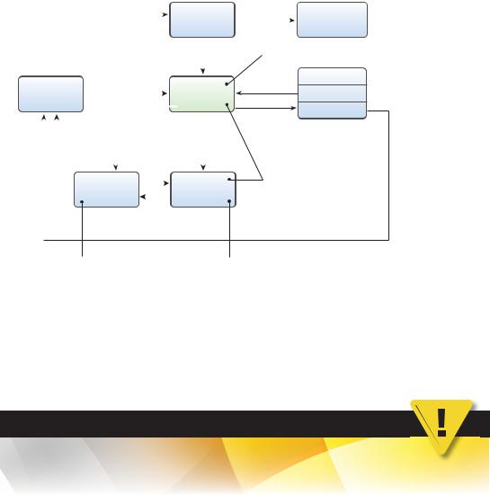
Figure 16-5 Major afferent and efferent connections of the striatal system. The striatum receives major input from three sources: the thalamus, neocortex, and substantia nigra. The striatum projects to the globus pallidus and substantia nigra. The globus pallidus is the effector nucleus of the striatal system; it projects to the thalamus and subthalamic nucleus. The substantia nigra also projects to the thalamus. The striatal motor system is expressed through the corticonuclear and corticospinal tracts. CM, centromedian nucleus; GABA, γ-aminobutyric acid; VA, ventral anterior nucleus; VL, ventral lateral nucleus.
124
Basal Nuclei and Extrapyramidal Motor System |
125 |
|
|
|
|
|
GLU |
|
|
|
||||||||
|
|
|
|
|
|
|
|
|
|
|
|
|
|
Neocortex |
||
|
|
|
|
|
|
|
|
|
|
|
|
|
|
|
|
GLU |
|
|
|
|
|
|
|
|
|
|
|
|
|
|
|
|
|
|
|
|
|
|
|
GLU |
|
|
|
|||||||
|
|
|
|
|
|
Striatum |
||||||||||
Thalamus |
|
|
||||||||||||||
|
|
|
|
|
|
|
|
|
|
|||||||
|
|
ACh |
|
|
|
|
|
|||||||||
|
|
|
|
|
|
|
|
|
|
|
GABA/ENK |
|
GABA/SP |
|||
|
|
|
|
|
|
|
|
|
|
|||||||
|
|
|
|
|
|
GLU |
|
|||||||||
|
|
|
|
|
|
|
||||||||||
GABA |
|
|
GABA |
|
|
|
||||||||||
|
|
|
|
Subthalamic |
GLU |
Globus |
||||||||||
|
|
|
|
|
|
|
|
|||||||||
|
|
|
|
|
|
|||||||||||
|
|
|
|
|
nucleus |
|
|
pallidus |
||||||||
|
|
|
|
|
|
|||||||||||
|
|
|
|
|
|
|
GABA |
|
|
|
||||||
|
|
|
|
|
|
|
|
|
|
|||||||
|
|
|
|
|
|
|
|
|
|
|
|
|
|
|
|
|
|
|
|
|
|
|
|
|
|
|
|
|
|
|
|
|
|
GLU |
Brain stem and |
|
|
|
spinal cord |
(Destruction results in Huntington’s disease)
S. nigra
Dopamine
Compacta  (Destruction results in
Parkinson’s disease)
Reticularis
GABA/SP
(Lesions found here in Wilson’s disease)
(Destruction results |
(Lesions found here |
in hemiballism) |
in kernicterus) |
Figure 16-6 Major neurotransmitters of the extrapyramidal motor system. Within the striatum, globus pallidus, and pars reticularis of the substantia nigra (S. nigra), γ-aminobutyric acid (GABA) is the predominant neurotransmitter. GABA may coexist in the same neuron with enkephalin (ENK) or substance P (SP). Dopamine-containing neurons are found in the pars compacta of the substantia nigra. Acetylcholine (ACh) is found in the local circuit neurons of the striatum. The subthalamic nucleus projects excitatory glutaminergic fibers to the globus pallidus. GLU, glutamate.
CASE 16-1
A 30-year-old man presents with dysarthria, dysphagia, stiffness, and slow ataxic gait. There is no history of schizophrenia or depression and no family history of any neurodegenerative disease.
Relevant Physical Exam Findings
●The patient scored a 20/26 on the mini-mental status exam. The patient showed increased tone in all extremities, with normal strength.
Relevant Lab Findings
●A generalized cerebral and cerebellar atrophy and a very small caudate nucleus were revealed on magnetic resonance imaging scans.
Diagnosis
●Huntington disease (chorea) is caused by a trinucleotide repeat in the gene coding for the Huntingtin protein. It is characterized by abnormal body movements and lack of coordination but can also affect
mental abilities.