

Spinal Cord |
49 |
CASE 6-1
A 46-year-old man was admitted with complaints of lower back pain that radiated down to his foot over the last 2 months. The pain was not relieved with medical therapy. What is your diagnosis?
Relevant Physical Exam Findings
●Absent right ankle jerk
●Weakness of dorsiflexion and plantar flexion
●Decreased pinprick over the dorsum of the foot
Relevant Lab Findings
●An anteroposterior myelogram demonstrated compression of the first sacral nerve root on the left at the level of the L5-S1 vertebrae.
Diagnosis
● Lumbar intervertebral disc herniation
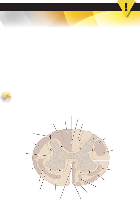
Tracts of the Spinal Cord
IPosterior (Dorsal) Column—Medial Lemniscus
Pathway (Figures 6-5 and 6-6)
Ascending tracts |
Descending tracts |
Fasciculus gracilis |
Fasciculus septomarginalis |
Fasciculus cuneatus |
Fasiculus interfascicularis |
Posterolateral tract |
|
(of Lissauer) |
|
Posterior |
Lat. corticospinal tract |
|
|
spinocerebellar tract |
|
|
Rubrospinal tract |
Spinospinal tract |
|
|
Spinospinal tract |
Anterior |
|
spinocerebellar tract |
Medullary |
|
|
|
reticulospinal tract |
Lat. spinothalamic tract |
Vestibulospinal tract |
|
|
|
Pontine reticulospinal tract |
Spinotectal tract |
Tectospinal tract |
|
|
Anterior spinothalamic tract |
|
Medial longitudinal fasciculus |
Anterior corticospinal tract |
Figure 6-5 The major ascending and descending pathways of the spinal cord. The ascending tracts are shown on the left, and the descending tracts are shown on the right.
50 |
Chapter 6 |
|
|
|
|
|
x |
|
|
|
|
rte |
|
|
|
|
|
icco |
|
|
|
|
|
t |
|
|
|
|
|
e |
|
|
|
|
|
h |
|
|
|
|
t |
|
|
|
m |
e |
s |
|
|
|
|
|
|
||
|
|
|
|
|
|
S |
o |
|
|
|
|
|
|
|
|
|
|
Thalamus
Internal capsule
Lentiform nucleus
Trigeminal nerve
Nucleus gracilis (neuron II)
Nucleus cuneatus (neuron II)
Internal arcuate fibers 
Cuneate fasciculus
Spinal ganglion cell 
Pacinian corpuscle
Meissner corpuscle

Postcentral gyrus
 Lower limb area
 Trunk area
 Upper limb area
 Head area
 Face area
Ventral posterolateral nucleus (of thalamus)
Medial lemniscus
Midbrain
Medial lemniscus
Pons
Medulla
Medial lemniscus
 Spinal trigeminal nucleus
 Decussation of
medial lemniscus
Gracile fasciculus

 Cuneate fasciculus
Cervical cord
Gracile fasciculus
Lumbosacral cord
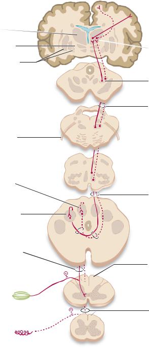
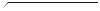
Figure 6-6 The posterior column—medial lemniscus pathway. Impulses conducted by this pathway mediate fine touch, conscious proprioception, and vibratory sense.

Spinal Cord |
51 |
A.Function. The posterior column—medial lemniscus pathway mediates fine touch, conscious proprioception, and vibratory sense.
B.Receptors include Pacinian and Meissner corpuscles, joint receptors, muscle spindles, and Golgi tendon organs.
C.First-order Neurons are located in the spinal ganglia at all levels. Central processes project to the spinal cord through the medial root entry zone. First-order neurons give rise to the:
1.Gracile fasciculus from the lower extremity.
2.Cuneate fasciculus from the upper extremity.
3.Collaterals for spinal reflexes (e.g., myotatic reflex).
D.Second-order Neurons are located in the gracile and cuneate nuclei of the caudal medulla. They give rise to axons and internal arcuate fibers that decussate and form a compact fiber bundle (i.e., medial lemniscus). The medial lemniscus ascends through the contralateral brain stem and terminates in the ventral posterolateral (VPL) nucleus of the thalamus.
E.Third-order Neurons are located in the VPL nucleus of the thalamus. They project through the posterior limb of the internal capsule to the postcentral gyrus—the primary somatosensory cortex (Brodmann areas 3, 1, and 2).
F.Transection of the Posterior Column—Medial Lemniscus Tract
1.Superior to the sensory decussation, transection results in contralateral loss of the posterior column modalities.
2.In the spinal cord, transection results in ipsilateral loss of the posterior column modalities.
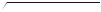
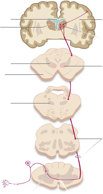
IIAnterolateral System (Figures 6-5 and 6-7)
A.Function. The anterolateral system is comprised of three main pathways: the spinothalamic tracts (lateral and medial) that mediate pain and temperature and crude touch, respectively; the spinoreticular tract, which carries pain to the reticular formation for arousal; and the spinotectal tract that mediates auditory and visual reflex orientation of the head and neck.
B.Receptors are free nerve endings. Pain ascends on both fastand slow-conducting pain fibers (i.e., A-δ and C, respectively).
C.First-order Neurons are found in the spinal ganglia at all levels. Central projections enter the spinal cord through the posterolateral tract (of Lissauer) (lateral root entry zone) to second-order neurons.
D.Second-order Neurons are found in the posterior horn. They give rise to axons that decussate in the anterior white commissure and ascend in anterior aspect of the contralateral lateral funiculus. Their axons terminate in the VPL nucleus of the thalamus.
E.Third-order Neurons are found in the VPL nucleus of the thalamus. They project through the posterior limb of the internal capsule to the primary somatosensory cortex (Brodmann areas 3, 1, and 2).
F.Transection of the Lateral Spinothalamic Tract of the anterolateral system results in contralateral loss of pain and temperature inferior to the level of the lesion.
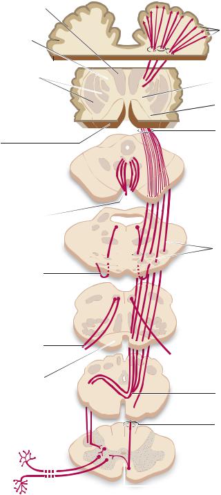
IIILateral Corticospinal Tract (Figures 6-5 and 6-8)
A.Function. The lateral corticospinal tract mediates voluntary motor activity, primarily of the upper limbs.
52 |
Chapter 6 |
|
|
|
|
|
|
|
x |
|
|
|
|
|
|
e |
|
|
|
|
|
|
rt |
|
|
|
|
|
|
o |
|
|
|
|
|
|
|
c |
|
|
|
|
|
|
|
y |
|
|
|
|
|
|
r |
|
|
|
|
|
|
|
o |
|
|
|
|
|
|
s |
|
|
|
|
|
S |
e |
n |
|
|
|
|
|
|
|
|
|
|
|
||
|
|
|
|
|
|
|
|
Corpus callosum
Thalamus
Internal capsule 
Medial lemniscus
Crus cerebri
Medial lemniscus
First-order neuron in spinal ganglion
Free nerve endings
Second-order neuron
Lower
limb
 Foot
Foot
Cerebral cortex (postcentral gyrus)
Upper
limb
Hand
Axons of neurons in posterior limb of internal capsule
Third-order neuron in ventral posterolateral nucleus (VPL)
Midbrain
Red nucleus
Pons
Medulla
Lateral spinothalamic tract
Anterior white commissure
Figure 6-7 The lateral spinothalamic tract. Impulses conducted by this tract mediate pain and thermal sense.
B.Origin and Termination
1.Origin. The lateral corticospinal tract arises in the premotor cortex (Brodmann area 6) and the primary motor cortex, or precentral gyrus (Brodmann area 4).
2.Termination. The lateral corticospinal tract terminates contralaterally, through interneurons, on
anterior horn motor neurons.
Thalamus
Posterior limb internal capsule
Lower
limb
Foot
Trunk
Spinal Cord |
53 |
Motor cortex (precentral gyrus)
Upper limb
Large pyramidal cells of Betz
Lenticular nucleus 
Caudate nucleus
(head)
Crus cerebri
CN III
CN VI
CN XII
Pyramid
Lateral corticospinal tract 
Motor end plates
Genu of internal capsule
Anterior limb of internal capsule
Corticospinal tract
Midbrain
Pons
Longitudinal fibers in basilar portion of pons
Medulla
Medulla
Pyramidal decussation
Anterior corticospinal tract
Spinal cord
Anterior white commissure
Figure 6-8 The lateral and anterior corticospinal (pyramidal) tracts. These major descending motor pathways mediate volitional motor activity.