

80 |
Chapter 9 |
VI |
The Abducent Nerve (CN VI) |
A.General Characteristics. The abducent nerve is a pure GSE nerve that innervates the lateral rectus, which abducts the eye.
1.It arises from the abducent nucleus that is found in the posteromedial tegmentum of the caudal pons.
2.Exiting intra-axial fibers pass through the corticospinal tract. A lesion results in alternating abducent hemiparesis.
3.It passes through the pontine cistern and cavernous sinus and enters the orbit through the superior orbital fissure.
B.Clinical Correlation. CN VI Paralysis is the most common isolated palsy that results from
the long peripheral course of the nerve. It is seen in patients with meningitis, subarachnoid hemorrhage, late-stage syphilis, and trauma. Abducent nerve paralysis results in the following defects:
1.Convergent (medial) strabismus (esotropia) with inability to abduct the eye.
2.Horizontal diplopia with maximum separation of the double images when looking toward the paretic lateral rectus.
VII The Facial Nerve (CN VII)
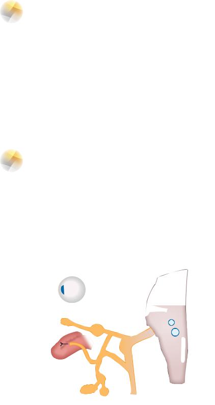
A. General Characteristics. The facial nerve is a GSA, general visceral afferent (GVA), SVA, GVE, and SVE nerve (Figures 9-5 and 9-6). It mediates facial movements, taste, salivation, lacrimation, and general sensation from the external ear. It is the nerve of the pharyngeal (branchial)
arch 2 (hyoid). It includes the facial nerve proper (motor division), which contains the SVE fibers that innervate the muscles of facial (mimetic) expression. CN VII includes the intermediate nerve,
which contains GSA, SVA, and GVE fibers. All first-order sensory neuronal cell bodies are found in the geniculate ganglion within the temporal bone.
1.Anatomy. The facial nerve exits the brain stem in the cerebellopontine angle. It enters the internal auditory meatus and the facial canal. It then exits the facial canal and skull through the stylomastoid foramen.
CN II
Lacrimal gland
Pterygopalatine ganglion
Nasal and 
 palatine glands


Tongue
(taste, anterior two-thirds) 
Lingual nerve
Submandibular ganglion 
Sublingual gland 

Submandibular gland
CN V2 |
|
|
Trigeminal ganglion |
||||
|
|
CN V3 |
|
|
|
|
|
|
|
CN V1 |
|
|
|
|
|
|
|
|
Pons |
|
|
Greater petrosal nerve |
|
|
|
|
|
|
|
||
|
|
|
|
|
|
|
Superior salivatory |
|
|
|
|
|
|
|
nucleus (GVE) |
|
|
C |
|
|
|
|
Motor nucleus of CN VII (SVE) |
|
|
|
|
|
|
||
|
|
|
|
|
|
|
|
B |
Geniculate |
|
|
|
|
Nucleus of solitary tract |
|
|
|
|
|||||
ganglion |
|
|
|
|
|
||
Chorda |
Stapedial |
|
|
|
|
Solitary tract (SVA) |
|
|
|
|
|
|
|||
tympani |
nerve |
|
|
|
|
|
|
A
Motor root of CN VII in stylomastoid foramen
Figure 9-5 The functional components of the facial nerve (cranial nerve [CN] VII).
Facial area of motor cortex
UMN lesion of corticonuclear tract 
(e.g., stroke of internal capsule)
Muscles of facial expression:
Frontalis
Orbicularis oculi
Buccinator
Orbicularis oris
Platysma
Cranial Nerves |
81 |
Facial motor nucleus of pons
Superior division
Inferior division
 LMN lesion of CN VII (e.g., Bell palsy)
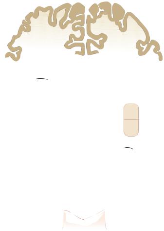
Figure 9-6 Corticonuclear innervation of the facial nerve (cranial nerve [CN] VII) nucleus. An upper motor neuron (UMN) lesion (e.g., stroke involving the internal capsule) results in contralateral weakness of the lower face, with sparing of the upper face. A lower motor neuron (LMN) lesion (e.g., Bell palsy) results in paralysis of the facial muscles in both the upper and lower face. (Redrawn from DeMyer WE. Technique of the Neurological Examination: A Programmed Text. 4th ed. New York: McGraw-Hill; 1994:177, with permission.)
2.The GSA component has cell bodies located in the geniculate ganglion. It innervates the posterior surface of the external ear through the posterior auricular branch of CN VII. It projects centrally to the spinal tract and nucleus of trigeminal nerve.
3.The GVA component has no clinical significance. The cell bodies are located in the geniculate ganglion. Fibers innervate the soft palate and the adjacent pharyngeal wall.
4.The SVA component (taste) has cell bodies located in the geniculate ganglion. It projects centrally to the solitary tract and nucleus. It innervates the taste buds from the anterior two-thirds of the tongue through:
a.The intermediate nerve.
b.The chorda tympani, which is located in the tympanic cavity medial to the tympanic membrane and malleus. It contains the SVA and GVE (parasympathetic) fibers.
c.The lingual nerve (a branch of CN V3).
d.The central gustatory pathway (see Figure 9-5). Taste fibers from CNs VII, IX, and X project through the solitary tract to the solitary nucleus. The solitary nucleus projects through the central tegmental tract to the ventral posteromedial nucleus (VPM) of the thalamus. The VPM projects to the gustatory cortex of the parietal lobe (parietal operculum).

82Chapter 9
5.The GVE component is a parasympathetic component that innervates the lacrimal, submandibular, and sublingual glands. It contains preganglionic parasympathetic neurons that are located in the superior salivatory nucleus of the caudal pons.
a.Lacrimal pathway (see Figure 9-5). The superior salivatory nucleus projects through the intermediate and greater petrosal nerves to the pterygopalatine ganglion. Postganglionic neurons from the pterygopalatine ganglion project through the inferior orbital fissure and travel
via the zygomatic nerve (a branch of V2) and lacrimal nerve (a branch of V1) to innervate the lacrimal gland.
b.Submandibular pathway (see Figure 9-5). The superior salivatory nucleus projects through the intermediate nerve and chorda tympani to the submandibular ganglion. Postganglionic neurons from the submandibular ganglion project to and innervate the submandibular and sublingual glands.
6.The SVE component arises from the facial motor nucleus, loops around the abducent nucleus of the caudal pons, and exits the brain stem in the cerebellopontine angle. It enters the internal auditory meatus, traverses the facial canal, sends a branch to the stapedius in the middle ear, and exits the skull through the stylomastoid foramen. It innervates the muscles of facial expression, the stylohyoid, the posterior belly of the digastric, and the stapedius.
B.Clinical Correlation. Lesions cause the following conditions:
1.Flaccid paralysis of the muscles of facial expression (upper and lower face).
2.Loss of the corneal (blink) reflex (efferent limb), which may lead to corneal ulceration (keratitis paralytica).
3.Loss of taste (ageusia) from the anterior two-thirds of the tongue, which may result from damage to the chorda tympani.
4.Hyperacusis (increased acuity to sounds) as a result of stapedius paralysis.
5.Bell palsy (peripheral facial paralysis), which is caused by a lower motor neuron lesion resulting from trauma or infection. This palsy involves the upper and lower face.
6.Crocodile tears syndrome (lacrimation during eating), which is a result of aberrant regeneration of GVE fibers after trauma. Regenerating preganglionic salivary fibers are misdirected to the pterygopalatine ganglion which projects to the lacrimal gland.
7.Supranuclear (central) facial palsy, which is caused by an upper motor neuron lesion and results in contralateral weakness of the lower face, with sparing of the upper face (see Figure 9-6).
8.Bilateral facial nerve palsies, which occur in Guillain–Barré syndrome.
9.Möbius syndrome, which consists of congenital facial diplegia (CN VII) and convergent strabismus (CN VI).
VIII The Vestibulocochlear Nerve (CN VIII) is an SSA nerve. It has two
functional divisions: the vestibular nerve, which maintains equilibrium and balance, and the cochlear nerve, which mediates hearing. It exits the brain stem at the cerebellopontine angle
and enters the internal auditory meatus. It is confined to the temporal bone.
A.Vestibular Nerve (see Figure 13-1)
1.General characteristics
a.It is associated functionally with the cerebellum (flocculonodular lobe) and ocular motor nuclei.
b.It regulates compensatory eye movements.
c.Its first-order neuronal cell bodies are located in the vestibular ganglion in the fundus of the internal auditory meatus.
d.It projects its peripheral processes to the hair cells of the cristae of the semicircular ducts and the hair cells of the utricle and saccule.
e.It projects its central processes to the four vestibular nuclei of the brain stem and the flocculonodular lobe of the cerebellum.
f.It conducts efferent fibers to the hair cells from the brain stem.
2.Clinical correlation. Lesions result in disequilibrium, vertigo, and nystagmus.

Cranial Nerves |
83 |
B.Cochlear Nerve (see Figure 12-1)
1.General characteristics
a.Its first-order neuronal cell bodies are located in the spiral (cochlear) ganglion of the modiolus of the cochlea, within the temporal bone.
b.It projects its peripheral processes to the hair cells of the organ of Corti.
c.It projects its central processes to the dorsal and ventral cochlear nuclei of the brain stem.
d.It conducts efferent fibers to the hair cells from the brain stem.
2.Clinical correlation. Destructive lesions cause hearing loss (sensorineural deafness). Irritative lesions can cause tinnitus (ear ringing). An acoustic neuroma (schwannoma) is a Schwann cell tumor of the cochlear nerve that causes deafness.
IX The Glossopharyngeal Nerve (CN IX) is a GSA, GVA, SVA, SVE,
and GVE nerve.
A.General Characteristics. The glossopharyngeal nerve is primarily a sensory nerve. Along with CNs VII and X, it mediates taste, salivation, and swallowing. It mediates input from the carotid
sinus, which contains baroreceptors that monitor arterial blood pressure. It also mediates input from the carotid body, which contains chemoreceptors that monitor the CO2 and O2 concentration of the
blood.
1.Anatomy. CN IX is the nerve of pharyngeal (branchial) arch 3. It exits the brain stem (medulla) from the postolivary sulcus with CNs X and XI. It exits the skull through the jugular foramen with CNs X and XI.
2.The GSA component innervates part of the external ear and the external auditory meatus through the auricular branch of the vagus nerve. It has cell bodies in the superior ganglion. It projects its central processes to the spinal tract and nucleus of trigeminal nerve.
3.The GVA component innervates structures that are derived from the endoderm (e.g., pharynx). It innervates the mucous membranes of the posterior one-third of the tongue, tonsil, upper pharynx, tympanic cavity, and auditory tube. It also innervates the carotid sinus (baroreceptors) and carotid body (chemoreceptors) through the sinus nerve. It has cell bodies in the inferior (petrosal) ganglion. It is the afferent limb of the gag reflex and the carotid sinus reflex.
4.The SVA component innervates the taste buds of the posterior one-third of the tongue. It has cell bodies in the inferior (petrosal) ganglion. It projects its central processes to the solitary tract and nucleus.
5.The SVE component innervates only the stylopharyngeus muscle. It arises from the nucleus ambiguus of the lateral medulla.
6.The GVE component is a parasympathetic component that innervates the parotid gland. Preganglionic parasympathetic neurons are located in the inferior salivatory nucleus of the medulla. They project through the tympanic and lesser petrosal nerves to the otic ganglion. Postganglionic fibers from the otic ganglion project to the parotid gland through the auriculotemporal nerve (CN V3).
B.Clinical Correlation. Lesions cause the following conditions:
1.Loss of the gag (pharyngeal) reflex (interruption of the afferent limb).
2.Hypersensitive carotid sinus reflex (syncope).
3.Loss of general sensation in the pharynx, tonsils, fauces, and back of the tongue.
4.Loss of taste from the posterior one-third of the tongue.
5.Glossopharyngeal neuralgia, which is characterized by severe stabbing pain in the root of the tongue.
84 |
Chapter 9 |
Motor cortex
Medulla
Medial lemniscus
Pyramid
UMN |
|
UMN |
|
 Corticonuclear tract
 Corticonuclear tract
Decussation
 Nucleus ambiguus
 UMN lesion
CN X (vagus nerve) |
|
LMN |
|
LMN
 LMN lesion

 Levator palati and palatal arches
A B
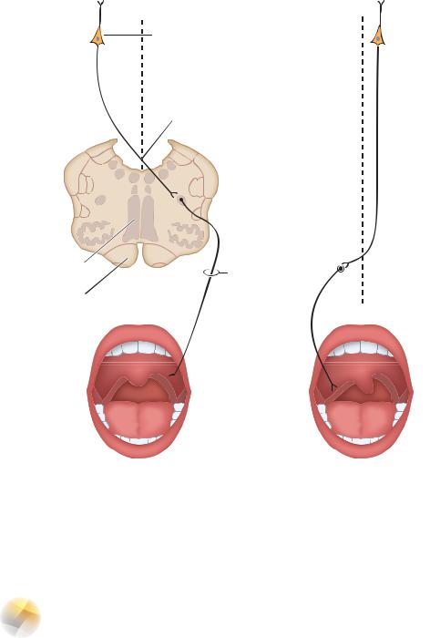
Figure 9-7 Innervation of the palatal arches and uvula. Sensory innervation is mediated by the glossopharyngeal nerve (cranial nerve [CN] IX). Motor innervation of the palatal arches and uvula is mediated by the vagus nerve (CN X). A. A normal palate and uvula in a person who is saying “Ah.” B. A patient with an upper motor neuron (UMN) lesion (left) and a lower motor neuron (LMN) lesion (right). When this patient says “Ah,” the palatal arches sag. The uvula deviates toward the intact (left) side.
XThe Vagal Nerve (CN X) is a GSA, GVA, SVA, SVE, and GVE nerve (see
Figure 9-7).
A. General Characteristics. The vagal nerve mediates phonation, swallowing (with CNs IX, XI, and XII), elevation of the palate, taste, and cutaneous sensation from the ear. It innervates the viscera of the neck, thorax, and abdomen.
1.Anatomy. The vagal nerve is the nerve of pharyngeal (branchial) arches 4 and 6. Pharyngeal arch 5 is either absent or rudimentary. It exits the brain stem (medulla) from the postolivary sulcus. It exits the skull through the jugular foramen with CNs IX and XI.

Cranial Nerves |
85 |
2.The GSA component innervates the infratentorial dura, external ear, external auditory meatus, and external surface of the tympanic membrane. It has cell bodies in the superior (jugular) ganglion, and it projects its central processes to the spinal tract and nucleus of trigeminal nerve.
3.The GVA component innervates the mucous membranes of the pharynx, larynx, esophagus, trachea, and thoracic and abdominal viscera (to the midtransverse colon). It has cell bodies in the inferior (nodose) ganglion. It projects its central processes to the solitary tract and nucleus.
4.The SVA component innervates the taste buds in the epiglottic region. It has cell bodies in the inferior (nodose) ganglion. It projects its central processes to the solitary tract and nucleus.
5.The SVE component innervates the pharyngeal (brachial) arch muscles of the larynx and pharynx, the striated muscle of the upper esophagus, musculus uvulae, levator palati, and palatoglossus. It arises from the nucleus ambiguus in the lateral medulla. The SVE component provides the efferent limb of the gag reflex.
6.The GVE component innervates the viscera of the neck and the thoracic (heart) and abdominal cavities as far as the midtransverse colon. Preganglionic parasympathetic neurons that are located in the dorsal motor nucleus of the medulla project to the terminal (intramural) ganglia of the visceral organs.
B.Clinical Correlation. Lesions and reflexes cause the following conditions:
1.Ipsilateral paralysis of the soft palate, pharynx, and larynx that leads to dysphonia (hoarseness), dyspnea, dysarthria, and dysphagia.
2.Loss of the gag (palatal) reflex (efferent limb).
3.Anesthesia of the pharynx and larynx that leads to unilateral loss of the cough reflex.
4.Aortic aneurysms and tumors of the neck and thorax that frequently compress the vagal nerve and can lead to cough, dyspnea, dysphagia, hoarseness, and chest/back pain.
5.Complete laryngeal paralysis, which can be rapidly fatal if it is bilateral (asphyxia).
6.Parasympathetic (vegetative) disturbances, including bradycardia (irritative lesion), tachycardia (destructive lesion), and dilation of the stomach.
7.The oculocardiac reflex, in which pressure on the eye slows the heart rate (afferent limb of CN V1 and efferent limb of CN X).
8.The carotid sinus reflex, in which pressure on the carotid sinus slows the heart rate (bradycardia; efferent limb of CN X).
XI The Accessory Nerve (CN XI), or spinal accessory nerve, is an SVE
nerve.
A.General Characteristics. The accessory nerve mediates head and shoulder movement.
It arises from the anterior horn of cervical spinal cord segments C1 through C6. The spinal roots exit
the spinal cord laterally between the anterior and posterior spinal roots, ascend through the foramen magnum, and exit the skull through the jugular foramen. It innervates the sternocleidomastoid and the trapezius.
B.Clinical Correlation. Lesions cause the following conditions:
1.Paralysis of the sternocleidomastoid results in difficulty in turning the head to the contralateral side.
2.Paralysis of the trapezius muscle results in shoulder droop and inability to shrug the shoulder.
XII The Hypoglossal Nerve (CN XII) is a GSE nerve.
A.General Characteristics. The hypoglossal nerve mediates tongue movement. It arises
from the hypoglossal motor nucleus of the medulla and exits the medulla in the preolivary sulcus. It exits the skull through the hypoglossal canal, and it innervates the intrinsic and extrinsic muscles
86 |
Chapter 9 |
Motor cortex |
|
UMN |
|
UMN |
|
|
Corticonuclear tract 
 Corticonuclear tract
Decussation
Hypoglossal
nucleus
|
|
|
|
|
UMN lesion |
|||||
|
|
|
|
|
||||||
|
|
|
|
|
||||||
Medulla |
C |
|
|
|
(spastic paralysis) |
|||||
|
|
|
|
|
|
|
|
|
|
|
|
G |
|
Decussation |
|
|
|
|
|
|
LMN |
Medial lemniscus |
P |
|
|
|
|
|
|
|
||
|
|
|
|
|
|
|
|
|||
|
|
|
|
|
|
|
|
|||
|
|
|
|
|
|
|
|
|
||
|
|
|
|
|
|
|
|
|
|
|
Pyramid |
|
|
Hypoglossal nerve |
|
|
|
|
|
|
|
|
|
|
|
|
|
|
|
|
||
|
|
|
LMN |
|
|
|
|
|
|
LMN lesion |
|
|
|
|
|
|
|
||||
|
|
|
|
|
|
|
||||
Genioglossus muscle |
|
|
|
|
|
|
|
|
|
(flaccid paralysis) |
A B
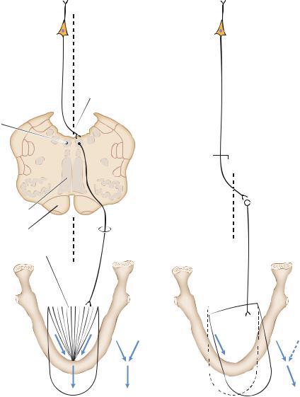
Figure 9-8 Motor innervation of the tongue. Corticonuclear fibers project predominantly to the contralateral hypoglossal motor nucleus. An upper motor neuron (UMN) lesion causes deviation of the protruded tongue to the weak (contralateral) side. A lower motor neuron (LMN) lesion causes deviation of the protruded tongue to the weak (ipsilateral) side. A. Normal tongue. B. Tongue with UMN and LMN lesions.
of the tongue. Extrinsic muscles are the genioglossus, styloglossus, and hyoglossus (palatoglossus is considered a muscle of the soft palate and is innervated by CN X).
B.Clinical Correlation
1.Transection results in hemiparalysis of the tongue.
2.Protrusion causes the tongue to point toward the lesioned (weak) side because of the unopposed action of the opposite genioglossus muscle (Figure 9-8).