


C H A P T E R 1 4
Visual System
Objectives
1.Outline the central and peripheral components of the visual pathway.
2.List the retinal layers and cell types found in each layer.
3.Describe the result of lesions at the optic nerve, optic chiasm, and optic tract.
4.Compare and contrast the pupillary reflexes, including direct versus consensual reflexes, dilation, convergence, and accommodation.
5.Summarize the various clinical correlates related to the visual system.
IIntroduction. The visual system is served by the optic nerve, which is a special
somatic afferent (SSA) nerve.
IIThe Visual Pathway (Figure 14-1) includes the following structures:
A.Ganglion Cells of the Retina form the optic nerve (cranial nerve [CN] II). They project from the nasal hemiretina to the contralateral lateral geniculate body and from the temporal hemiretina to the ipsilateral lateral geniculate body.
B.The optic nerve projects from the lamina cribrosa of the scleral canal, through the optic canal, to the optic chiasm (Figure 14-2).
1.Transection of the optic nerve causes ipsilateral blindness, with no direct pupillary light reflex.
2.A lesion of the optic nerve at the optic chiasm transects all fibers from the ipsilateral retina and fibers from the contralateral inferior nasal quadrant that loop into the optic nerve. This lesion causes ipsilateral blindness and a contralateral upper temporal quadrant defect (junction scotoma).
C.The optic chiasm contains decussating fibers from the two nasal hemiretinas. It contains noncrossing fibers from the two temporal hemiretinas and projects fibers to the suprachiasmatic nucleus of the hypothalamus.
1.Midsagittal transection or pressure (often from a pituitary tumor) causes bitemporal hemianopia.
2.Bilateral lateral compression causes binasal hemianopia (often from calcified internal carotid arteries).
D.The optic tract contains fibers from the ipsilateral temporal hemiretina and the contralateral nasal hemiretina. It projects to the ipsilateral lateral geniculate body, pretectal nuclei, and superior colliculus. Transection causes contralateral hemianopia.
108
|
|
|
Visual System |
109 |
|
Temporal |
Nasal Nasal |
Temporal |
|
1 |
|
Visual |
|
|
|
|
|
|
|
|
|
fields |
|
|
2 |
Left eye |
|
Right eye |
|
|
|
|
|
|
|
|
|
Retina |
|
|
|
Optic chiasm |
|
|
3 |
1 |
|
Optic nerve |
|
|
2 |
2 |
Optic tract |
|
|
4 |
3 |
|
|
|
|
|
||
4 |
|
|
|
|
5 |
|
Meyer’s loop |
|
|
|
|
|
||
5 |
|
|
|
|
|
6 |
|
Lateral |
|
|
|
|
|
|
6 |
|
7 |
geniculate nucleus |
|
|
Visual radiation to lingual gyrus |
|
||
|
|
|
|
|
|
|
|
Visual radiation to cuneus |
|
7 |
|
|
|
|
|
|
|
Visual cortex area 17 |
|
Figure 14-1 The visual pathway from the retina to the visual cortex showing visual field defects. (1) Ipsilateral blindness. (2) Binasal hemianopia. (3) Bitemporal hemianopia. (4) Right hemianopia. (5) Right upper quadrantanopia. (6) Right lower quadrantanopia. (7) Right hemianopia with macular sparing. (8) Left constricted field as a result of end-stage glaucoma. Bilateral constricted fields may be seen in hysteria. (9) Left central scotoma as seen in optic (retrobulbar) neuritis in multiple sclerosis. (10) Upper altitudinal hemianopia as a result of bilateral destruction of the lingual gyri. (11) Lower altitudinal hemianopia as a result of bilateral destruction of the cunei.
E.The lateral geniculate body is a six-layered nucleus. Layers 1, 4, and 6 receive crossed fibers; layers 2, 3, and 5 receive uncrossed fibers. The lateral geniculate body receives input from layer VI of the striate cortex (Brodmann area 17). It also receives fibers from the ipsilateral temporal hemiretina and the contralateral nasal hemiretina. It projects through the geniculocalcarine tract to layer IV of the primary visual cortex (Brodmann area 17).
F.The geniculocalcarine tract (visual radiation) projects through two divisions to the visual cortex.
1.The upper division (Figure 14-3) projects to the upper bank of the calcarine sulcus, the cuneus. It contains input from the superior retinal quadrants, which represent the inferior visual field quadrants.
a.Transection causes a contralateral lower quadrantanopia.
b.Lesions that involve both cunei cause a lower altitudinal hemianopia (altitudinopia).
2.The lower division (see Figure 14-3) loops from the lateral geniculate body anteriorly (Meyer loop), then posteriorly, to terminate in the lower bank of the calcarine sulcus, the lingual gyrus. It contains input from the inferior retinal quadrants, which represent the superior visual field quadrants.
a.Transection causes a contralateral upper quadrantanopia (“pie in the sky”).
b.Transection of both lingual gyri causes an upper altitudinal hemianopia (altitudinopia).
G.The visual cortex (Brodmann area 17) is located on the banks of the calcarine fissure. The cuneus is the upper bank. The lingual gyrus is the lower bank. Lesions cause contralateral hemianopia with macular sparing. The visual cortex has a retinotopic organization:
1.The posterior area receives macular input (central vision).
2.The intermediate area receives paramacular input (peripheral input).
3.The anterior area receives monocular input.

110 Chapter 14
|
|
|
|
|
|
|
|
|
|
|
|
|
|
|
Choroid |
||||
|
|
|
|
|
|
|
|
|
|
|
|
|
|
|
|||||
|
|
|
|
|
|
|
|
|
|
|
|
|
|
|
Choriocapillaris |
||||
|
|
|
|
|
|
|
|
|
PEL |
|
|
|
|
|
Tight junction |
||||
|
|
|
|
|
|
|
|
|
|
|
|
|
|
|
|||||
|
|
|
|
|
|
|
|
|
|
|
|
|
|
|
(blood–retina barrier) |
||||
|
|
|
|
|
|
|
|
|
LRC |
Cone |
Rod |
||||||||
|
|
|
|
|
|
|
|
|
|
|
|
|
|
|
|
|
|
|
|
|
|
|
|
|
|
|
|
|
OLM |
|
|
|
|
|
|
|
|
|
|
|
|
|
|
|
|
|
|
|
|
|
|
|
|
|
|
|
|
|
|
|
|
|
|
|
|
|
|
|
|
|
|
|
|
|
|
|
|
|
|
Visual cortex (area 17) |
ONL |
|
|
|
|
|
|
|
|
|
|
||||||||
|
|
|
|
|
|
|
|
|
Pedicle |
|
|
|
Sphericle |
||||||
|
|
|
|
|
|
|
|
|
|
|
|
|
|
|
Capillary plexus |
||||
|
|
|
|
|
|
|
|
|
OPL |
|
|
|
|
|
|
|
|
|
|
|
|
Lateral geniculate body |
|
|
|
|
|
|
|
|
Horizontal cell |
||||||||
|
|
|
|
|
|
|
|
|
|
||||||||||
|
|
|
|
|
|
|
|
|
|
|
|
|
|
|
|
|
|
Bipolar cell |
|
|
|
|
|
|
|
|
|
|
|
|
|
|
|
|
|
|
|
||
Optic |
|
|
|
|
|
Optic tract |
INL |
|
|
|
|
|
|
|
|
|
Amacrine cell |
||
|
|
|
|
|
|
|
|
|
|
|
|
|
|
||||||
|
|
|
|
|
|
|
|
|
|
|
|
|
|
|
|||||
|
|
|
|
Optic chiasm |
|
|
|
|
|
|
|
|
|
|
Müller cell |
||||
nerve |
|
|
|
|
|
|
|
|
|
|
|
|
|
|
|
||||
|
|
|
|
|
|
|
|
|
|
|
|
|
|
|
|||||
|
|
|
|
|
Oligodendrocytes |
|
|
|
|
|
|
|
|
|
|
||||
|
|
|
|
|
|
|
|
|
|
|
|
|
|
|
|
||||
|
|
|
|
|
|
|
|
|
IPL |
|
|
|
|
|
|
|
|
|
|
|
|
|
|
Scleral canal |
|
|
|
|
|
|
|
|
|
|
Ganglion cell |
||||
|
|
|
|
GCL |
|
|
|
|
|
|
|
|
|
|
|||||
|
|
|
|
|
|
|
NFL |
|
|
|
|
|
|
|
|
|
Basement |
||
|
|
|
|
|
|
|
|
|
|
|
|
|
|
|
|
|
|||
Optic disk |
|
|
|
|
|
|
|
|
|
|
|
|
|
|
|
|
|
membrane |
|
|
|
|
|
|
|
|
|
|
|
|
|
|
|
|
|
|
|||
|
|
|
|
|
|
|
|
|
|
|
|
|
|
|
|
|
|
|
Central |
|
|
|
|
|
|
|
|
|
|
|
|
|
|
|
|
|
|
|
artery of |
|
|
|
|
|
|
|
ILM |
|
|
|
|
|
|
|
|
|
|
retina |
|
|
|
|
|
|
|
|
|
Vitreous body |
|||||||||||
Light
Figure 14-2 Histology of the retina. The retina has ten layers: (1) pigment epithelium layer (PEL), (2) layer of rods and cones (LRC), (3) outer limiting membrane (OLM), (4) outer nuclear layer (ONL), (5) outer plexiform layer (OPL), (6) inner nuclear layer (INL), (7) inner plexiform layer (IPL), (8) ganglion cell layer (GCL), (9) nerve fiber layer (NFL), and (10) inner limiting layer (ILL). The tight junctions binding the pigment epithelial cells make up the blood–retina barrier. Retinal detachment usually occurs between the pigment layer and the layer of rods and cones. The central artery of the retina perfuses the retina to the outer plexiform layer, and the choriocapillaris supplies the outer five layers of the retina. The Müller cells are radial glial cells that have support function. Myelin of the central nervous system (CNS) is produced by oligodendrocytes, which are not normally found in the retina. (Adapted from Dudek RW. High-Yield Histology. Baltimore, MD: Williams & Wilkins; 1997:64, with permission.)

Visual System |
111 |
Lesion A of visual radiations to sup. bank of calcarine sulcus
Lat. geniculate body
Field defects
 A
A
Lower r. homonymous quadrantanopia
 Calcarine sulcus
B
Meyer’s loop |
Upper r. homonymous quadrantanopia |
Lesion B of visual radiations to inf. bank of calcarine sulcus
Figure 14-3 Relations of the left upper and left lower divisions of the geniculocalcarine tract to the lateral ventricle and calcarine sulcus. Transection of the upper division (A) results in right lower homonymous quadrantanopia. Transection of the lower division (B) results in right upper homonymous quadrantanopia. (Reprinted from Fix JD. BRS Neuroanatomy. Baltimore, MD: Williams & Wilkins; 1997:261, with permission.)
III The Pupillary Light Reflex Pathway (Figure 14-4) has an afferent
limb (CN II) and an efferent limb (CN III). It includes the following structures:
A.Ganglion Cells of the Retina, which project bilaterally to the pretectal nuclei.
B.The pretectal nucleus of the midbrain, which projects (through the posterior commissure) crossed and uncrossed fibers to the accessory oculomotor (Edinger–Westphal) nucleus.
C.The accessory oculomotor (Edinger–Westphal) nucleus of CN III, which gives rise to preganglionic parasympathetic fibers. These fibers exit the midbrain with CN III and synapse with postganglionic parasympathetic neurons of the ciliary ganglion.
D.The ciliary ganglion, which gives rise to postganglionic parasympathetic fibers. These fibers innervate the sphincter pupillae.
IV The Pupillary Dilation Pathway (Figure 14-5) is mediated by the
sympathetic division of the autonomic nervous system. Interruption of this pathway at any level causes ipsilateral Horner syndrome. It includes the following structures:
A.The hypothalamus. Hypothalamic neurons of the paraventricular nucleus project directly to the ciliospinal center (T1–T2) of the intermediolateral cell column of the spinal cord.
B.The ciliospinal center of Budge (T1–T2) projects preganglionic sympathetic fibers through the sympathetic trunk to the superior cervical ganglion.
C.The superior cervical ganglion projects postganglionic sympathetic fibers through the peri-
vascular plexus of the carotid system to the dilator muscle of the iris. Postganglionic sympathetic fibers pass through the tympanic cavity and cavernous sinus and enter the orbit through the superior orbital fissure.
112 Chapter 14
Pretectal nucleus
Brachium of superior colliculus
Accessory oculomotor nucleus
Red nucleus
Posterior commissure
Cerebral aqueduct
 Thalamus
 Medial geniculate nucleus
 Lateral geniculate nucleus
Crus cerebri
 Optic tract
CN III

 Ciliary ganglion
CN II
Optic chiasm
 Retinal ganglionic cell


 Sphincter pupillae
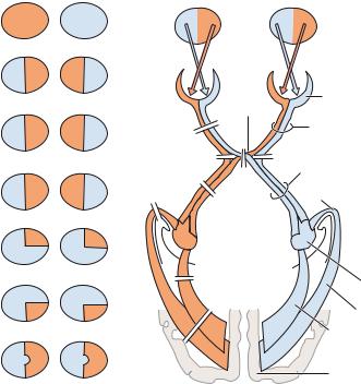
Figure 14-4 The pupillary light pathway. Light shined into one eye causes both pupils to constrict. The response in the stimulated eye is called the direct pupillary light reflex. The response in the opposite eye is called the consensual pupillary light reflex. CN, cranial nerve.
VThe Near Reflex and Accommodation Pathway
A.The cortical visual pathway projects from the primary visual cortex (Brodmann area 17) to the visual association cortex (Brodmann area 19).
B.The visual association cortex (Brodmann area 19) projects through the corticotectal tract to the superior colliculus and pretectal nucleus.
C.The superior colliculus and pretectal nucleus project to the oculomotor complex of the midbrain. This complex includes the following structures:
1.The rostral accessory oculomotor (Edinger–Westphal) nucleus, which mediates pupillary constriction through the ciliary ganglion.
2.The caudal accessory oculomotor (Edinger–Westphal) nucleus, which mediates contraction of the ciliary muscle. This contraction increases the refractive power of the lens.
3.The medial rectus subnucleus of CN III (nucleus of Perlia) which mediates convergence.