

C H A P T E R 5
Meninges, Ventricles,
and Cerebrospinal Fluid
Objectives
1.Describe the location and identifying characteristics of the dura mater, arachnoid, and pia mater.
2.Explain where cerebrospinal fluid (CSF) is created and the route it takes to the systemic circulation.
3.Identify the ventricles and list the subdivisions and characteristics of each.
4.Describe the flow of CSF through the ventricular system.
5.Identify the meningeal spaces and include a description of what is found in each, and whether the spaces are real or potential.
6.Describe the various types of hydrocephalus and meningitis, along with their causes and findings.
IMeninges—three connective tissue membranes that surround the spinal cord and
brain.
A.Consist of the pia mater, arachnoid mater, and dura mater.
1.Pia mater—delicate, highly vascular layer of connective tissue. Closely covers the surface of the brain and spinal cord.
2.Arachnoid mater—delicate, avascular connective tissue membrane. Located between the dura mater and the pia mater. Loosely adhered to the dura mater by dural border cells and the pressure of the underlying cerebrospinal fluid (CSF).
3.Dura mater—outer layer of meninges. Consists of dense connective tissue that is divided into an outer periosteal (endosteal) layer and an inner meningeal layer. In the cranial vault, the meningeal layer forms dural folds and forms the dura mater of the spinal cord. Dural venous sinuses are located between periosteal and meningeal layers of dura mater.
B.Meningeal Spaces
1.Subarachnoid space (Figure 5-1) lies between the pia mater and the arachnoid.
a.Terminates at the level of the second sacral vertebra.
b.Contains the CSF.
2.Subdural space
a.In the cranium, the subdural space is traversed by “bridging” veins.
b.In the spinal cord, it is a clinically insignificant potential space.
3.Epidural space
a.Cranial epidural space is a potential space. It contains the meningeal arteries and veins.
b.Spinal epidural space contains fatty areolar tissue, lymphatics, and venous plexuses. The epidural space may be injected with a local anesthetic to produce a paravertebral (“saddle”) nerve block.
36
|
Meninges, Ventricles, and Cerebrospinal Fluid |
37 |
|
Arachnoid granulation |
|
Superior sagittal sinus |
|
|
|
Great cerebral vein |
|
|
(of Galen) |
|
|
Superior cistern |
|
|
Straight sinus in |
|
Interventricular |
tentorium cerebelli |
|
|
|
|
foramen (of Monro) |
Confluence of |
|
Third ventricle |
|
|
the sinuses |
|
Interpeduncular cistern
Cerebral aqueduct |
|
Fourth ventricle |
|
|
|
Pontine cistern |
|
|
|
|
Median aperture |
|
|
Cerebellomedullary cistern (cisterna magna) |
|
|
Subarachnoid space |
|
|
Central canal 
 Conus medullaris
Lumbar cistern 
Filum terminale
Figure 5-1 The subarachnoid spaces and cisterns of the brain and spinal cord. Note that the conus medullaris terminates at L-1. The lumbar cistern ends at S-2. (Reprinted from Noback CR, Strominger NL, Demarest R. The Human Nervous System. 4th ed. Baltimore, MD: Williams & Wilkins; 1991:68, with permission.)
C.Meningeal Tumors
1.Meningiomas—benign, well-circumscribed, slow-growing tumors. They account for 15% of primary intracranial tumors and are more common in women than in men (3:2). Ninety percent of meningiomas are supratentorial.
2.Subdural and epidural hematomas
a.Subdural hematoma—caused by laceration of the superior cerebral (bridging) veins.
b.Epidural hematoma—caused by laceration of a cerebral artery, most commonly the middle meningeal artery.
D.Meningitis—inflammation of the pia-arachnoid area of the brain, the spinal cord, or both.
1.Bacterial meningitis—characterized clinically by fever, headache, nuchal rigidity, and Kernig
sign. (With the patient supine, the examiner flexes the patient’s hip but cannot extend the knee without causing pain. (Remember: Kernig = knee.) More than 70% of cases occur in children younger than 5 years. The disease may cause cranial nerve palsies and hydrocephalus.

38Chapter 5
a.Common causes
i.Newborns (younger than 1 month), bacterial meningitis is most frequently caused by group B streptococci (Streptococcus agalactiae), Escherichia coli, and Listeria monocytogenes.
ii.Older infants and young children (1 to 23 months)—most commonly caused by Streptococcus pneumoniae.
iii.Young adults (2 to 18 years)—most frequently caused by Neisseria meningitidis.
iv.Older adults (19 years and older)—most frequently caused by S. pneumoniae. Immunization against Haemophilus influenzae has significantly reduced this type of meningitis.
b.CSF findings
i.Numerous polymorphonuclear leukocytes
ii.Decreased glucose levels
iii. Increased protein levels
2.Viral Meningitis—also known as aseptic meningitis. Is characterized clinically by fever, headache, nuchal rigidity, and Kernig sign.
a.Common causes—many viruses are associated with viral meningitis, including mumps, echovirus, Coxsackievirus, Epstein–Barr virus, and herpes simplex type 2.
b.CSF findings
i.Numerous lymphocytes
ii.Normal glucose levels
iii. Moderately increased protein levels
IIVentricular System
A.Choroid Plexus—a specialized structure that projects into all four ventricles of the brain. Consists of infoldings of blood vessels of the pia mater that are covered by modified ciliated ependymal cells. It secretes CSF.
B.Ventricles Contain CSF and Choroid Plexus
1.Two lateral ventricles communicate with the third ventricle through the interventricular foramina (of Monro).
2.Third ventricle—located between the medial walls of the diencephalon. Communicates with the fourth ventricle through the cerebral aqueduct.
3.Cerebral aqueduct (of Sylvius) connects the third and fourth ventricles. Blockage of the cerebral aqueduct results in noncommunicating hydrocephalus.
4.Fourth ventricle communicates with the subarachnoid space through three outlet foramina: two lateral foramina (of Luschka) and one median foramen (of Magendie).
C.Hydrocephalus—dilation of the cerebral ventricles caused by blockage of the CSF pathways. Characterized by excessive accumulation of CSF in the ventricles or subarachnoid space.
1.Noncommunicating hydrocephalus results from obstruction within the ventricles (e.g., congenital aqueductal stenosis).
2.Communicating hydrocephalus results from blockage within the subarachnoid space (e.g., adhesions after meningitis).
3.Normal-pressure hydrocephalus occurs when the CSF is not absorbed by the arachnoid
villi. May occur secondary to posttraumatic meningeal hemorrhage. Characterized by the triad of progressive dementia, ataxic gait, and urinary incontinence. (Remember: wacky, wobbly, and wet.)
4.Hydrocephalus ex vacuo results from a loss of cells in the caudate nucleus (e.g., Huntington disease).
5.Pseudotumor cerebri (benign intracranial hypertension) results from increased resistance to CSF outflow at the arachnoid villi. Occurs in obese young women and is characterized by papilledema without mass, elevated CSF pressure, and deteriorating vision. The ventricles may be slit-like.

Meninges, Ventricles, and Cerebrospinal Fluid |
39 |
Table 5-1: Cerebrospinal Fluid Profiles in Subarachnoid Hemorrhage, Bacterial Meningitis, and Viral Encephalitis
Cerebrospinal Fluid |
Normal |
Subarachnoid Hemorrhage |
Bacterial Meningitis |
Viral Encephalitis |
Color |
Clear |
Bloody |
Cloudy |
Clear, cloudy |
Cell count/mm3 |
<5 lymphocytes |
Red blood cells present |
>1,000 polymorpho- |
25–500 |
|
|
|
nuclear leukocytes |
lymphocytes |
Protein |
<45 mg/dL |
Normal to slightly elevated |
Elevated, >100 mg/dL |
Slightly elevated |
Glucose 66% of |
>45 mg/dL |
Normal |
Reduced |
Normal |
blood (80–120 mg/dL) |
|
|
|
|
III Cerebrospinal Fluid—a colorless acellular fluid. Flows through the ventricles and into the subarachnoid space.
A.Function
1.CSF supports the central nervous system (CNS) and protects it against concussive injury.
2.Transports hormones and hormone-releasing factors.
3.Removes metabolic waste products through absorption.
B.Formation and Absorption—CSF is formed by the choroid plexus. Absorption is primarily through the arachnoid villi into the superior sagittal sinus.
C.Composition of CSF is clinically relevant (Table 5-1).
1.The normal number of mononuclear cells is fewer than 5/μL.
2.Red blood cells in the CSF indicate subarachnoid hemorrhage (e.g., caused by trauma or a ruptured berry aneurysm).
3.CSF glucose levels are normally 50 to 75 mg/dL (66% of the blood glucose level). Glucose levels are normal in patients with viral meningitis and decreased in patients with bacterial meningitis.
4.Total protein levels are normally between 15 and 45 mg/dL in the lumbar cistern. Protein levels are increased in patients with bacterial meningitis and normal or slightly increased in patients with viral meningitis.
5.Normal CSF pressure in the lateral recumbent position ranges from 80 to 180 mm H2O.
IV Herniation (Figures 5-2 to 5-7)
A.Transtentorial (Uncal) Herniation is protrusion of part of the brain through the tentorial incisure. May result in oculomotor paresis and contralateral hemiplegia.
B.Transforaminal (Tonsillar) Herniation—protrusion of the brainstem and cerebellum through the foramen magnum. Clinical complications include obtundation and death.
C.Subfalcine (Cingulate) Herniation—herniation below the falx cerebri (cerebral falx). Does not necessarily result in severe clinical symptoms. Can present as headache. Compression of the anterior cerebral artery may result in contralateral lower limb weakness.
40 |
Chapter 5 |
1
 2
2
3
8
4 |
5 |
7 |
|
6 |
|||
|
|
9
Figure 5-2 Coronal section of a tumor in the supratentorial compartment. (1) Anterior cerebral artery; (2) subfalcine herniation; (3) shifting of the lateral ventricles; (4) posterior cerebral artery (compression results in contralateral hemianopia); (5) uncal (transtentorial) herniation; (6) Kernohan notch (contralateral cerebral peduncle), with damaged corticospinal and corticonuclear fibers; (7) tentorium cerebelli; (8) pyramidal cells that give rise to the corticospinal tract; (9) tonsillar (transforaminal) herniation, which damages vital medullary centers. (Adapted with permission from Leech RW, Shumann RM. Neuropathology. New York: Harper & Row; 1982:16.)
4 |
5 |
|
3 |
6 |
|
7 |
||
2 |
||
8 |
||
|
||
1 |
7 |
|
|
Figure 5-3 Axial section through the midbrain and the herniating parahippocampal gyrus. The left oculomotor nerve is being stretched (dilated pupil). The left posterior cerebral artery is compressed, resulting in a contralateral hemianopia. The right crus cerebri is damaged (Kernohan notch) by the free edge of the tentorial incisure, resulting in a contralateral hemiparesis. Kernohan notch results in a false localizing sign. The caudal displacement of the brainstem causes rupture of the paramedian arteries of the basilar artery. The posterior cerebral arteries lie superior to the oculomotor nerves. (1) Parahippocampal gyrus; (2) crus cerebri; (3) posterior cerebral artery; (4) optic nerve; (5) optic chiasma; (6) oculomotor nerve;
(7) free edge of tentorium; (8) Kernohan notch. (Adapted from Leech RW, Shumann RM. Neuropathology. New York: Harper & Row; 1982:19, with permission.)
Meninges, Ventricles, and Cerebrospinal Fluid |
41 |
B
C
A
D
E
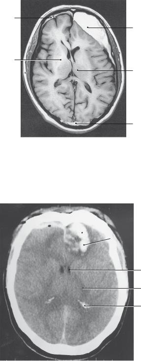
Figure 5-4 Magnetic resonance imaging scan showing brain trauma. (A) Internal capsule; (B) subdural hematoma; (C) subdural hematoma; (D) thalamus; (E) epidural hematoma. The hyperintense signals are caused by methemoglobin. This is a T1-weighted image.
 A
A
B
C
D
Figure 5-5 Computed tomography axial section showing an intraparenchymal hemorrhage in the left frontal lobe. (A) Intraparenchymal hemorrhage; (B) lateral ventricle; (C) internal capsule; (D) calcified glomus in the trigone region of the lateral ventricle.
42 |
Chapter 5 |
A
B
C
D
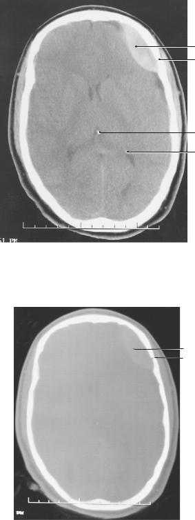
Figure 5-6 Computed tomography axial section showing an epidural hematoma and a skull fracture. (A) Epidural hematoma; (B) skull fracture; (C) calcified pineal gland; (D) calcified glomus in the trigone region of the lateral ventricle. The epidural hematoma is a classic biconvex, or lentiform, shape.
A
B
Figure 5-7 Computed tomography (CT) axial section showing a skull fracture (A) on the left side. An epidural hematoma (B) underlies the fracture. The CT scan shows a bone window.

Meninges, Ventricles, and Cerebrospinal Fluid |
43 |
CASE 5-1
The patient is a 68-year-old man with alcoholic cirrhosis. He fell 4 weeks ago. He has a history of progressive weakness on the right side. What is the most likely diagnosis?
Relevant Physical Exam Findings
●Hemiparesis
●Reflex asymmetry
Relevant Lab Findings
●Computed tomography scan of the head showed a crescent-shaped hypodense area between the cortex and skull. This mass has resulted in massive midline shift to the right, with subfalcine and uncal herniation.
Diagnosis
●Subdural hematoma results from bleeding between the dura mater and the arachnoid membrane from the bridging veins that connect the cerebral cortex to the dural venous sinuses. Subdural hematoma is common after acute deceleration injury from a fall or motor vehicle accident but rarely is associated with skull fracture.