


C H A P T E R 1 2
Auditory System
Objectives
1.Describe the central and peripheral components of the auditory pathway.
2.Compare and contrast conduction and nerve deafness and describe the clinical diagnostic tests for each.
3.Outline the clinical testing and relevance of brain stem auditory evoked response (BAER).
IIntroduction. The auditory system is an exteroceptive special somatic afferent system
that can detect sound frequencies from 20 Hz to 20,000 Hz. It is served by the vestibulocochlear nerve (CN VIII). It is derived from the otic vesicle, which is a derivative of the otic placode, a thickening of the surface ectoderm.
IIThe Auditory Pathway (Figure 12-1) consists of the following structures:
A.The hair cells of the organ of Corti are innervated by the peripheral processes of bipolar cells of the spiral ganglion. They are stimulated by vibrations of the basilar membrane.
1.Inner hair cells (IHCs) are the chief sensory elements; they synapse with dendrites of myelinated neurons whose axons make up 90% of the cochlear nerve.
2.Outer hair cells (OHCs) synapse with dendrites of unmyelinated neurons whose axons make up 10% of the cochlear nerve. The OHCs reduce the threshold of the IHCs.
B.The bipolar cells of the spiral (cochlear) ganglion project peripherally to the hair cells of the organ of Corti. They project centrally as the cochlear nerve to the cochlear nuclei.
C.The cochlear nerve (cranial nerve [CN] VIII) extends from the spiral ganglion to the cerebellopontine angle, where it enters the brain stem.
D.The cochlear nuclei receive input from the cochlear nerve. They project contralaterally to the superior olivary nucleus and lateral lemniscus.
E.The superior olivary nucleus, which plays a role in sound localization, receives bilateral input from the cochlear nuclei. It projects to the lateral lemniscus.
F.The trapezoid body is located in the pons. It contains decussating fibers from the anterior cochlear nuclei.
G.The lateral lemniscus receives input from the contralateral cochlear nuclei and superior olivary nuclei.
100
|
|
|
|
|
|
|
|
|
|
|
|
Auditory System |
101 |
|||
Thalamus |
|
|
|
Caudate nucleus |
|
|
|
|||||||||
|
|
|
|
|
|
|
|
|
Internal capsule |
|
|
|
||||
|
|
|
|
|
|
|
|
|
Putamen |
|
Lentiform nucleus |
|
||||
|
|
|
|
|
|
|
|
|
|
|
|
|
|
|
|
|
|
|
|
|
|
|
|
|
|
Globus pallidus |
|
|
|||||
|
|
|
|
|
|
|
|
|
|
|
|
|||||
|
|
|
|
|
|
|
|
|
|
|
|
Superior temporal gyrus |
|
|||
|
|
|
|
|
|
|
|
|
|
|
|
|||||
|
|
|
|
|
|
|
|
|
|
|
|
Auditory radiations in sublenticular |
|
|||
|
|
|
|
|
|
|
|
|
|
|
|
|||||
|
|
|
|
|
|
|
|
|
|
|
|
part of internal capsule |
|
|||
Brachium of |
|
|
|
|
|
|
|
Medial geniculate body |
|
|||||||
inferior colliculus |
|
|
|
|
|
|
|
|
|
|
Commissure of |
|
|
|
||
|
|
|
|
|
|
|
|
|
|
|
|
|||||
|
|
|
|
|
|
|
|
|
|
|
|
|
|
|
||
|
|
|
|
|
|
|
|
|
|
|
|
inferior colliculus |
|
|||
Midbrain |
|
|
|
|
|
|
|
|
|
|
|
Nucleus of inferior colliculus |
|
|||
|
|
|
|
|
|
|
|
|
|
|
|
|||||
|
|
|
|
|
|
|
|
|
Lateral lemniscus |
|
||||||
|
|
|
|
|
|
|
|
|
||||||||
|
|
|
|
|
|
|
|
|
|
|
|
|
||||
|
|
|
|
|
|
|
|
|
|
|
|
|
||||
|
|
|
|
|
|
|
|
|
|
|
|
|
||||
|
|
|
|
|
|
|
|
|
|
|
|
Nucleus and commissure |
|
|||
|
|
|
|
|
|
|
|
|
|
|
|
|
||||
|
|
|
|
|
|
|
|
|
|
|
|
of lateral lemniscus |
|
|||
Posterior and |
|
|
|
|
|
|
|
|
|
|
|
|
||||
anterior cochlear |
|
|
|
|
|
|
|
Inner |
Tectorial membrane |
|
||||||
nuclei |
|
|
|
|
|
|
|
|
||||||||
|
|
|
|
|
|
|
|
|
|
|
|
|||||
|
|
hair cells |
Outer |
|
|
|
|
||
Superior |
hair cells |
|||
|
||||
olivary nucleus |
|
|
Basilar membrane |
|
|
||||
Trapezoid body |
||||
Spiral ganglion |
||||
|
Pyramidal tract |
|||
|
|
|||
|
|
Cochlear nerve (CN VIII) |
||
|
Base of pons |
|
||
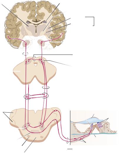
Figure 12-1 Peripheral and central connections of the auditory system. This system arises from the hair cells of the organ of Corti and terminates in the transverse temporal gyri of Heschl of the superior temporal gyrus. It is characterized by the bilaterality of projections and the tonotopic localization of pitch at all levels. For example, high pitch (20,000 Hz) is localized at the base of the cochlea and in the posteromedial part of the transverse temporal gyri. CN, cranial nerve.
H.The nucleus of inferior colliculus receives input from the lateral lemniscus. It projects through the brachium of the inferior colliculus to the medial geniculate body.
I.The medial geniculate body receives input from the nucleus of the inferior colliculus. It projects through the internal capsule as the auditory radiation to the primary auditory cortex, the superior temporal gyrus (transverse temporal gyri of Heschl).
J.The superior temporal gyrus (transverse temporal gyri of Heschl) contains the primary auditory cortex (Brodmann areas 41 and 42). The gyri are located in the depths of the lateral sulcus.

102 Chapter 12
IIIHearing Defects
A.Conduction Deafness is caused by interruption of the passage of sound waves through the external or middle ear. It may be caused by obstruction (e.g., wax), otosclerosis, or otitis media and is often reversible.
B.Nerve Deafness (Sensorineural, or Perceptive, Deafness) is typically permanent
and is caused by disease of the cochlea, cochlear nerve (acoustic neuroma), or central auditory connections. It is usually caused by presbycusis that results from degenerative disease of the organ of Corti in the first few millimeters of the basal coil of the cochlea (high-frequency loss of 4,000 to 8,000 Hz).
IV Auditory Tests
A.Tuning Fork Tests (Table 12-1)
1.Weber test is performed by placing a vibrating tuning fork on the vertex of the skull. Normally, a patient hears equally on both sides.
a.A patient who has unilateral conduction deafness hears the vibration more loudly in the affected ear.
b.A patient who has unilateral partial nerve deafness hears the vibration more loudly in the normal ear.
2.The Rinne test compares air and bone conduction. It is performed by placing a vibrating tuning fork on the mastoid process until the vibration is no longer heard; then the fork is held in front of
the ear. Normally, a patient hears the vibration in the air after bone conduction is gone. Note that a positive Rinne test means that sound conduction is normal (air conduction [AC] is greater than bone conduction [BC]), whereas a negative Rinne test indicates conduction loss, with BC greater than AC (Table 12-1).
a.A patient who has unilateral conduction deafness does not hear the vibration in the air after bone conduction is gone.
b.A patient who has unilateral partial nerve deafness hears the vibration in the air after bone conduction is gone.
B.Brain Stem Auditory Evoked Response (BAER)
1.Testing method. Clicks are presented to one ear, then to the other. Scalp electrodes and a computer generate a series of seven waves. The waves are associated with specific areas of the auditory pathway.
2.Diagnostic value. This method is valuable for diagnosing brain stem lesions (multiple sclerosis) and posterior fossa tumors (acoustic neuromas). It is also useful for assessing hearing in infants. Approximately 50% of patients with multiple sclerosis have abnormal BAERs.
Table 12-1: Tuning Fork Test Results
Otologic Finding |
Weber Test |
Rinne Test |
Conduction deafness (left ear) |
Lateralizes to left ear |
BC > AC on left |
|
|
AC > BC on right |
Conduction deafness (right ear) |
Lateralizes to right ear |
BC > AC on right |
|
|
AC > BC on left |
Nerve deafness (left ear) |
Lateralizes to right ear |
AC > BC both ears |
Nerve deafness (right ear) |
Lateralizes to left ear |
AC > BC both ears |
Normal ears |
No lateralization |
AC > BC both ears |
AC, air conduction; BC, bone conduction.

Auditory System |
103 |
CASE 12-1
A 45-year-old woman presents with a 10-year history of auditory decline in her left ear. The problem began after her first pregnancy. There is no history of otologic infection or trauma. What is the most likely diagnosis?
Relevant Physical Exam Findings
●The external auditory meatus and tympanic membrane were benign bilaterally.
●The Weber test lateralized to the left side at 512 Hz, and the Rinne test was negative at 512 Hz on the left and was positive on the right.
Diagnosis
● Otosclerosis