C H A P T E R 1 9
Cross-Sectional Anatomy
of the Brain
Objective
1.Identify the major structures of the brain from the three orthogonal planes, including sagittal, coronal, and axial sections.
IIntroduction. Thick, stained brain slices in this chapter are accompanied by
corresponding magnetic resonance imaging scans. Together they represent a mini-atlas of brain slices in the three orthogonal planes (i.e., midsagittal, coronal, and axial). An insert on each figure shows the level of the slice. The most commonly tested structures are labeled.
142

Central sulcus
Precuneus
Pineal body
Parieto-occipital sulcus
Thalamus |
Cerebral aqueduct |
|
Fornix
Septum pellucidum
Anterior commissure
Pericallosal artery
Cuneus
Calcarine sulcus
Lingual gyrus
Decussation of superior cerebellar peduncles
Medial longitudinal fasciculus
Fourth ventricle
 Tonsil
Hypothalamus
Mamillary body |
Basilar artery Posterior commissure |
Figure 19-1 Midsagittal section of the brain with meninges and blood vessels intact. Arachnoid granulations are seen along the crest of the hemisphere. The posterior commissure, decussation of the superior cerebellar peduncles, and medial longitudinal fasciculus are well demonstrated. (Modified from Roberts M, Hanaway J, Morest DK. Atlas of the Human Brain in Section. 2nd ed. Philadelphia, PA: Lea & Febiger; 1987:85.)
143

144
Thalamus |
Central sulcus |
|
Corpus callosum (body) |
Choroid plexus |
|
|
|
|
Lateral ventricle |
|
|
|
|
Corpus callosum (splenium) |
Cingulate gyrus/cingulum |
|
Calcarine sulcus |
|
|
|
Caudate nucleus |
|
|
Corpus callosum |
|
Superior cerebellar peduncle |
|
|
|
(genu) |
|
|
Corpus callosum |
|
|
(rostrum) |
|
|
Mamillothalamic tract |
|
Medial lemniscus |
|
|
|
Optic nerve (CN II) |
|
|
Red nucleus |
|
Inferior olivary nucleus |
|
|
|
Substantia nigra |
|
|
|
Abducent nerve (CN VI) |
Corticospinal fibers |
Figure 19-2 Parasagittal section through the red nucleus, medial lemniscus, and inferior olivary nucleus. The corticospinal fibers can be traced from the crus cerebri to the spinal cord. The abducent nerve (CN VI) is seen exiting from the junction of the pons and medulla. (Modified from Roberts M, Hanaway J, Morest DK. Atlas of the Human Brain in Section. 2nd ed. Philadelphia, PA: Lea & Febiger; 1987:81.)

Cingulate gyrus
Superior frontal gyrus |
Anterior cerebral artery Crista galli
Basilar artery
Sphenoid sinus 
Clivus 
Nasopharynx 
C2 
Cross-Sectional Anatomy of the Brain |
145 |
Paracentral lobule
Precuneus
Superior sagittal sinus
Parietooccipital fissure
Great cerebral vein (of Galen)
Cuneus
Straight sinus |
Calcarine sulcus
Lingual gyrus
 Diploe
Cerebellum
Cerebellomedullary cistern (Cistern magna)
Figure 19-3 Midsagittal magnetic resonance imaging section through the brain and brain stem showing the important structures surrounding the third and fourth ventricles. This is a T1-weighted image. The gray matter appears gray (hypointense), whereas the white matter appears white (hyperintense).
|
|
|
|
|
Fornix |
Thalamus |
||||
Corpus callosum |
|
|
|
|
|
|
|
|
|
Great cerebral vein |
|
|
|
||||||||
Lateral ventricle |
|
|
|
|
|
|
|
|
|
(of Galen) |
|
|
|
|
|
|
|
|
Pineal gland |
||
|
|
|
|
|||||||
|
|
|
|
|
|
|
|
|
|
|
Anterior cerebral artery |
|
|
|
|
|
|
|
|
Superior and inferior colliculi |
|
|
|
|
|
|||||||
Optic chiasm |
|
|
|
|
|
|
|
|
|
|
|
|
|
|
|
|
|
|
|
||
|
|
|
|
|
|
|
Fourth ventricle |
|||
|
|
|
|
|
||||||
|
|
|
|
|
|
|
|
|
|
|
Hypophysis/infundibulum |
|
|
|
|
|
|
Cerebellar vermis |
|||
|
|
|
|
|||||||
|
|
|
|
|
|
|
|
|
|
|
Mamillary body |
|
|
|
|
||||||
|
|
|
|
|
|
|
|
|
|
Cerebellomedullary cistern |
|
|
|
|
|
|
|
|
|
|
|
Cerebral aqueduct |
|
|
|
(Cisterna magna) |
||||||
|
|
|
|
|||||||
|
|
|
|
|
|
|
|
|
|
Spinal cord |
|
|
|
|
|
|
|
|
|
|
|
|
|
|
|
|
|
|
|
|
|
Subarachnoid space |
|
|
|
|
|
|
|
|
|
|
|
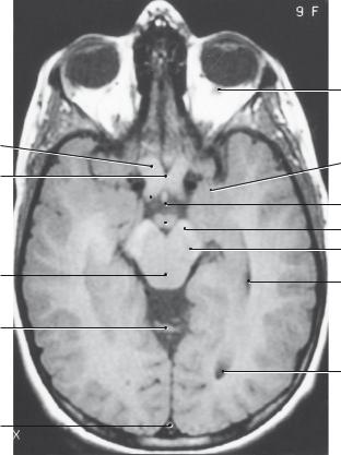
Figure 19-4 Midsagittal magnetic resonance imaging section through the brain stem and diencephalon. Note the cerebrospinal fluid tract: lateral ventricle, cerebral aqueduct, fourth ventricle, cerebellomedullary cistern (cisterna magna), and spinal subarachnoid space. Note also the relation between the optic chiasm, infundibulum, and hypophysis (pituitary gland).

146
Internal capsule
Caudate nucleus
Subthalamic nucleus
Anterior commissure
Olfactory tract
Optic nerve (CN II)
|
Central sulcus |
|
Cinculate sulcus |
Cingulate sulcus |
(marginal branch) |
Thalamus
Medial geniculate nucleus
Parieto-occipital sulcus
 Calcarine sulcus
Basis pedunculi (cerebral peduncle)
 Dentate nucleus
Inferior cerebellar peduncle
Pontine nuclei
Olive
Abducent nerve (CN VI)
Substantia nigra
Figure 19-5 Parasagittal section through the caudate nucleus, subthalamic nucleus, substantia nigra, and dentate nucleus. (Modified from Roberts M, Hanaway J, Morest DK. Atlas of the Human Brain in Section. 2nd ed. Philadelphia, PA: Lea & Febiger; 1987:79.)
Longitudinal fissure
Septum pellucidum
Lateral ventricle (frontal horn)
Claustrum 
|
Nucleus of |
Anterior commissure |
diagonal band |
Amygdala |
Optic chiasm |
|
Corpus callosum
Septal nucleus (lateral)
Caudate nucleus (head)
Internal capsule
Globus pallidus
Insular cortex
Putamen
 Lateral sulcus
Anterior perforated substance
Middle cerebral artery
Internal carotid artery
Figure 19-6 Coronal section through the anterior commissure, amygdala, septal nuclei, and optic chiasm. (Modified from Roberts M, Hanaway J, Morest DK. Atlas of the Human Brain in Section. 2nd ed. Philadelphia, PA: Lea & Febiger; 1987:9.)
147

148 Chapter 19
Septum pellucidum
Internal capsule
Amygdala |
Hypophysis |
Cavernous sinus |
Sphenoid sinus |
Nasopharynx
Longitudinal cerebral fissure
Cingulate gyrus
Corpus callosum
Lateral ventricle
 Caudate nucleus
Third ventricle
Optic chiasm
Infundibulum
Interior carotid artery
Figure 19-7 Coronal magnetic resonance imaging section through the amygdala, optic chiasm, infundibulum, and internal capsule. The cavernous sinus encircles the sella turcica and contains the following structures: cranial nerves (CN) III, IV, VI, V1, and V2; postganglionic sympathetic fibers; and the internal carotid artery. This is a T1-weighted image.

Corpus callosum
Anterior nucleus
Internal capsule
Putamen
Optic tract
Amygdala
Lateral ventricle (temporal horn)
Hippocampal formation
Third ventricle
Mamillary nucleus
Interpeduncular fossa
Base of pons
Fornix
Massa intermedia
Caudate nucleus
Ventral lateral nucleus
Mamillothalamic tract (MTT)
Claustrum
Globus pallidus
Subthalamic nucleus
Substantia nigra
Crus cerebri
Figure 19-8 Coronal section through the posterior limb of the internal capsule, mammillothalamic tract, mammillary body, and hippocampal formation. The optic tracts are visible bilaterally. (Modified from Roberts M, Hanaway J, Morest DK. Atlas of the Human Brain in Section. 2nd ed. Philadelphia, PA: Lea & Febiger; 1987:19.)
149

150 Chapter 19
Corpus callosum
Caudate nucleus
Putamen |
|
|
|
Thalamus |
|
|
Internal capsule
Globus pallidus
Substantia nigra
Hippocampus
Interpeduncular fossa
Crus cerebri |
|
Base of pons |
|
Pyramid of medulla
Figure 19-9 Coronal magnetic resonance imaging section of the brain and brainstem at the level of the thalamus, and hippocampal formation. Note that the posterior limb of the internal capsule lies between the thalamus and the lentiform nucleus (putamen and globus pallidus). This is a T1-weighted postcontrast image.

Corpus callosum |
Thalamus |
|
Fornix |
||
Lateral ventricle |
||
|
Caudate nucleus
Internal capsule
Claustrum
VPL
Putamen
VPM
 Globus pallidus
Stria terminalis |
Red nucleus
Caudate nucleus
Hippocampal formation
Substantia nigra |
Optic tract |
|
|
Interpeduncular nucleus |
Crus cerebri |
Pontine nuclei |
Figure 19-10 Coronal section through the thalamus, ventral posteromedial nucleus (VPM) and the ventral posterolateral nucleus (VPL), posterior limb of the internal capsule, substantia nigra, and red nucleus. (Modified from Roberts M, Hanaway J, Morest DK. Atlas of the Human Brain in Section. 2nd ed. Philadelphia, PA: Lea & Febiger; 1987:23.)
151

152
Medial dorsal nucleus
Medial geniculate nucleus
Putamen
Lateral geniculate nucleus
Caudate nucleus
Hippocampal formation
Lateral lemniscus
Cingulate gyrus
Corpus callosum
Fornix
Caudate nucleus
Third ventricle
Lateral posterior nucleus
Posterior commissure
Internal capsule
Cerebral aqueduct
Medial lemniscus
Medial longitudinal fasciculus
Decussation of superior cerebellar peduncles
Middle cerebellar peduncle
Inferior olivary nucleus
Figure 19-11 Coronal section through the lateral and medial lemnisci, lateral and medial geniculate nuclei, and hippocampal formation. (Modified from Roberts M, Hanaway J, Morest DK. Atlas of the Human Brain in Section. 2nd ed. Philadelphia, PA: Lea & Febiger; 1987:25.)

Fornix |
Corpus callosum (splenium) |
|
|
|
Choroid plexus |
Lateral ventricle
Pineal gland
Superior colliculus
Pulvinar nucleus
Fornix
Caudate nucleus (tail)
Hippocampal formation
Hippocampal formation |
|
|
Inferior colliculus |
Trochlear nerve (CN IV) |
|
Inferior cerebellar peduncle |
Superior cerebellar peduncle |
|
Abducent nucleus |
Figure 19-12 Coronal section through the pulvinar, pineal gland (epiphysis), superior and inferior colliculi, and trochlear nerve (CN IV). (Modified from Roberts M, Hanaway J, Morest DK. Atlas of the Human Brain in Section. 2nd ed. Philadelphia, PA: Lea & Febiger; 1987:29.)
153

154
Internal capsule (ant. limb)
Globus pallidus
Insular cortex 
Internal capsule
(post. limb)
Stria medullaris
Choroid plexus
Pulvinar nucleus
Fornix
Anterior commissure
Third ventricle
Caudate nucleus
Putamen
Internal capsule (genu)
Claustrum
Massa intermedia
Medial dorsal nucleus
Caudate nucleus
Fornix
Optic radiations
Figure 19-13 Axial section through the internal capsule, anterior commissure, and pulvinar nucleus. (Modified from Roberts M, Hanaway J, Morest DK. Atlas of the Human Brain in Section. 2nd ed. Philadelphia, PA: Lea & Febiger; 1987:51.)

Cross-Sectional Anatomy of the Brain |
155 |
Lateral ventricle
Septum pellucidum
Putamen
Globus pallidus
Insula
External capsule 
Velum interpositum
Superior sagittal sinus
Corpus callosum (genu)
Caudate nucleus
Internal capsule
(anterior limb)
Internal capsule (genu)
Internal capsule (posterior limb)
Third ventricle and thalamus
 Trigone
(of lateral ventricle)
Corpus callosum (splenium)
Optic radiations
 Visual cortex
Figure 19-14 Axial magnetic resonance imaging section at the level of the internal capsule and basal nuclei (ganglia). Note that the caudate nucleus bulges into the frontal horn of the lateral ventricle. In Huntington’s disease, there is a massive loss of γ-aminobutyric acid (GABA)-ergic neurons in the caudate nucleus that results in hydrocephalus ex vacuo. A lesion of the genu of the internal capsule results in a contralateral weak lower face with sparing of the upper face. This is a T1-weighted image.
156
Gyrus rectus
Hypothalamus
Optic tract
Mamillary body
Fornix (fimbria)
Hippocampal formation
Basis pedunculi (cerebral peduncle)
Anterior perforated substance
Amygdala
Substantia nigra
Lateral geniculate nucleus
Caudate nucleus (tail)
Lateral ventricle (temporal horn)
Medial geniculate nucleus
Red nucleus
Medial longitudinal fasciculus
Superior colliculus
Cerebral aqueduct
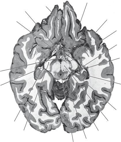
Figure 19-15 Axial section through the mammillary nuclei and the superior colliculi. (Modified from Roberts M, Hanaway J, Morest DK. Atlas of the Human Brain in Section. 2nd ed. Philadelphia, PA: Lea & Febiger; 1987:57.)
157
Gyrus rectus
Optic tract
Optic chiasm
Basis pedunculi
Amygdala
Anterior commissure
Lateral ventricle (temporal horn)
Mamillary body
Hippocampal formation
Substantia nigra
 Medial longitudinal fasciculus
Parahippocampal gyrus
Lateral lemniscus
Cerebral aqueduct
Inferior colliculus |
Cerebellum |
|
Figure 19-16 Axial section through the mammillary nuclei, optic chiasm, and inferior colliculi. (Modified from Roberts M, Hanaway J, Morest DK. Atlas of the Human Brain in Section. 2nd ed. Philadelphia, PA: Lea & Febiger; 1987:59.)

 Uncus / amygdala