


Thank you
for purchasing this e-book.
To receive special offers and news about our latest products,
sign up below.
 Sign Up
Or visit LWW.com

TM
FIFTH Neuroanatomy
EDITION
TM
FIFTH EDITION
Neuroanatomy
Douglas J. Gould, PhD
Professor and Vice Chair
Department of Biomedical Sciences
William Beaumont School of Medicine
Oakland University
Rochester, Michigan
Jennifer K. Brueckner-Collins, PhD
Professor and Vice Chair
Department of Anatomical Sciences and Neurobiology
University of Louisville School of Medicine
Louisville, Kentucky
Author of First-Fourth Editions:
James D. Fix, PhD
(1931–2010)

Acquisitions Editor: Crystal Taylor
Product Development Editor: Stephanie Roulias
Director of Medical Marketing: Lisa Zoks
Production Project Manager: Bridgett Dougherty
Design Coordinator: Teresa Mallon
Manufacturing Coordinator: Margie Orzech
Prepress Vendor: Aptara, Inc.
Fifth edition
Copyright © 2016 Wolters Kluwer.
Copyright © 2009 Wolters Kluwer Health / Lippincott Williams & Wilkins. Copyright © 2005 Lippincott Williams & Wilkins, a Wolters Kluwer business. Copyright © 2000 by Lippincott Williams & Wilkins. Copyright © 1995 by Lippincott-Raven Publishers. All rights reserved. This book is protected by copyright. No part of this book may be reproduced or transmitted in any form or by any means, including as photocopies or scanned-in or other electronic copies, or utilized by any information storage and retrieval system without written permission from the copyright owner, except for brief quotations embodied in critical articles and reviews. Materials appearing in this book prepared by individuals as part of their official duties as U.S. government employees are not covered by the above-mentioned copyright. To request permission, please contact Wolters Kluwer at Two Commerce Square, 2001 Market Street, Philadelphia, PA 19103, via email at permissions@lww.com, or via our website at lww.com (products and services).
9 8 7 6 5 4 3 2 1
Printed in China
978-1-4511-9343-5
Library of Congress Cataloging-in-Publication Data available upon request
This work is provided “as is,” and the publisher disclaims any and all warranties, express or implied, including any warranties as to accuracy, comprehensiveness, or currency of the content of this work.
This work is no substitute for individual patient assessment based upon healthcare professionals’ examination of each patient and consideration of, among other things, age, weight, gender, current or prior medical conditions, medication history, laboratory data and other factors unique to the patient. The publisher does not provide medical advice or guidance and this work is merely a reference tool. Healthcare professionals, and not the publisher, are solely responsible for the use of this work including all medical judgments and for any resulting diagnosis and treatments.
Given continuous, rapid advances in medical science and health information, independent professional verification of medical diagnoses, indications, appropriate pharmaceutical selections and dosages, and treatment options should be made and healthcare professionals should consult a variety of sources. When prescribing medication, healthcare professionals are advised to consult the product information sheet (the manufacturer’s package insert) accompanying
each drug to verify, among other things, conditions of use, warnings and side effects and identify any changes in dosage schedule or contraindications, particularly if the medication to be administered is new, infrequently used or has a narrow therapeutic range. To the maximum extent permitted under applicable law, no responsibility is assumed by
the publisher for any injury and/or damage to persons or property, as a matter of products liability, negligence law or otherwise, or from any reference to or use by any person of this work.
LWW.com
I dedicate this work to my beloved wife, Marie. Your strength, courage, and love are the engine that moves our family forward and provides the foundation for our girls to grow into proud, strong women. I love you. Thank you.
Douglas J. Gould
I dedicate my contributions to this book to my son, Lincoln. You are the light of my life and you make each and every day meaningful and fun! You have already taught me a lifetime of lessons about love, life, and the importance of play in the short 9 1/2 months that we have had together and I am eternally grateful to you for that. I hope that we will have the blessed opportunity to share many more years learning from and loving each other. I love you to the moon and back, my sweet bunny.
Jennifer K. Brueckner-Collins

P R E F A C E
Based on your feedback on previous editions of this text, the fifth edition has been reorganized and updated significantly in order to provide an accurate and quick review of important clinical aspects of neuroanatomy for the future physician. New features include the replacement of the “key concepts” with more focused “objectives” for each chapter, driving the content, order, and level of detail. The chapters have been reordered and recombined to group “like” topic more closely. A new Gross Structure chapter has been incorporated to lay the foundation for understanding the sectional anatomy in the Atlas chapter. The fourth edition’s Thalamus and Hypothalamus chapters are now integrated in the fifth edition as a new Diencephalon chapter; the previous Spinal cord, Spinal cord tracts, and Spinal cord lesions chapters are combined in a centralized Spinal Cord chapter; and the former Brainstem and Brainstem lesions chapters are united in a new Brainstem chapter. Terminology updates have been included to ensure consistency with Terminologica Anatomica.
We would appreciate receiving your comments and/or suggestions concerning High-Yield™ Neuroanatomy Fifth Edition especially after you have taken the USMLE Step 1 examination. Your suggestions will find their way into the sixth edition. You may contact us at djgould@oakland.edu or jkbrue02@louisville.edu.
vii

C O N T E N T S
Preface vii
1 |
GROSS STRUCTURE OF THE BRAIN 1 |
||||||
|
I |
Divisions of the Brain 1 |
|
|
|
|
|
2 |
DEVELOPMENT OF THE NERVOUS SYSTEM 10 |
||||||
|
I |
The Neural Tube |
10 |
|
|
|
|
|
II |
The Neural Crest |
10 |
|
|
|
|
|
III |
The Cranial Neuropore |
12 |
|
|
|
|
|
IV |
The Caudal Neuropore |
12 |
|
|
|
|
|
V |
Microglia 12 |
|
|
|
|
|
|
VI |
Myelination 12 |
|
|
|
|
|
|
VII |
The Optic Nerve and Chiasma |
12 |
|
|||
|
VIII |
The Hypophysis (pituitary gland) |
12 |
|
|||
|
IX |
Congenital Malformations of the CNS |
13 |
||||
3 |
NEUROHISTOLOGY |
17 |
|
|
|||
|
I |
Neurons 17 |
|
|
|
|
|
|
II |
Nissl Substance |
17 |
|
|
|
|
|
III |
Axonal Transport |
17 |
|
|
|
|
|
IV |
Anterograde (Wallerian) Degeneration |
18 |
||||
|
V |
Chromatolysis 18 |
|
|
|
|
|
|
VI |
Regeneration of Nerve Cells |
18 |
|
|||
|
VII |
Neuroglia 19 |
|
|
|
|
|
|
VIII |
The Blood–Brain Barrier |
19 |
|
|
|
|
|
IX |
The Blood–CSF Barrier |
19 |
|
|
|
|
|
X |
Pigments and Inclusions |
20 |
|
|
|
|
|
XI |
Classification of Nerve Fibers |
21 |
|
|||
|
XII |
Tumors of the CNS and PNS |
21 |
|
|||
|
XIII |
Cutaneous Receptors 23 |
|
|
|
||
4 |
BLOOD SUPPLY |
25 |
|
|
|
|
|
|
I |
The Spinal Cord and Caudal Brainstem |
25 |
||||
|
II |
The Internal Carotid System 25 |
|
|
|||
|
III |
The Vertebrobasilar System |
27 |
|
|
||
|
IV |
The Blood Supply of the Internal Capsule 28 |
|||||
|
V |
Veins of the Brain |
28 |
|
|
|
|
viii
Contents ix
VI |
Venous Dural Sinuses 29 |
VII |
Angiography 29 |
VIII |
The Middle Meningeal Artery 29 |
5MENINGES, VENTRICLES, AND
CEREBROSPINAL FLUID 36
|
I |
Meninges 36 |
|
|
|
|
|
|
|
|
||
|
II |
Ventricular System |
38 |
|
|
|
|
|
||||
|
III |
Cerebrospinal Fluid |
|
39 |
|
|
|
|
|
|||
|
IV |
Herniation |
39 |
|
|
|
|
|
|
|
||
6 |
SPINAL CORD |
44 |
|
|
|
|
|
|||||
|
I |
Gray and White Rami Communicans |
44 |
|
|
|||||||
|
II |
Spinal Nerves |
44 |
|
|
|
|
|
|
|
||
|
III |
Conus Medullaris |
44 |
|
|
|
|
|
||||
|
IV |
Location of the Major Motor and Sensory Nuclei of the Spinal Cord 45 |
||||||||||
|
V |
The Cauda Equina |
47 |
|
|
|
|
|
||||
|
VI |
The Myotatic Reflex |
|
47 |
|
|
|
|
|
|||
|
Case 6-1 |
49 |
|
|
|
|
|
|
|
|
|
|
|
I |
Posterior (Dorsal) Column—Medial Lemniscus Pathway |
49 |
|||||||||
|
II |
Anterolateral System |
51 |
|
|
|
|
|
||||
|
III |
Lateral Corticospinal Tract |
51 |
|
|
|
|
|||||
|
Case 6-2 |
54 |
|
|
|
|
|
|
|
|
|
|
|
I |
Diseases of the Motor Neurons and Corticospinal Tracts |
54 |
|||||||||
|
II |
Sensory Pathway Lesions |
55 |
|
|
|
|
|||||
|
III |
Combined Motor and Sensory Lesions |
|
55 |
|
|
||||||
|
IV |
Peripheral Nervous System (PNS) Lesions |
57 |
|
||||||||
|
V |
Intervertebral Disk Herniation 57 |
|
|
|
|
||||||
|
VI |
Cauda Equina Syndrome (Spinal Roots L3 to C0) 57 |
|
|||||||||
|
VII |
Conus Medullaris Syndrome (Cord Segments S3 to C0) |
58 |
|||||||||
7 |
BRAINSTEM |
|
59 |
|
|
|
|
|
|
|
||
|
I |
Introduction |
59 |
|
|
|
|
|
|
|
||
|
II |
Cross Section Through the Caudal Medulla |
59 |
|
||||||||
|
III |
Cross Section Through the Mid-Medulla |
59 |
|
|
|||||||
|
IV |
Cross Section Through the Rostral Medulla |
61 |
|
||||||||
|
V |
Cross Section Through the Caudal Pons |
62 |
|
|
|||||||
|
VI |
Cross Section Through the Mid-Pons |
63 |
|
|
|||||||
|
VII |
Cross Section Through the Rostral Pons |
63 |
|
|
|||||||
|
VIII |
Cross Section Through the Caudal Midbrain |
64 |
|
||||||||
|
IX |
Cross Section Through the Rostral Medulla |
64 |
|
||||||||
|
X |
Corticonuclear Fibers |
64 |
|
|
|
|
|
||||
xContents
Lesions of the Brainstem |
65 |
|
|
I |
Lesions of the Medulla |
65 |
|
II |
Lesions of the Pons 65 |
|
|
III |
Lesions of the Midbrain |
66 |
|
IV |
Acoustic Neuroma (Schwannoma) 67 |
||
V |
Jugular Foramen Syndrome |
67 |
|
VI |
“Locked-in” Syndrome |
68 |
|
VII |
Central Pontine Myelinolysis |
68 |
|
VIII |
“Top of the Basilar” Syndrome 68 |
||
IX |
Subclavian Steal Syndrome |
68 |
|
X |
The Cerebellopontine Angle |
68 |
|
8 AUTONOMIC NERVOUS SYSTEM 70
I |
Introduction 70 |
|
II |
Cranial Nerves (CN) With Parasympathetic Components 71 |
|
III |
Communicating Rami 73 |
|
IV |
Neurotransmitters |
73 |
V |
Clinical Correlation |
73 |
9 CRANIAL NERVES 75
I |
The Olfactory Nerve 75 |
|
|
|
II |
The Optic Nerve (CN II) |
75 |
|
|
III |
The Oculomotor Nerve (CN III) |
76 |
|
|
IV |
The Trochlear Nerve (CN IV) 77 |
|
||
V |
The Trigeminal Nerve (CN V) |
78 |
|
|
VI |
The Abducent Nerve (CN VI) |
80 |
|
|
VII |
The Facial Nerve (CN VII) |
80 |
|
|
VIII |
The Vestibulocochlear Nerve (CN VIII) |
82 |
||
IX |
The Glossopharyngeal Nerve (CN IX) |
83 |
||
X |
The Vagal Nerve (CN X) |
84 |
|
|
XI |
The Accessory Nerve (CN XI) |
85 |
|
|
XII |
The Hypoglossal Nerve (CN XII) 85 |
|
||
10 TRIGEMINAL SYSTEM 87
I |
Introduction 87 |
|
II |
The Trigeminal Ganglion 87 |
|
III |
Trigeminothalamic Pathways 88 |
|
IV |
Trigeminal Reflexes |
89 |
V |
The Cavernous Sinus |
90 |
11 DIENCEPHALON |
92 |
|
I |
Introduction |
92 |
II |
The Thalamus |
92 |
Contents xi
|
III |
Blood Supply |
94 |
|
|
|
IV |
The Internal Capsule |
94 |
|
|
|
V |
The hypothalamus |
95 |
|
|
12 |
AUDITORY SYSTEM |
100 |
|||
|
I |
Introduction |
100 |
|
|
|
II |
The Auditory Pathway 100 |
|||
|
III |
Hearing Defects 102 |
|
||
|
IV |
Auditory Tests |
102 |
|
|
13 |
VESTIBULAR SYSTEM |
104 |
|||
|
I |
Introduction |
104 |
|
|
|
II |
The Labyrinth |
104 |
|
|
|
III |
The Vestibular Pathways 104 |
|||
|
IV |
Vestibulo-ocular Reflexes |
106 |
||
14 |
VISUAL SYSTEM |
108 |
|||
|
I |
Introduction |
108 |
|
|
|
II |
The Visual Pathway |
108 |
|
|
|
III |
The Pupillary Light Reflex Pathway 111 |
|||
|
IV |
The Pupillary Dilation Pathway 111 |
|||
|
V |
The Near Reflex and Accommodation Pathway 112 |
|||
|
VI |
Cortical and Subcortical Centers for Ocular Motility 113 |
|||
|
VII |
Clinical Correlation |
114 |
|
|
15 |
LIMBIC SYSTEM |
116 |
|||
|
I |
Introduction |
116 |
|
|
|
II |
Major Components |
116 |
|
|
|
III |
The Papez Circuit 116 |
|
||
|
IV |
Clinical Correlations |
118 |
|
|
16 BASAL NUCLEI AND EXTRAPYRAMIDAL MOTOR SYSTEM 120
I |
Basal Nuclei (Ganglia) 120 |
II |
The Extrapyramidal (Striatal) Motor System 120 |
III |
Clinical Correlation 121 |
17 CEREBELLUM 126
I |
Function |
126 |
|
II |
Anatomy |
127 |
|
III |
The Deep Cerebellar Nuclei |
128 |
|
IV |
The Major Cerebellar Circuit |
128 |
|
xii |
Contents |
|
|
V |
Cerebellar Dysfunction 129 |
|
VI |
Cerebellar Syndromes and Tumors 129 |
18 CEREBRAL CORTEX |
131 |
||||
I |
Introduction |
131 |
|
|
|
II |
The Six-Layered Neocortex |
131 |
|||
III |
Functional Areas 132 |
|
|
||
IV |
Focal Destructive Hemispheric Lesions and Symptoms 135 |
||||
V |
Cerebral Dominance |
135 |
|
||
VI |
Split Brain Syndrome |
137 |
|||
VII |
Other Lesions of the Corpus Callosum 138 |
||||
VIII |
Brain and Spinal Cord Tumors 138 |
||||
IX |
Apraxia |
138 |
|
|
|
X |
Aphasia |
139 |
|
|
|
XI |
Dysprosodies |
140 |
|
|
|
19 CROSS-SECTIONAL ANATOMY OF THE BRAIN 142
I Introduction 142
20 NEUROTRANSMITTERS 161
I |
Major Neurotransmitters 161 |
II |
Functional and Clinical Considerations 165 |
Appendix I: Table of Cranial Nerves 167
Appendix II: Table of Common Neurological Disease States 170
Glossary 173
Index 183
C H A P T E R 1
Gross Structure
of the Brain
Objectives
1.Describe the telencephalon including the lobes of the cerebral hemispheres and the major gyri, sulci and lobules of each.
2.Differentiate the structures of the limbic and olfactory senses from other parts of the brain.
3.List the different parts of the diencephalon, brainstem, and cerebellum.
IDivisions of the Brain. The brain consists of five divisions: telencephalon,
diencephalon, mesencephalon, metencephalon, and myelencephalon.
A.Telencephalon consists of the cerebral hemispheres and the basal nuclei. The cerebral hemispheres contain the lateral ventricles.
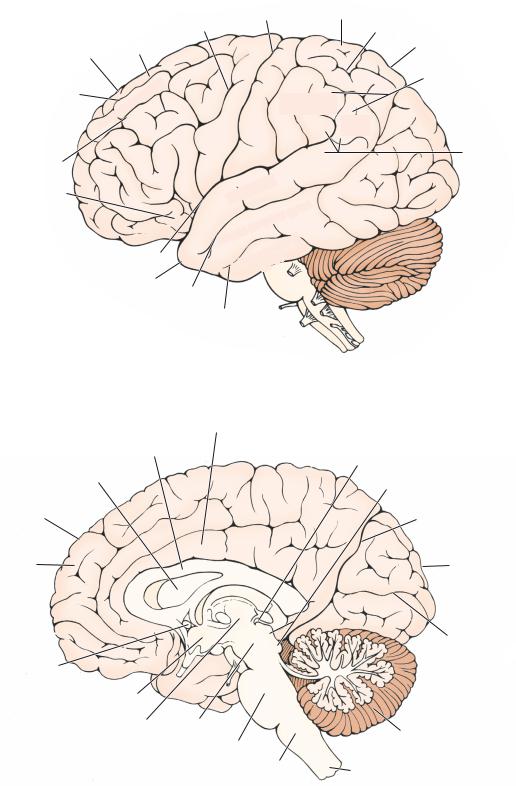
1.Cerebral hemispheres (Figures 1-1 to 1-3) consist of six lobes and the olfactory structures:
a.Frontal lobe extends from the central sulcus to the frontal pole and lies superior to the lateral sulcus. It contains:
●Precentral gyrus—consists of the primary motor area (area 4).
●Superior frontal gyrus—contains supplementary motor cortex on the medial surface (area 6).
●Middle frontal gyrus—contains the frontal eye field (area 8).
●Inferior frontal gyrus—contains the Broca speech area in the dominant hemisphere (areas 44 and 45).
●Gyrus rectus and orbital gyri—separated by the olfactory sulcus.
●Anterior paracentral lobule—found on the medial surface between the superior frontal gyrus (paracentral sulcus) and the central sulcus.
b.Parietal lobe extends from the central sulcus to the occipital lobe and lies superior to the temporal lobe.
●Postcentral gyrus—the primary somatosensory area of the cerebral cortex (areas 3, 1, and 2).
●Superior parietal lobule comprises association areas involved in somatosensory functions (areas 5 and 7).
●Inferior parietal lobule consists of the supramarginal gyrus, which interrelates somatosensory, auditory, and visual inputs (area 40) and the angular gyrus (area 39) that receives impulses from primary visual cortex.
●Precuneus—located between the paracentral lobule and the cuneus.
●Posterior paracentral lobule—located on the medial surface between the central sulcus and the precuneus.
1
2Chapter 1
Precentral Central sulcus |
Superior parietal |
|
lobule |
Interparietal |
|
sulcus |
|
|
|
sulcus |
|
|
|
Superior frontal |
|||||
Frontal lobe |
|
|
|
gyrus |
|
|
|
|
|
|
|
|
|
||
Superior frontal |
|
|
|
|
frontal |
gyrus |
|
|
|
|
|
|
|
||
sulcus |
|
|
|
|
|
|
|
|
|
|
|
|
|
|
|
|
|
Middle |
|
|
|
||
|
|
|
|
|
|||
Inferior frontal |
|
|
|
Inferior frontal gyrus |
|||
|
|
|
|
|
|
|
|
sulcus |
|
|
|
|
|
|
|
Broca's motor |
|
|
|
|
|
|
|
speech area |
|
|
|
|
|
|
|
|
|
|
|
|
|
||
|
|
|
Orbital gyrus |
||||
|
Lateral (Sylvian) |
||||||
|
|
|
|
sulcus |
|
|
|
gyrus |
Postcentral |
gyrus |
|
|
Precentral |
|
|
Angular |
|
|
|
Supramarginal |
||
|
|
|
gyrus |
|
|
|
|
|
gyrus |
|
|
gyrus |
|
|
temporal |
|
|
||
Superior |
|
|
gyrus |
|
temporal |
|
|||
|
|
|||
Middle |
temporal |
|
||
|
|
|||
Inferior |
|
|
|
|
|
gyrus |
|
|
|
Parietal lobe
Inferior parietal lobule
Wernicke's
area
 Occipital
lobe
Temporal lobe
Superior temporal sulcus
Middle temporal sulcus
Figure 1-1 Lateral surface of the brain showing the principal gyri and sulci.
|
|
|
Cingulate gyrus |
Parietal lobe |
|||
|
|
|
|
||||
|
|
Corpus callosum |
|
Posterior commissure |
|||
|
|
|
|||||
|
|
|
|
|
|||
|
Septum pellucidum |
|
|
Superior and |
|||
|
|
|
|
|
inferior collilculi |
||
Frontal |
|
|
Parieto-occipital |
||||
lobe |
|
|
|||||
|
|
|
sulcus |
||||
|
|
|
|
|
|
||
Superior |
|
|
|
|
|
||
frontal |
|
|
|
Occipital |
|||
gyrus |
|
|
|
||||
|
|
|
|
lobe |
|||
|
|
|
|
|
|
|
|
|
|
|
|
|
|
|
Occipital |
|
|
|
|
|
|
|
|
|
|
|
|
|
|
|
pole |
Frontal |
|
|
|
|
|
Calcarine |
|
|
|
||||||
pole |
|
|
|
|
fissure |
||
Anterior |
|
|
|
|
(sulcus) |
||
|
|
|
|
|
|||
commissure |
|
|
|
|
|
||
|
|
Hypothalamus |
|
|
|
|
|
|
|
Thalamus |
Midbrain |
|
|
|
|
|
|
|
|
Cerebellum |
|||
|
|
|
|
|
|||
|
|
|
Pons |
|
|
|
|
|
|
|
Medulla |
|
Spinal cord |
||
|
|
|
|
|
|||
Figure 1-2 Midsagittal section of the brain and brainstem showing the structures surrounding the third and fourth ventricles.
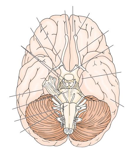
Medial olfactory stria
Lateral olfactory stria
Mammillary bodies
Middle temporal  gyrus
gyrus
Inferior temporal  gyrus
gyrus
Inferior temporal  sulcus
Collateral
sulcus
Pons
Olive
Pyramid
Gross Structure of the Brain |
3 |
Prefrontal cortex
Gyrus rectus
Olfactory bulb
Olfactory tract
Optic chiasm
Uncus
 Parahippocampal
gyrus
Occipitotemporal
gyrus
Figure 1-3 Inferior surface of the brain showing the principal gyri and sulci.
c.Temporal lobe extends from the temporal pole to the occipital lobe, inferior to the lateral sulcus. It contains:
●Transverse temporal gyrus (of Heschl)—found within the lateral sulcus. It contains the primary auditory areas of the cerebral cortex (areas 41 and 42).
●Superior temporal gyrus—associated with auditory functions and contains the Wernicke speech area in the dominant hemisphere (area 22).
●Middle temporal gyrus
●Inferior temporal gyrus
●Lateral occipitotemporal gyrus (fusiform gyrus)—lies between the inferior temporal sulcus and the collateral sulcus.
d.Occipital lobe lies posterior to a line connecting the parieto-occipital sulcus and the preoccipital notch. It contains two structures:
●Cuneus—situated between the parieto-occipital sulcus and the calcarine sulcus and contains the visual cortex (areas 17, 18, and 19).
●Lingual gyrus lies inferior to the calcarine sulcus and contains the visual cortex (areas 17, 18, and 19).
e.Insular lobe (insula) lies within the lateral sulcus.
4 |
Chapter 1 |
|
|
|
|
|
|
|
|
|
Postcommissural |
|
|
|
|
Cingulate gyrus |
fornix |
|
||
|
Precommissural |
|
Corpus callosum |
Anterior |
|
|
|
|
|
||||
|
|
nucleus |
Fornix |
|||
|
fornix |
|
|
|
|
|
|
|
|
|
|
|
|
|
|
|
|
|
|
|
|
|
|
|
|
|
|
Anterior commissure
Septal
area
Medial hypothalamus
Amygdala
Stria Hippocampal
Mammillary terminalis formation body
Figure 1-4 Midsagittal section of the brain showing the components of the limbic lobe.
f.Limbic lobe (Figure 1-4)—a C-shaped collection of structures found on the medial hemispheric surface that encircles the corpus callosum and the lateral aspect of the midbrain. It includes:
●Paraterminal gyrus and subcallosal area—located anterior to the lamina terminalis and inferior to the rostrum of the corpus callosum.
●Cingulate gyrus lies parallel and superior to the corpus callosum and merges with the parahippocampal gyrus.
●Parahippocampal gyrus lies between the hippocampal and collateral sulci and terminates in the uncus.
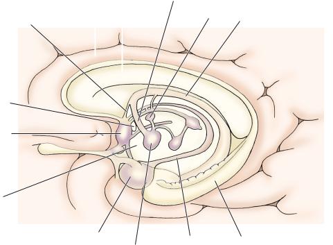
●Hippocampal formation (Figure 1-5)—connected to the hypothalamus and septal area via the fornix.
g.Olfactory structures—found on the orbital (inferior) surface of the brain and include the following:
●Olfactory bulb and tract represent an outpouching of the telencephalon. The olfactory bulb receives the olfactory nerve (CN I).
●Olfactory trigone and striae
●Anterior perforated substance created by penetrating striate arteries.
●Diagonal band of Broca interconnects the amygdaloid nucleus and the septal area.
2.Basal nuclei (ganglia) (Figure 1-6) constitute the subcortical nuclei of the telencephalon and include:
a.Caudate nucleus—part of the striatum, together with the putamen.
b.Putamen—part of the striatum, together with the caudate nucleus and part of the lentiform nucleus along with the globus pallidus.
c.Globus pallidus—part of the lentiform nucleus, together with the putamen.
d.Subthalamic nucleus—part of the diencephalon that functions with the basal nuclei.
|
|
|
|
|
Gross Structure of the Brain |
5 |
|
|
|
|
Septum pellucidum |
|
|
|
|
Corpus callosum |
|
|
Head of |
|
|
|
|
|
|
||
|
|
(genu) |
|
|
caudate nucleus |
|
|
|
|
|
|
Internal capsule |
|
Anterior horn |
|
|
(anterior limb) |
|
||
|
|
|
|
|||
(lateral ventricle) |
|
|
Interventricular foramen |
|
||
|
|
|
|
|
|
|
|
|
|
|
|
(of Monro) |
|
|
|
|
|
|
Putamen |
|
|
|
|
|
|
|
|
Amygdala |
|
|
|
Internal capsule |
|
|
|
|
|
(genu) |
|
||
|
|
|
|
|
|
|
|
|
|
|
|
Globus |
|
Hippocampus |
|
|
|
pallidus |
|
|
|
|
|
|
|||
|
|
|
|
|
Internal capsule |
|
|
|
|
|
|
(posterior limb) |
|
|
|
|
|
|
Third ventricle |
|
Fornix |
|
|
Thalamus |
|
||
|
|
|
|
|||
|
|
|
|
|
Tail of |
|
|
|
|
|
|
caudate nucleus |
|
Corpus callosum |
|
|
|
|
||
(splenium) |
|
|
|
|
||
Figure 1-5 Horizontal section of the brain showing the components of the internal capsule.
Body of caudate nucleus
Stria terminalis
Thalamus
Head of caudate nucleus
Putamen
Globus pallidus
Amygdala
Tail of caudate nucleus
Figure 1-6 Schematic diagram of basal nuclei.

6Chapter 1
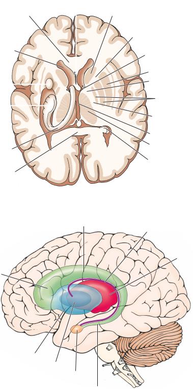
3.Lateral ventricles (see Figure 1-5)—ependyma-lined cavities of the cerebral hemispheres that contain CSF and choroid plexus. They communicate with the third ventricle via two interventricular foramina (of Monro) and are separated from each other by the septum pellucidum.
4.Cerebral cortex consists of a thin layer or mantle of gray matter that covers the surface of each cerebral hemisphere and is folded into gyri that are separated by sulci.
5.White matter includes the cerebral commissures and the internal capsule.
a.Cerebral commissures (see Figure 1-2) interconnect the cerebral hemispheres and include the following structures:
●Corpus callosum—the largest commissure of the brain and it interconnects the two hemispheres. It has four parts, including the rostrum, genu, body, and splenium.
●Anterior commissure—interconnects the olfactory bulbs with the middle and inferior temporal lobes.
●Hippocampal commissure (commissure of the fornix)—located between the fornices and inferior to the splenium of the corpus callosum.
b.Internal capsule (see Figure 1-5) consists of the white matter located between the basal nuclei and the thalamus. It has five parts:
●Anterior limb—located between the caudate nucleus and putamen and contains a mixture of ascending and descending fibers.
●Genu—located between the anterior and posterior limbs and contains primarily the corticonuclear (corticobulbar) fibers.
●Posterior limb—located between the thalamus and lentiform nucleus (comprising the putamen and the globus pallidus) and is primarily made up of corticospinal fibers.
●Retrolenticular portion—located posterior to the lentiform nucleus and contains the optic radiations.
Third ventricle |
|
|
Internal capsule |
Caudate nucleus (head) |
|
Stria medullaris |
Ant. nucleus (thalamus) |
|
Habenular trigone |
Stria terminalis |
|
|
||
|
Lenticular nucleus |
|
|
Pulvinar (thalamus) |
|
Pineal body |
|
|
Sup. colliculus |
Medial geniculate body |
|
|
||
Brachium of inf. colliculus |
Lat. geniculate body |
|
Cerebral peduncle |
||
Inf. colliculus |
||
Ant. medullary velum |
||
Trochlear nerve (CN IV) |
||
|
||
Sup. cerebellar peduncle |
Posterior median sulcus |
|
Median eminence |
||
(brachium conjunctivum) |
||
Middle cerebellar peduncle |
Facial colliculus |
|
Sulcus limitans |
||
(brachium pontis) |
||
Inf. cerebellar peduncle |
Vestibular area |
|
|
||
(restiform body) |
Striae medullares |
|
|
Hypoglossal trigone |
|
Cuneate tubercle |
Vagal trigone |
|
|
||
Gracile tubercle |
Obex |
|
|
||
Tuberculum cinereum |
Posterior median sulcus |
|
Lat. funiculus |
||
Posterior intermediate sulcus |
||
Fasciculus cuneatus |
||
|
||
Fasciculus gracilis |
Posterolateral suclus |
|
Figure 1-7 Posterior surface anatomy of the brainstem. |
|
Gross Structure of the Brain |
7 |
●Sublenticular portion—located inferior to the lentiform nucleus and contains auditory radiations.
B.Diencephalon (see Figures 1-2 and 1-7) receives the optic nerve (CN II) and consists of the following structures:
1.Epithalamus
2.(Dorsal) Thalamus—separated from the hypothalamus by the hypothalamic sulcus.
3.Hypothalamus (Figure 1-8)
4.Subthalamus (ventral thalamus)—inferior to the thalamus and lateral to the hypothalamus.
5.Third ventricle and associated structures.
C.Mesencephalon (Midbrain) (see Figures 1-7 and 1-8)—located between the diencephalon and the pons and contains the cerebral aqueduct interconnecting the third and fourth ventricles.
1.Anterior surface
a.Cerebral peduncle
b.Interpeduncular fossa
i.Oculomotor nerve (CN III)
ii.Posterior perforated substance—created by the penetrating branches of the posterior cerebral and posterior communicating arteries.
2.Posterior surface
a.Superior colliculus (visual system)
b.Brachium of the superior colliculus
Optic nerve |
Optic chiasm |
|
Caudate nucleus |
Internal capsule |
|
Lenticular nucleus |
Optic tract |
|
|
||
Ant. perforated substance |
|
|
|
Tuber cinereum |
|
Interpeduncular fossa |
|
|
(post. perforated |
|
|
substance) |
Mamillary body |
|
|
||
Cerebral peduncle |
CN III |
|
(crus cerebri) |
||
|
||
|
CN IV |
|
|
CN V (motor root) |
|
Pons |
CN V (sensory root) |
|
|
||
|
CN VI |
|
Middle cerebellar peduncle |
|
|
CN VIII |
CN VII |
|
CN VII (nervus intermedius) |
||
CN XII |
CN IX |
|
|
||
Olive |
CN X |
|
Pyramid |
CN XI |
|
|
||
First cervical n. |
Pyramidal decussation |
Figure 1-8 Anterior surface anatomy of the brainstem.

8Chapter 1
c.Inferior colliculus (auditory system)
d.Brachium of the inferior colliculus
e.Trochlear nerve (CN IV)—the only cranial nerve to exit the brainstem from the posterior aspect.
D.Pons (see Figures 1-7 and 1-8)—located between the midbrain and the medulla.
1.Anterior surface
a.Base of the pons
b.Cranial nerves, including trigeminal nerve (CN V), abducent nerve (CN VI), facial nerve (CN VII), and vestibulocochlear nerve (CN VIII)
2.Posterior surface (rhomboid fossa)
a.Locus ceruleus contains the largest collection of norepinephrinergic neurons in the CNS.
b.Facial colliculus contains the abducent nucleus and internal genu of the facial nerve.
c.Sulcus limitans separates the alar plate from the basal plate.
d.Striae medullares of the rhomboid fossa divides the rhomboid fossa into the superior pontine portion and the inferior medullary portion.
E.Medulla Oblongata (myelencephalon) (see Figures 1-7 and 1-8)—located between the pons and the spinal cord.
1.Anterior surface
a.Pyramid contains descending tracts.
b.Olive contains the inferior olivary nucleus.
Anterior lobe
Midbrain
Pons |
|
|
|
|
|
|
|||
Flocculonodular |
Primary |
|||
lobe |
fissure |
|||
Posterolateral |
Posterior lobe |
|||
fissure |
||||
|
||||
A
Anterior lobe |
Vermis |
Primary
fissure  Flocculonodular lobe
Lateral 
hemisphere
Posterior lobe
Posterolateral
fissure
B
Figure 1-9 Surface features of the cerebellum from a lateral view
(A) and a posterior view (B).
Gross Structure of the Brain |
9 |
c.Cranial nerves, including glossopharyngeal nerve (CN IX), vagus nerve (CN X), (spinal) accessory nerve (CN XI), and hypoglossal nerve (CN XII)
2.Posterior surface
a.Gracile tubercle
b.Cuneate tubercle
c.Rhomboid fossa
i.Striae medullares of the rhomboid fossa
ii.Vagal trigone
iii. Hypoglossal trigone
iv.Sulcus limitans
v.Area postrema (vomiting center)
F.Cerebellum (Figures 1-7 and 1-9)—located in the posterior cranial fossa, attached to the brain-
stem by three cerebellar peduncles. It forms the roof of the fourth ventricle. It is separated from the occipital and temporal lobes by the tentorium cerebelli and contains the following surface structures/ parts:
1.Hemispheres consist of two lateral lobes.
2.Vermis
3.Flocculus and vermal nodulus form the flocculonodular lobule.
4.Tonsil is a rounded lobule on the inferior surface of each cerebellar hemisphere. With increased intracranial pressure, it may herniate through the foramen magnum.
5.Superior cerebellar peduncle connects the cerebellum to the pons and midbrain.
6.Middle cerebellar peduncle connects the cerebellum to the pons.
7.Inferior cerebellar peduncle connects the cerebellum to the pons and medulla.
8.Anterior lobe lies anterior to the primary fissure.
9.Posterior lobe is located between the primary and posterolateral fissures.
10.Flocculonodular lobe lies posterior to the posterolateral fissure.