

136 |
4 Renal Tumors in Children |
|
|
•Factors that increase the risk of recurrence and metastasis include:
–Cellular variant.
–Older age at presentation.
–Positive surgical margins.
•Metastases to distant organs such as the brain, bone, and lungs have been reported with the cellular type.
•There is also a variant of mesoblastic nephroma which is cystic. This is called cystic mesoblastic nephroma which can be confused with other congenital cystic lesions of the kidney.
4.3.5Clinical Features
•The diagnosis of mesoblastic nephroma may be made antenatally on ultrasound performed as early as 18–20 weeks’ gestation.
•Fetal ultrasonography is useful in differentiating a renal mass from hydronephrosis but if the mass is large it may be difficult to determine the origin of the tumor.
•Fetal magnetic resonance imaging (MRI) may be more helpful in determining the organ of origin because of the ability to take images in multiple planes.
•Prematurity and polyhyramnios with or without hydrops fetalis are known to be associated with mesoblastic nephroma.
•Postnatally, although abdominal ultrasound and CT-scan are useful in diagnosing mesoblastic nephroma, MRI is the most accurate imaging modality, as it most accurately depicts the local and regional extent of the tumor.
•These radiological investigations although they may suggest the most likely diagnosis but cannot definitively differentiate a mesoblastic nephroma from other renal tumors and histologic examination is the only definitive diagnosis.
•Polyhydramnios is reported in 71 % of pregnancies associated with mesoblastic nephroma.
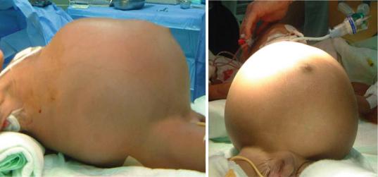
•The most common clinical presentation is an asymptomatic abdominal mass.
•The mass can grow rapidly and attain a large size (Figs. 4.72, 4.73, 4.74, and 4.75).
•Hematuria is a rare manifestation of mesoblastic nephroma.
•Paraneoplastic syndromes such as hypertension or hypercalcemia may be present.
•Hypertension is thought to be secondary to increased renin production by the trapped glomeruli in the tumor.
•Hypercalcemia: This is due to prostaglandin secretion from the tumor cells.
Figs. 4.72 and 4.73 Clinical photographs showing marked abdominal distension secondary to a very large mesoblastic nephroma
4.3 Mesoblastic Nephroma |
137 |
|
|
Figs. 4.74 and 4.75 Clinical photographs of a very large mesoblastic nephroma that was completely excised. In spite of the large size there were no secondaries
•Potential intrauterine complications with large mesoblastic nephroma:
–Abdominal dystocia at birth
–Arterio-venous shunting with subsequent development of hydrops fetalis.
4.3.6Investigations
•Complete blood count
•Serum electrolyte, BUN and creatinine
•Live function tests
•Plain radiograph: This may demonstrate a soft tissue mass displacing bowel. Calcification is very rarely seen (Figs. 4.76).
•Abdominal Ultrasound:
–This reveals a well-defined mass with lowlevel homogeneous echoes.
–The presence of concentric echogenic and hypoechoic rings can be a helpful diagnostic feature.
–A more complex pattern due to hemorrhage, cyst formation and necrosis can also be seen and tends to favor the cellular variant.
–Color Doppler ultrasound may show increased vascularity.
–Antenatal ultrasound may also show evidence of associated polyhydramnios.
•Abdominal CT-scan (Figs. 4.77 and 4.78):
– This shows a solid hypoattenuating renal
Fig. 4.76 Plain x-ray showing a large soft tissue density occupying almost the whole abdomen
lesion with variable contrast enhancement.
–Cystic areas, necrosis, and hemorrhage are uncommon but can be seen in the cellular variant.
•Abdominal MRI:
–Antenatal abdominal MRI is the most useful investigation to assess and diagnose mesoblastic nephroma. This reveals a homogenous hypointense renal mass.