

биохимия атеросклероза
.pdf148 Dean Gilham and Richard Lehner
151.Lehner R, Verger R: Purification and characterization of a porcine liver microsomal triacylglycerol hydrolase. Biochemistry 36: 1861–1868, 1997.
152.Lehner R, Cui Z, Vance DE: Subcellullar localization, developmental expression and characterization of a liver triacylglycerol hydrolase. Biochem J 338(Pt 3): 761–768, 1999.
153.Matsushima M, Inoue H, Ichinose M, Tsukada S, Miki K, Kurokawa K,
Takahashi T, Takahashi K: The nucleotide and deduced amino acid sequences of porcine liver proline-β-naphthylamidase. Evidence for the identity with carboxylesterase. FEBS Lett 293: 37–41, 1991.
154.Dolinsky VW, Gilham D, Alam M, Vance DE, Lehner R: Triacylglycerol hydrolase: role in intracellular lipid metabolism. Cell Mol Life Sci 61: 1633–1651, 2004.
155.Lehner R, Vance DE: Cloning and expression of a cDNA encoding a hepatic microsomal lipase that mobilizes stored triacylglycerol. Biochem J 343(Pt 1): 1–10, 1999.
156.Dolinsky VW, Sipione S, Lehner R, Vance DE: The cloning and expression of a murine triacylglycerol hydrolase cDNA and the structure of its corresponding gene. Biochim Biophys Acta 1532: 162–172, 2001.
157.Alam M, Ho S, Vance DE, Lehner R: Heterologous expression, purification, and characterization of human triacylglycerol hydrolase. Protein Expr Purif 24: 33–42, 2002.
158.Douglas DN, Dolinsky VW, Lehner R, Vance DE: A role for Sp1 in the transcriptional regulation of hepatic triacylglycerol hydrolase in the mouse. J Biol Chem 276: 25621–25630, 2001.
159.Gilham D, Lehner R: The physiological role of triacylglycerol hydrolase in lipid metabolism. Rev Endocr Metab Disord 5: 303–309, 2004.
160.Rusinol AE, Cui Z, Chen MH, Vance JE: A unique mitochondria-associated membrane fraction from rat liver has a high capacity for lipid synthesis and contains pre-Golgi secretory proteins including nascent lipoproteins. J Biol Chem 269: 27494–27502, 1994.
161.Gaustad R, Berg T, Fonnum F: Heterogeneity of carboxylesterases in rat liver cells. Biochem Pharmacol 44: 827–829, 1992.
162.Gilham D, Ho S, Rasouli M, Martres P, Vance DE, Lehner R: Inhibitors of hepatic microsomal triacylglycerol hydrolase decrease very low density lipoprotein secretion. FASEB J 17: 1685–1687, 2003.
163.Soni KG, Lehner R, Metalnikov P, O’Donnell P, Semache M, Gao W, Ashman K, Pshezhetsky AV, Mitchell GA: Carboxylesterase 3 (EC 3.1.1.1) is a major adipocyte lipase. J Biol Chem 279: 40683–40689, 2004.
164.Gilham D, Alam M, Gao W, Vance DE, Lehner R: Triacylglycerol hydrolase is localized to the endoplasmic reticulum by an unusual retrieval sequence where it participates in VLDL assembly without utilizing VLDL lipids as substrates. Mol Biol Cell 16: 984–996, 2005.
165.Durrington PN, Newton RS, Weinstein DB, Steinberg D: Effects of insulin and glucose on very low density lipoprotein triglyceride secretion by cultured rat hepatocytes. J Clin Invest 70: 63–73, 1982.
166.Brown AM, Wiggins D, Gibbons GF: Glucose phosphorylation is essential for the turnover of neutral lipid and the second stage assembly of triacylglycerolrich apoB-containing lipoproteins in primary hepatocyte cultures. Arterioscler Thromb Vasc Biol 19: 321–329, 1999.
Chapter 7. Provision of Lipids for VLDL Assembly |
149 |
167.Dolinsky VW, Gilham D, Hatch GM, Agellon LB, Lehner R, Vance DE: Regulation of triacylglycerol hydrolase expression by dietary fatty acids and peroxisomal proliferator-activated receptors. Biochim Biophys Acta 1635: 20–28, 2003.
168.Becker A, Bottcher A, Lackner KJ, Fehringer P, Notka F, Aslanidis C, Schmitz G: Purification, cloning, and expression of a human enzyme with acyl coenzyme A: cholesterol acyltransferase activity, which is identical to liver carboxylesterase. Arterioscler Thromb 14: 1346–1355, 1994.
169.Langmann T, Becker A, Aslanidis C, Notka F, Ullrich H, Schwer H, Schmitz G: Structural organization and characterization of the promoter region of a human carboxylesterase gene. Biochim Biophys Acta 1350: 65–74, 1997.
170.Huang GS, Yang SM, Hong MY, Yang PC, Liu YC: Differential gene expression of livers from apoE deficient mice. Life Sci 68: 19–28, 2000.
Biochemistry of Atherosclerosis edited by S.K. Cheema, Springer, New York, 2006
8
Oxidatively Modified Low-Density
Lipoproteins and Thrombosis
GARRY X. SHEN
Abstract
The atherothrombogenecity of low-density lipoprotein (LDL) is considerably increased following oxidative modification by a group of chemical, physical, or biological agents. Multiple lines of evidence suggest the presence of oxidized LDL (oxLDL) in human tissues and circulation. Elevated levels of oxLDL or its antibodies were detected in patients with atherosclerotic coronary artery disease or venous thrombosis. OxLDL or minimally modified LDL, including glycated LDL, upregulates the expression of factors promoting coagulation, antifibrinolysis, or platelet aggregation in vascular cells. The prothrombotic effects of oxidatively modified LDL may partially result from their stimulatory effects on inflammatory regulators or stress response mediators. Large clinical trials demonstrated that 3-hydroxy-3-methylglutaryl coenzyme A (HMG-CoA) reductase inhibitors, statins, effectively reduced ischemic events or deaths in cardiac or diabetic patients with or without hypercholesterolemia. The cardiac beneficial effect of statins may be partially due to the pleiotropic effects of statins on thrombosis-related factors or oxidative stress. Angiotensin-converting enzymes (ACE) inhibitors and angiotensin II (AII) receptor antagonists inhibit the oxidation of LDL, which may contribute to their effects on reducing platelet activation, thrombosis, and endothelial dysfunction. Accumulating data suggest the crucial role of oxidatively modified LDL in the development of thrombosis in addition to atherosclerosis. Pharmacological treatment with statins or AII antagonists potentially prevents thrombosis through their negative impact on the oxidation of LDL.
Keywords: angiotensin II antagonists; endothelial dysfunction; oxidized LDL; inflammation; statins; thrombosis
Abbreviations: LDL: low-density lipoprotein, IHD: ischemic heart disease, oxLDL: oxidized LDL, SR: scavenger receptor, SMC: smooth muscle cells, EC: endothelial cells, ACS: acute coronary syndrome, MI: myocardial infarction, HDL: high-density lipoprotein, APS: antiphospholipid syndrome, SLE: systematic lupus erythematosus, TF: tissue factor, TFPI: tissue factor pathway inhibitor, APC: activated protein C, EPCR: endothelial cell protein C receptor, tPA: tissue plasminogen activator, uPA: urokinase plasminogen activator, PAI-1: plasminogen activator inhibitor-1, VLDL: very low-density lipoprotein, vWF: von Willebrand factor, NO: nitric oxide, MCP-1: monocyte chemoattractant protein-1, TNF-α: tumor necrosis factor-α, ICAM-1: intracellular adhesion molecule-1, VCAM-1: vascular cell adhesion
150
Chapter 8. Oxidatively Modified LDL and Thrombosis |
151 |
molecule-1, HSP: heat shock protein, HMG-CoA: 3-hydroxy-3-methylglutaryl coenzyme A, Statins: HMG-CoA reductase inhibitors, F1+2: prothrombin fragment 1+2, AII: angiotension II, ACE: angiotensin-converting enzyme, AT1: AII receptor type 1
Introduction
Low-density lipoprotein (LDL) is an independent risk factor for ischemic heart disease (IHD) [1]. LDL is removed from blood circulation via the LDL receptor [2]. LDL may be oxidatively modified by a group of biological factors or cells in vitro or in vivo. Particles of oxidized LDL (oxLDL) are recognized by various scavenger receptors (SR) on the surface of vascular smooth muscle cells (SMC), endothelial cells (EC), and macrophages [3, 4]. Oxidative modification substantially increases the atherogenecity of LDL [5]. Growing amounts of evidence suggest that oxLDL also affects the processes of coagulation, fibrinolysis, platelet activation, and thrombosis [6, 7]. This chapter summarizes up-to-date information on the role of oxLDL in thrombogenesis and potential pharmacological management.
Thrombosis and Cardiovascular Events
Thrombosis at the sites of atherosclerotic lesions is a common cause of acute coronary syndrome (ACS). ACS is the leading cause of morbidity and mortality in North America [8]. Thrombus has been frequently detected in coronary arteries of patients with acute myocardial infarction (MI), unstable angina, or sudden death [9]. Vulnerable plaques have been considered as triggers for thrombogenesis and ACS [10]. Exposure of blood components to rough surfaces of plaques may trigger platelet aggregation, coagulation, and the formation of fibrin clots. Under normal condition, fibrin clots are quickly cleared through fibrinolysis. When the activity of fibrinolysis is reduced, fibrin clots may develop to thrombus [11]. Intravascular thrombosis may cause ischemia in tissues or organs where blood supply relies on the involved artery.
LDL and Cardiovascular Diseases
LDL is the major carrier of cholesterol in blood circulation. Apolipoprotein B-100 is the sole apolipoprotein in LDL. The lipid core of LDL is composed of cholesterol, cholesteryl esters, triglycerides, and phospholipids. LDL may be subclassed to small dense and large LDL based on their sizes, density, and lipid components. Small dense LDL contains relatively abundant amounts of triglycerides compared to large LDL. The levels of small dense LDL are closely associated with the incidences of IHD and diabetes [12]. LDL transports cholesterol to peripheral tissues for the synthesis of cellular
152 Garry X. Shen
membrane and steroid hormones. Peripheral cells internalize LDL through the LDL receptors and clathrin vesicle-dependent endocytosis [13]. The expression of the LDL receptor is negatively regulated by intracellular cholesterol [14]. Increased levels of LDL and accelerated development of atherosclerosis were found in humans and animals with deficiency or mutation of LDL receptor [15, 16].
Oxidative Modification of LDL
LDL modified by copper ion or acetylation may be incorporated into macrophages or SMC [17]. Biological products (hypochloride, peroxynitrite, or glucose), prolonged exposure to cells (EC, SMC, or monocytes), or physical factors (ultraviolet or radiation) may also oxidatively modify LDL [18, 19]. Increased peroxidation products are detected in glycated or cell-modified LDL, which have been known as minimally modified LDL [20, 21]. Glycation increases the susceptibility of LDL to oxidation [22]. Increased levels of glycated LDL have been detected in diabetic patients [23]. Modified LDL particles are recognized and incorporated into macrophages, SMC, or EC via SR or other receptors. SR-AI/II mediates the uptake of oxLDL, but not acetyl-LDL. SR-BI mediates the uptake of oxLDL, acetyl-LDL, and high-density lipoprotein (HDL) [24]. CD36 is a type of SR-B, but it is expressed in EC in addition to macrophages [25]. Lectin-like oxLDL recep- tor-1 is another type of receptor mediating the uptake of oxLDL into EC [26]. Receptors specifically mediating the uptake of glycated LDL have not been identified. The oxidative derivative of glycated LDL is potentially recognized by SR or receptor for advanced glycation end products [27].
Existence of OxLDL in Human Body
The presence of oxLDL in tissue or blood circulation of humans has been a controversial issue for decades. Antigens of oxLDL were detected in dietinduced atherosclerotic animals and human atherosclerotic lesions in late 1980s [28]. OxLDL is associated with SMCor macrophage-derived foam cells and necrotic lipid core in atherosclerotic lesions [29]. Increased levels of autoantibody against malondialdehyde-LDL were detected in plasma of patients with IHD [30]. Increased levels of oxLDL antigen were recently detected in blood circulation of patients with cardiac events [31].
OxLDL and Thrombosis
OxLDL has been considered as a risk factor for atherothrombosis [32]. In comparison to atherosclerosis, the role of oxLDL in thrombosis has been less well understood. Since increased levels of LDL and oxidative stress often
Chapter 8. Oxidatively Modified LDL and Thrombosis |
153 |
coexist with other risk factors (diabetes, hypercholesterolemia, hyperglycemia, hypertriglyceridemia, and low HDL-cholesterol), it is often difficult to separate the effect of oxLDL from the other risk factors. High incidences of thrombosis are characteristic in patients with antiphospholipid syndrome (APS) or systemic lupus erythematosus (SLE). Elevated levels of autoantibody against oxLDL were correlated with venous thrombosis in SLE patients in a cohort study [33]. Significantly increased levels of oxLDL and its antibody were detected in APS patients compared to healthy subjects, or in APS patients with thrombosis compared to those without [34]. Circulating oxLDL may form complexes with β2-glycoprotein I. High levels of oxLDL- β2-glycoprotein I complex were associated with arterial thrombosis in APS patients [35]. These findings suggest that oxLDL may cause thrombosis in patients with immunological disorders. The effects of oxLDL on thrombosisrelated activities are discussed in the following sections.
Effects of oxLDL on Coagulation
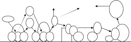
The activation of tissue factor (TF) is crucial for triggering the extrinsic coagulation cascade, which leads to the formation of thrombin and fibrin clot. TF may activate factor X and IX. This process is inhibited by tissue factor pathway inhibitor (TFPI). Thrombomodulin is a binding site on EC surface for thrombin. Both oxLDL and minimally modified LDL stimulate the expression of thrombomodulin in EC [36, 37], which may partially compensate the increased activity of coagulation. OxLDL increases the activity of protein C in vascular EC [38]. Protein C is activated by the complex of thrombin and thrombomodulin to generate activated protein C (APC) [39]. The formation of APC is increased when protein C is bound to endothelial cell protein C receptor (EPCR). APC becomes anticoagulant after it dissociates from EPCR and binds to protein S. Protein S–APC complex inactivates the activated forms of factor V and VIII, which inhibits coagulation (Fig. 8.1). OxLDL enhances the expression of TF in monocytes induced by endotoxin
[40].Both LDL and oxLDL induce the expression of TF in vascular SMC
[41].OxLDL increases the binding sites for factor VIII, and the activation of factor VIII and IX on the surface of macrophages and SMC [42]. OxLDL does not affect the expression of TFPI but promotes the degradation of TFPI in EC [43]. These findings suggest that oxLDL activates both extrinsic and intrinsic coagulation pathways.
Role of oxLDL in Platelet Activation
Treatment with oxLDL increases the aggregation and adhesion of platelets [44], and reduces the membrane fluidity of platelets [45]. OxLDL enhances the synthesis of thromboxane A2, one of the strongest biological agonists for platelet aggregation, in platelets [46]. OxLDL increases the production of prostacyclin, a strong inhibitor for platelet aggregation after a short period of incubation with EC, but inhibits the production of prostacyclin following
154 |
Garry X. Shen |
|
|
|
|
|
|
Blood |
|
|
|
|
|
|
|
|
|
|
Platelet aggregation |
Coagulation |
(−) |
Vi |
|
|
|
|
|
||||
|
IXa |
|
|
(+) |
|
|
or |
|
|
(+) |
|
|
VIIIi |
||
|
|
|
|
|
|
||
TFPI |
|
|
|
T |
|
|
|
|
Xa |
|
|
|
|
|
|
(−) |
|
Va, |
|
|
|
|
|
TF |
|
|
APC |
|
Va |
||
|
|
VIIIa |
|
|
|||
|
|
|
T PC |
|
|||
|
|
|
|
|
|
|
APC or |
VIIa IX X |
Xa |
IXa PT |
TM |
EPCR EPCR |
|
VIIIa |
|
|
|
|
|
|
|
|
S |
Plasma membrane |
|
|
|
|
|
|
|
FIGURE 8.1. Simplified scheme for coagulation and protein C pathway. TF, tissue factor; TFPI, tissue factor pathway inhibitor; VIIa, activated factor VII; X, factor X; Xa, activated X; IX, factor IX; IXa, activated IX; PT, prothrombin; Va, activated factor V; T, thrombin; TM, thrombomodulin; PC, protein C; EPCR, endothelial protein C receptor; V, factor V; VIII, factor VIII; Va, activated V; VIIIa, activated VIII; APC, activated protein C; S, protein S; Vi, inactivated factor V; VIIIi, inactivated factor VIII. (+), stimulation; (−), inhibition.
longer-term incubations [47, 48]. Glycated LDL promotes platelet aggregation that is associated with increased intracellular calcium concentration and nitric oxide (NO) production in platelets [49]. Hypochlorite-modified LDL induces greater platelet aggregation than copper ion-oxLDL [50]. The phosphorylation of p38 kinase mediates the activation of platelet by oxLDL or hypochloride-modified LDL [51]. CD36 mediates oxLDL-induced platelet activation [52]. Thus, oxidatively modified LDL may activate platelets through multiple mechanisms.
Impact of oxLDL on Fibrinolysis
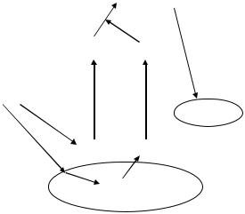
Imbalance between coagulation and fibrinolysis leads to intravascular thrombosis. The biologically active product of fibrinolytic system is plasmin, which functions in dissolving fibrin clot and maintains the fluency of blood flow. The precursor of plasmin is plasminogen, which is synthesized in liver and is abundant in most biological fluids. The generation of plasmin is mainly regulated by tissue and urokinase plasminogen activators (tPA and uPA) (Fig. 8.2). The activity of tPA and uPA is regulated by their major physiological inhibitor, plasminogen activator inhibitor-1 (PAI-1) [53, 54]. Tremoli et al. [55] reported that LDL and oxLDL increased the release of PAI-1 from EC via a LDL receptor-independent pathway. The activity and mRNA levels of PAI-1 in EC are also elevated by oxLDL treatment [56]. The release of tPA from EC is attenuated by incubation with oxLDL [57]. LDL increases the generation of PAI-1 and decreases the release of tPA from EC. Augmentation of the formation of conjugated diene is detected in previously unmodified
Chapter 8. Oxidatively Modified LDL and Thrombosis |
155 |
Plasminogen  Plasmin
(+)
(−)
tPA PAI-1
Blood
oxLDL, LDL
|
(−) |
|
fibrin clot |
|
|
|
|
(+) |
tPA |
PAI-1 |
EC |
|
|||
|
|
PAI-1 gene |
|
FIGURE 8.2. Simplified scheme for fibrinolytic system and effect of oxLDL and LDL. tPA, tissue plasminogen activator; PAI-1, plasminogen activator inhibitor-1; EC, endothelial cells; LDL, low-density lipoprotein; oxLDL, oxidized LDL. (+), stimulation; (−), inhibition.
LDL following prolonged exposure to EC [58]. The activation of protein kinase C-β is required for oxLDL-induced overproduction of PAI-1 in EC [59]. Glycation enhances the effects of LDL on PAI-1 production and further reduces the release of tPA from EC [58]. LDL isolated from type 2 diabetic patients with suboptimal glucose control stimulates the transcription and generation of PAI-1 from EC [60]. Beside LDL and its modified forms, very low-density lipoprotein (VLDL) and lipoprotein(a) also stimulate the generation of PAI-1 from EC [56, 61, 62]. A VLDL-responsive element has been detected in −672/−657 bp of the PAI-1 promoter [63]. OxLDL and LDL activate a responsive element independent from the VLDL-responsive element in the PAI-1 promoter [64]. The findings from above studies suggest that oxidatively modified LDL may reduce fibrinolytic activity in blood circulation by enhancing the production of PAI-1 and decreasing the generation of tPA in vascular EC.
Endothelial Dysfunction and Thrombosis
Intact endothelium provides an anticoagulant and antiplatelet deposition surface, which is critical for preventing thrombosis in vascular lumen. Endothelial dysfunction may be assessed from abnormal secretion of endothelial products, vascular tone, or the loss of EC [65]. EC synthesize a large numbers of products, including protein C, protein S, tPA, PAI-1,
156 Garry X. Shen
prostacyclin, TF, TFPI, von Willebrand factor (vWF), thrombomodulin, heparin, and factor V. vWF is a coagulation factor mainly produced by EC. OxLDL stimulates the release of vWF from EC [66]. Results from epidemiological studies suggest that elevated level of vWF is a predictor of the progression of atherosclerosis [67]. Prostacyclin is predominantly produced in EC. It is not only an inhibitor of platelet aggregation, but also a potent vasodilator [68]. Extensive studies on endothelium-derived relaxing factor or NO provides additional insight into the importance of endothelium in the regulation of vascular tone [69]. OxLDL reduces the generation of NO and impairs vascular relaxation [70, 71]. Vasoconstriction results in turbulence of blood flow, which may activate platelet and coagulation cascade. Flowmediated dilation may be detected in brachial artery using 2D ultrasound scan with pharmacological provocation [72]. This technique provides a noninvasive tool to assess endothelial vasodilation in vivo, although the accuracy and reproducibility of the method requires to be improved. Severe endothelial injury may increase the amounts of EC in blood circulation. Circulating EC in blood may be assessed using antibody against CD146 [73]. Increased levels of circulating EC have been detected in clinical situations associated with endothelial injury, including ACS, peripheral vascular disease, sickle cell anemia, and inflammatory diseases [74–77]. Impairment of endothelial layer leads to a prothrombotic surface in vascular lumen. Clinical significance of increased levels of circulating EC remains unclear.
Inflammation and Thrombosis
Inflammation plays a crucial role at the initial stage of atherosclerosis. Monocytes and neutrophils migrate from blood circulation and penetrate into endothelium via adhesion molecules, including P-selectin, E-selectin, monocyte chemoattractant protein-1 (MCP-1), tumor necrosis factor-α (TNF-α), intracellular adhesion molecule-1 (ICAM-1), and vascular cell adhesion mol- ecule-1 (VCAM-1) [78, 79]. Inflammation creates a procoagulant surface on vascular intima, which activates coagulation cascade and promotes the generation of thrombin. Thrombin elicits multiple inflammatory responses, including the upregulation of the expression of inflammatory mediators [80]. OxLDL is a strong agonist for the expression of many inflammatory mediators, including P-selectin [81], ICAM-1, VCAM-1 [82], MCP-1 [83], and TNF-α [84]. Anticoagulation complex, APC–EPCR, has anti-inflammatory effects in vivo [85], oxLDL increases the formation of APC [38]. OxLDL plays multifunctional roles in the modulation of inflammation, which indirectly promote thrombogenesis.
Stress and Thrombosis
Stress-mediated response regulates the folding and processing of a number of cellular proteins during various biological stresses, including heat shock,
Chapter 8. Oxidatively Modified LDL and Thrombosis |
157 |
inflammation, oxidative, shear, and restrain stresses. Heat shock factor is activated during many stresses, which mediates the transcription of heat shock protein (HSP) genes [86]. OxLDL induces the expression of HSP-70 in EC [87]. Hyperthemia increases the expression of PAI-1 in EC [88]. Restrain stress induced the expression of PAI-1 in mice in a tissueand cell-type- specific manner. Increased PAI-1 mRNA and thrombosis was more evident in aged mice than in young mice [89]. The findings suggest that stress may promote thrombosis partially resulting from its inhibition on fibrinolysis. The relationship between oxLDL, stress and the expression of thrombosisrelated factors remains to be exploited.
Thrombosis, Endothelial Functions, and Diabetes
Increased thrombotic vascular diseases were detected in patients with diabetes [90]. Elevated levels of vWF, factor VII, TF, fibrinogen or PAI-1, and decreased levels of antithrombin III, protein C, or tPA were associated with diabetes [91, 92]. Increased platelet activation was found in diabetic patients [93]. Endothelial injury and oxidative stress are potential underlying mechanisms for the increased thrombosis in diabetes. Hyperglycemia, advanced glycation end products, and dyslipidemia may impair endothelial function and increase oxidative stress. LDL from type 2 diabetic patients or glycated LDL increase the production of PAI-1 in EC and decrease the release of tPA from EC compared to unmodified LDL from healthy donors [58, 60]. Endothelial dysfunction is an early finding of diabetic vascular abnormalities. Reduction in flow-mediated dilation has been detected in children with diabetes [94]. Hyperglycemia increases susceptibility of LDL to oxidation. Substantial increases in the generation of hydrogen peroxide are detected in EC treated with glycated LDL or oxLDL for 1–2 h [95]. Insulin resistance is one of the underlying mechanisms for the pathogenesis of type 2 diabetes. Insulin resistance is associated with an upregulation of CD36, a receptor for oxLDL, in macrophages of experimental obesity [96]. Increased uptake of oxLDL in vascular wall may elicit the production of oxidative stress, impairs endothelium, and promote thrombosis-related processes in diabetic patients.
Effects of Statins on Thrombosis
3-Hydroxy-3-methylglutaryl coenzyme A (HMG-CoA) reductase is a ratelimiting enzyme for cholesterol synthesis, which regulates the generation of mevalonate from acetyl CoA. Mevalonate is a precursor of a group of active intracellular mediators (farnesyl and geranyl pyrophosphates) in addition to cholesterol. HMG-CoA reductase inhibitors (statins) were designed for lowering cholesterol synthesis [97]. Results from a recent meta-analysis demonstrated that statin treatment reduced coronary event by 27%, stroke by 18%, and all-causes of mortality by 15% in hypercholesterolemic patients in comparison to placebo controls [98]. The beneficial effects of statins have been