- •Contents
- •1. Cell Biology and Epithelia
- •2. Connective Tissue
- •3. Cartilage and bone
- •5. Nervous Tissue
- •6. Immune Tissues
- •7. Respiratory System
- •8. Gastrointestinal System
- •12. Integument
- •1. Gonad Development
- •4. Embryonic Period (Weeks 3-8)
- •1. Back and Autonomic Nervous System
- •2. Thorax
- •5. Lower Limb
- •4. The Spinal Cord
- •5. The Brain Stem
- •7. Basal Ganglia
- •11. Limbic System
- •Index

Immune Tissues |
6 |
LYMPH AND LYMPHATIC DRAINAGE
The lymphatic systemdrains lymph, excess fluid found in the extravascular space of most tissues. Lymphatic capillaries begin as blind tubes lined by endothelial cells with incomplete basal lamina and without tight junctions. The capillaries drain into largerlymphaticvessels, which have one-wayvalves, somewhat analo gous to those ofveins. Larger lymphatics have some surrounding smooth muscle.
Lymph Transport
Lymph is first transported to lymph nodes, then by lymphatic vessels from one lymph node to the next, and then back into the systemic venous circula tion, through the thoracic duct into the origin ofthe left brachiocephalic vein or through right lymphatic duct into the origin of the right brachiocephalic vein. Lymphatic vessels are not found in CNS, cartilage, bone, bone marrow, thymus, placenta, cornea, teeth and fingernails.
Most lymph is filtered through multiple lymph nodes on its way to the blood stream. The lymph nodes, mucosal associated lymphoid tissue (MALT), and spleen act as filters which can collect circulating lymphocytes, antigen sources, antigen-presenting cells, and macrophages from the lymph, providing further opportunities for lymphocyte antigen-interaction, lymphocyte proliferation, and maturation.
Normally, red blood cells are not seen in lymphatics, and lymph is clear and rela tivelycell-poor. Somelymph, such as that draining from the small intestine after a meal, canhave a high lipid content (chylomicrons) and appear opaque (chylous).
MEDICAL 65


Chapter 6 • Immune Tissues
Lymph Circulation
In lymph nodes, the B and T cells have exited the blood circulation, having come initially from bone marrow or thymus, respectively, but continue to circulate from one site to another for long periods, up to years. The lymph nodes also contain antigen-presenting cells and provide opportunities for lymphocytes to encounter antigens that they have been programmed to recognize, either for the first time or after prior encounter with resulting proliferation, maturation, and migration from another part ofthe immune system.
Afferent lymphatics carry lymph from peripheral tissues or other lymph nodes. Afferent lymphatics enter lymph nodes bypiercing the capsule ofthe node around its periphery. The lymph drains into a peripheral which empties into smaller paratrabecular sinuses that pass through the cortex ofthe node. In the cortex are resident T and B lymphocytes, macrophages and antigen presenting cells, and stromalreticulin cells, as well as smallblood vessels, includingpost-cap illaryvenules withhigh (cuboidalrather thansquamous) endothelialcells
HEVs are specialized to allow lymphocytes to exit the bloodstream in nodes or other extranodal lymphoid sites.
Efferent lymphatics convey T-cell progeny and B memory cells from the node at the medulla at the hilum ofthe node and travel via lymphatics to other lymph
nodes or the bloodstream. Some B cells give rise to plasma cells that populate the medullary cords deep to the cortex and secrete antibody into blood and lymph, whichis then carried to the rest ofthebody. Normallyfewifanyplasmacells circu late in the peripheral blood, in contrast to other lymphocyte cell types.
MUCOSA-ASSOCIATED LYMPHOID TISSUE OF THE GUT (GALT)
Inmucosa-associatedlymphoid tissue ofthe gut (GALT) the overlying epithe lium can be specialized to transport antigens through the epithelial layer to the underlying lymphoid cells. The ileum has a high concentration of GALT orga nized into Peyer's patches, which are partially covered on their luminal surface by special microfold or M cells. M cells are epithelial cells that can pick up anti gens from the ileal lumen and present them to B cells, which have surface immu noglobulin that recognizes the antigens. The B cells that recognize antigen from the M cells then present the antigen to T-helper cells via follicular dendritic cells.
MEDICAL 69

Section I • Histology and Cell Biology
Copyright McGraw-Hi/I Companies. Used with permission.
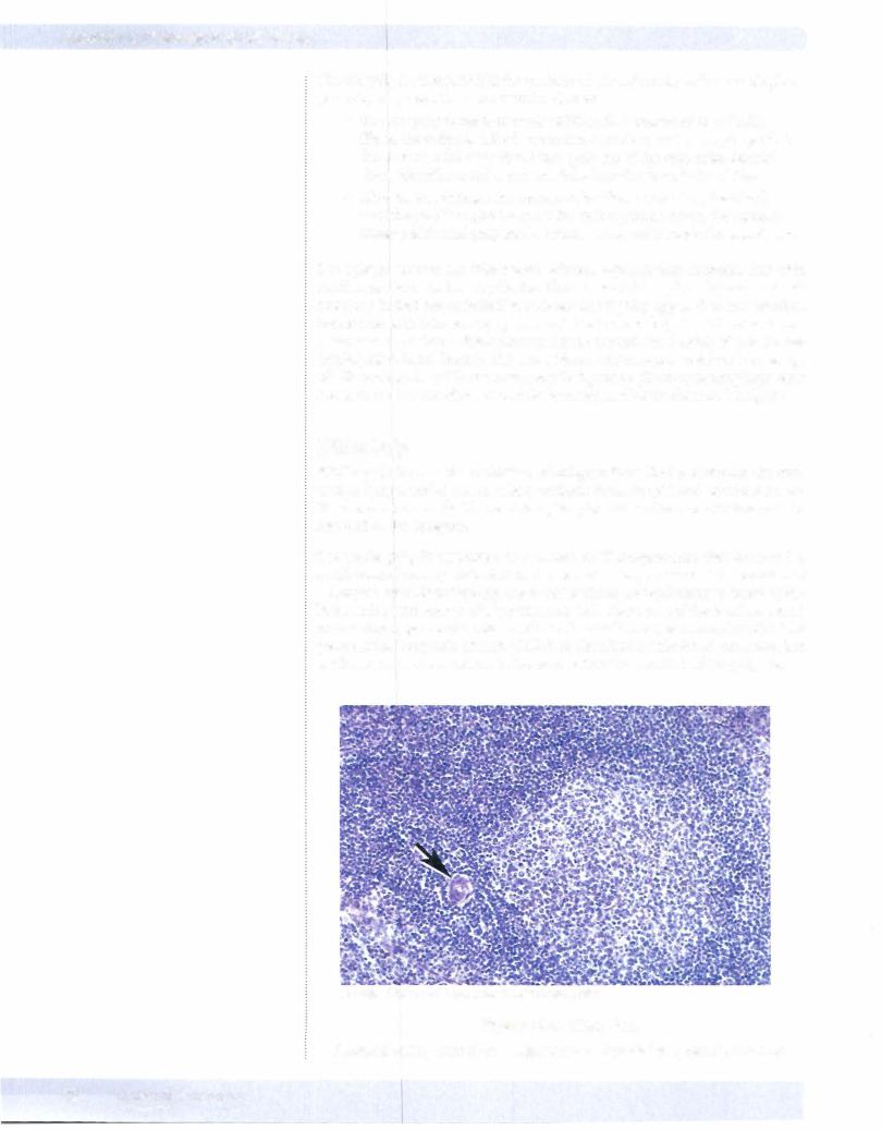
Figure 1-6-3. Cortex of a Lymph Node with Several Germinal Centers
Afferent lymphatics drain into the subcapsular sinus (arrow).
Copyright McGraw-HillCompanies. Used with permission.
Figure 1-6-4. Medulla of a Node with Medullary Cords (A) and Medullary Sinuses (8)
SPLEEN
In contrast to lymph nodes and thymus that have distinct layers (cortex and medulla) in which different functions are segregated, the white pulp of the spleen is arranged in periarteriolar aggregates of lymphocytes and antigen processing cells surrounded by red pulp.
70 MEDICAL

Chapter 6 • Immune Tissues
Trabecular artery
Periarterial sheath
(T cells) Central artery
Marginal
Sinuses
Figure 1-6-5. Spleen Schematic
CopyrightMcGraw-Hill Companies. Used with permission.
Figure 1-6-6. Spleen that lacks a distinct cortex and medulla.
Areas of white pulp (A) are randomly interspersed with red pulp (B) and connective tissue trabeculae (C). There is a dense connective tissue capsule (arrow), a covering layer of mesothelium
(curved arrow) and central artery (arrowhead).
Red Pulp
Red pulp filters and clears the bloodstream ofdamaged or aging red blood cells, particulate matter, large antigens including bacteria and unwanted cells. It carries this function out in a distinct stroma with macrophages. The red pulp consists of cords and sinuses.
MEDICAL 71


After passing through the white pulp, the central artery gives rise to smaller peni cillar arteries that enter the red pulp. Other radial artery and arteriolar branches of the central arteries empty into marginal located at the margin or outer zone of the white pulp, at its interface with red pulp. This provides an opportunity for blood-borne antigens to be removed from the blood by neighboring macro phages, making them available to antigen-processing cells, T and B lymphocytes of the white pulp, and initiating an immune response. These marginal sinuses eventually drain into splenic veins.
Chapter 6 • Immune Tissues
Note
95% of developingT cells die in the thymic cortex.
THYMUS
Like the lymph node, the thymus is divided into a distinct cortex and medulla. The thymus serves as a site ofmaturation ofT-lymphocyteprecursorsafter they exit from the bone marrow and arrive at the thymus via blood circulation. As these T cells develop, they are in intimate contact with epithelial cells of the thymus, also connected by desmosomes called dendritic epithelial cells because they have extensive thin branches oftheir cytoplasm extending out from the cell bodies. Because of the extensive branching of the epithelial cytoplasm there is extensive surface interaction possible between the developing T lymphocytes and thymic epithelial cells.
The thymic epithelial cells ofthe cortex are involved in clonal selection ofT cells. As the cells complete this process, the second set of dendritic epithelial cells in volved in clonal deletion of self-reactive T cells is located deeper in the medulla. The cortex is recognized by its darker staining. It has a high concentration of close-packed small lymphocytes with little cytoplasm and only a few epithelial cells and other cells. The medulla appears paler due to higher content of epithe lial and other cells with more cytoplasm separating the lymphocyte nuclei. Med ullary thymic epithelial cells form small aggregates of squamous epithelial cells called Hassall's corpuscles. Hassall's corpuscles are a reliable histologic marker for thymic medulla.
Another aspect ofthe thymus is that in the early stages ofT-cell maturation when positive and negative selection are taking place, there is a functional blood-thy mic barrier created by cortical thymic epithelial cells. These cells interact with the endothelium of thymic cortical blood vessels that "protects" the developing thymic lymphocytes from exposure to foreign antigens.
Note
The blood-thymus barrier consists of cortical thymic reticular cells joined by desmosomes, a dual basal lamina, and endothelial cells joined by tight junctions.
MEDICAL 73

Section I • Histology and Cell Biology
Note
Hassall's corpuscles secrete lymphopoietin that stimulates T-cell maturation in the medulla.
CopyrightMcGraw-Hill Companies. Used with permission.
Figure 1-6-8. Thymus with dark-staining cortex, lighter-staining medulla and connective tissue capsule (arrow)
Copyright McGraw-Hill Companies. Used withpermission.
Figure 1-6-9. Thymic cortex (A), medulla (B), and
Hassall's corpuscle (arrow)
•In the infant thymus there will be a well-developed outer cortex and a more central and pale medulla.
•In the thymus from an adult, most T-lymphocyte development is over and the thymus has undergone regression. This is characterized by a decrease in lymphocyte population, infiltration by adipose tissue, and loss of a clear distinction between cortex and medulla. Hassall's corpus cles are still present and often show central cystic degeneration.
74 MEDICAL

Chapter 6 • Immune Tissues
•In both infant and adult thymus there are prominent extensions of the fibrous capsule (trabecula) that extend deep into the thymus, separating incomplete lobules of thymic tissue. This is a distinctive feature of thymus compared to other lymphoid organs such as spleen and lymph node, as they are not as well divided into lobules.
•
•
•
ChapterSummary
The thymus contains trabeculae and has a cortical and medullary region.
Epithelial reticular cells and Hassall's corpuscles are located within the medulla. The cortex lacks germinal centers. The thymus protects developing T cells by the blood-thymus barrier.
The lymph node has 3 layers: outer cortial, inner cortical (paracortical), and medullary. The outer cortical layer contains most ofthe nodules and germinal centers. Most ofthe B lymphocytes reside here, whereas T lymphocytes reside in the paracortical layer. Dendritic cells within lymph nodes are antigen-presenting cells. High endothelial venules are the site of repopulation of lymph nodes and are located within the paracortical zones.
The spleen is very vascular and has red and white pulp. White pulp is composed of lymphoid tissue. T lymphocytes are located in the periarterial sheaths, while peripheral white pulp and germinal centers contain B lymphocytes. Red pulp consists of splenic cords and venous sinusoids. Its function is to delay passage of defective red blood cells to enable their elimination through phagocytosis by macrophages.
MEDICAL 75

