Добавил:
Опубликованный материал нарушает ваши авторские права? Сообщите нам.
Вуз: Предмет: Файл:
Kaplan USMLE-1 (2013) - Anatomy.pdf
Скачиваний:
82
Добавлен:
02.06.2020
Размер:
64.44 Mб
Скачать

Lower Limb 5

LUMBOSACRAL PLEXUS

The lumbosacral plexus provides the motor and sensory innervation to the lower limb and is formed by ventral rami of the L2 through S3 spinal nerves.

The major nerves of the plexus are the:

-

Femoral nerve


posteriordivisions of L2 through L4

-


v


t

ant rior divisions of L2 through L4

Obturator


-


nerve


Tibialner e

an-erioredivisions of L4 through S3


Common

fibular nerve-posterior divisions of L4 through S2


-


-

Superior gluteal nerve posterior divisions of L4 through S 1

Inferiorgluteal nerve -posterior divisions of LS through S2


-

The tibial nerve and common fibular nerve travel together through the gluteal region and thigh in a common connective tissue sheath and together are called the sciatic nerve.

The common fibular nerve divides in the proximal leg into the superfi­ cial and deep fibularnerves.

Sciatic nerve

Figure 111-5-1 . Lumbosacral Plexus

MEDICAL 301

Section Ill • Gross Anatomy

Terminal Nerves of Lumbosacral Plexus

The terminal nerves ofthe lumbosacral plexus are described in Table IIl-5- 1 .

Table 111-5-1. Terminal Nerves of Lumbosacral Plexus

Terminal Nerve

Origin

Muscles Innervated

Femoral nerve

L2 through L4

Anterior compartment of


posterior divisions

thigh (quadriceps femoris,


sartorius, pectineus)

Obturator nerve

L2 through L4

Medial com partment of


anterior divisions

thigh (gracilis, adductor


longus, adductor brevis,


anterior portion of adductor


magnus)

Primary Actions

Extend knee

Flex hip

Adduct thigh

Medially rotate thigh

Tibial nerve

L4 through 53

Posterior compartment of


anterior divisions

thigh (semimembranosus,


semitendinosus, long head


of biceps femoris, posterior


portion of adductor magnus)


Posterior compartment of


leg (gastrocnemius, soleus,


flexor digitorum longus,


flexor hallucis longus,


tibialis posterior)


Plantar muscles of foot

Common fibular nerve

L4 through 52

Short head of biceps femoris


posterior divisions


Superficial fibular nerve


Lateral compartment of leg


(fibularis longus, fibularis


brevis)

Flex knee

Extend thigh

Plantar flex foot (51-2)

Flex digits

Inversion

Flex knee

Eversion

Deep fibular nerve

Anterior compartment of leg


(tibialis anterior, extensor


hallucis, extensor digitorum,


fibularis tertius)

Dorsiflexfoot (L4-5)

Extend digits

Inversion

302 MEDICAL

Section Ill • Gross Anatomy

Clinical Correlate

The common fibular nerve crosses the lateral aspect of the knee at the neck ofthe fibula, where it is the most frequently damaged nerve ofthe lower limb. Patients will present with loss of dorsiflexion at the ankle (foot drop), loss of eversion, and sensory loss on the lateral surface ofthe leg and the dorsum ofthe foot.

Clinical Correlate

The common fibular nerve may be compressed by the piriformis muscle when the nerve passes through the piriformis instead of inferior to the muscle with the tibial nerve. Piriformis syndrome results in motor and sensory loss to the lateral and anterior compartments ofthe leg.

Clinical Correlate

The sciatic nerve is often damaged following posterior hip dislocation. A complete sciatic nerve lesion results in sensory and motor deficits in the posterior compartment ofthe thigh and all functions below the knee.

304 MEDICAL

NERVE INJURIES AND ABNORMALITIES OF GAIT

Superior Gluteal Nerve

Weakness in abduction ofthe hip

Impairment ofgait; patient cannot keep pelvis level when standing on one leg.

Sign is "Trendelenburg gait."

Inferior Gluteal Nerve

Weakened hip extension

Difficulty rising from a sitting position or climbing stairs

Femoral Nerve

Weakened hip flexion

Weakened extension ofthe knee

Sensory loss on the anterior thigh, medial leg, and foot

Obturator Nerve

Loss ofadduction ofthe thigh as well as sensoryloss on medial thigh

Sciatic Nerve

Weakened extension ofthe thigh

Loss of flexion ofthe knee

Loss ofallfunctions below the knee

Sensory loss on the posterior thigh, leg (except medial side), and foot

Tibial nerve only

Weakness in flexion ofthe knee

Weakness in plantar flexion

Weakened inversion

Sensory loss on the leg (except medial) and plantar foot

Commonfibularnerve (neckoffibula)

Produces a combination of deficits oflesions of the deep and superficial fibular nerves

Deepfibularnerve

Weakened inversion

Loss ofextension ofthe digits

Loss of dorsiflexion ("foot drop")

Sensory loss limited to skin ofthe first web space between the great and second toes

Chapter 5 • Lower Limb

RADIOLOGY

Lateral


Patella

femoral


Medial

condyle


femoral condyle


Lateral


tibial condyle


Medial tibial


condyle

Fibular head


lntercondylar


Fibular neck

©2010

eminence


Group, Inc. All

DxR Development

From the IMC,


rights reserved.

Figure 111-5-8. Lower Extremities: Anteroposterior View of Knee

Patella

Fibular

Head

From the IMC, ©2010 DxR Development

Group, Inc. All rights reserved.

Figure 111-5-9. Lower Extremities: Lateral Knee

MEDICAL 311

Section Ill • Gross Anatomy

Clinical Correlate

Robin sequence presents with a triad of poor mandibular growth, cleft palate, and a posteriorly placed tongue.

Treacher Collins syndrome also presents with mandibular hypoplasia, zygomatic hypoplasia, down-slanted palpebral fissures, colobomas, and malformed ears.

Clinical Correlate

Cribriform plate fractures may result in dysosmia and rhinorrhea (CSF).

Clinical Correlate

First arch syndrome results from abnormal formation of pharyngeal arch 1 because of faulty migration of neural crest cells, causing facial anomalies. Two well-described syndromes are Treacher Collins syndrome and Pierre Robin sequence. Both defects involve neural crest cells.

Pharyngeal fistula occurs when pouch 2 and groove 2 persist, thereby forming a fistula generally found along the anterior border of the sternocleidomastoid muscle.

Pharyngeal cyst occurs when pharyngeal grooves that are normally obliterated persist, forming a cyst usually located at the angle of the mandible.

Ectopic thyroid, parathyroid, or thymus results from abnormal migration of these glands from their embryonic position to their adult anatomic position. Ectopic thyroid tissue is found along the midline ofthe neck. Ectopic parathyroid or thymus tissue is generally found along the lateral aspect ofthe neck. May be an important issue during neck surgery.

Thyroglossal duct cyst or fistula occurs when parts of the thyroglossal duct persist, generally in the midline near the hyoid bone. The cyst may also be found at the base ofthe tongue (lingual cyst).

DiGeorge sequence occurs when pharyngeal pouches 3 and 4 fail to differentiate into the parathyroid glands and thymus. Neural crest cells are involved.

CRANIUM

Cranial Fossae

Anterior

Middle

Posterior

Cribriform plate (I)

Optic canal (II and ophthalmic artery)

Superior orbital fissure (Ill, IV, VI, V1 and ophthalmic veins)

Foramen lacerum

Internal auditory meatus (VII and VIII)

Jugular foramen (IX, X, and XI)

Hypoglossal canal (XII)

Foramen magnum (XI, spinal cord, vertebral arteries)

Figure 111-6-8. Foramina: Cranial Fossae

320 MEDICAL

Section Ill • Gross Anatomy

Names of the Major Dural Sinuses

1 . Superior sagittal*

2.Inferior sagittal

3.Straight*

4.Transverse* (2)

5.Sigmoid (2)

6.Cavernous (2)

7.Superior petrosal (2)

* Drain into the confluence of sinuses located at the inion.

Folds (Duplications) of Dura Mater

A.Faix Cerebri

B.Tentorium Cerebelli

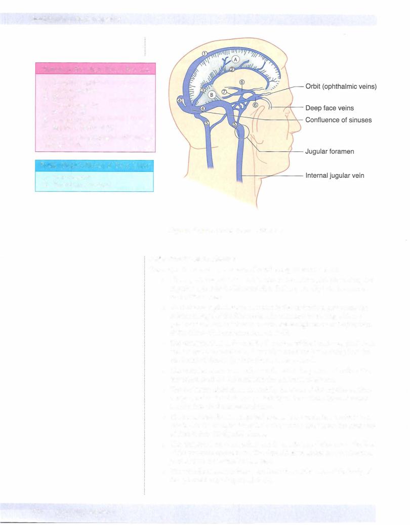
Figure 111-6-1 2. Dural Venous Sinuses

Major dural venous sinuses

The major dural venous sinuses are the following (Figure III-6- 12):

The superior sagittal sinus is located in the midsagittal plane along the superior aspect of the falx cerebri. It drains primarily into the conflu­ ence of the sinuses.

The inferior sagittal sinus is located in the midsagittal plane near the inferior margin of the falx cerebri. It terminates by joining with the great cerebral vein (of Galen) to form the straight sinus at the junction of the falx cerebri and tentorium cerebelli.

The straight sinus is formed by the union of the inferior sagittal sinus and the great cerebral vein. It usually terminates by draining into the confluens of sinuses (or into the transverse sinus) .

The occipital sinus is a small sinus found in the posterior border of the tentorium cerebelli. It drains into the confluens of sinuses.

The confluens ofsinuses is formed by the union of the superior sagittal,

straight, and occipital sinuses posteriorly at the occipital bone. It drains laterally into the 2 transverse sinuses.

The transversesinuses are paired sinuses in the tentorium cerebelli and attached to the occipital bone that drain venous blood from the confluens of sinuses into the sigmoid sinuses.

The sigmoidsinuses are paired and form a S-shaped channel in the floor of the posterior cranial fossa. The sigmoid sinus drains into the internal jugularvein at the jugular foramen.

The paired cavernous sinuses are located on either side of the body of the sphenoid bone (Figure III-6- 1 3) .

324 MEDICAL

Section Ill • Gross Anatomy

INTRACRANIAL HEMORRHAGE

Epidural Hematoma

An epiduralhematoma (Figure III-6- 14A) results from trauma to the lateral aspect of the skull which lacerates the middle meningeal artery. Arterial hemorrhage occurs rapidly in the epidural space between the periosteal dura and the skull.

Epidural hemorrhage forms a lens-shaped (biconvex) hematoma at the lateral hemisphere.

Epidural hematoma is associated with a momentary loss of conscious­ ness followed by a lucid (asymptomatic) period of up to 48 hours.

Patients then develop symptoms of elevated intracranial pressure such as headache, nausea, and vomiting, combined with neurological signs such as hemiparesis.

Herniation of the temporal lobe, coma, and death may occur rapidly if the arterial blood is not evacuated.

Subdural Hematoma

A subdural hematoma (Figure III-6- 14B) results from head trauma that tears superficial ("bridging") cerebral veins at the point where they enter the superior sagittal sinus. A subdural hemorrhage occurs between the meningeal dura and the arachnoid.

Subdural hemorrhage forms a crescent-shaped hematoma at the lateral hemisphere.

Large subdural hematomas result in signs of elevated intracranial pres­ sure such as headache and nausea.

Small or chronic hematomas are often seen in elderly or chronic alco­ holic patients.

Over time, herniation of the temporal lobe, coma, and death may result if the venous blood is not evacuated.

326 MEDICAL

Chapter 6 • Head and Neck

A. Epidural Hematoma*

B. Subdural Hematoma*

Figure 111-6-14. lntracranial Hemorrhage

Subarachnoid Hemorrhage

A subarachnoid hemorrhage results from a rupture of a berry aneurysm in the circle ofWillis. The most common site is in the anterior part ofthe circle ofWillis at the branch point ofthe anterior cerebral and anterior communicating arteries. Other common sites are in the proximal part of the middle cerebral artery or at the junction ofthe internal carotid and posterior communicating arteries.

Typical presentation associated with a subarachnoid hemorrhage is the onset of a severe headache.

ORBITAL MUSCLES AND THEIR INNERVATION

In the orbit, thereare 6 ex.traocular muscles thatmovethe eyeball (Figure III-6-16).

A seventh muscle, the levator palpebrae superioris, elevates the upper eyelid.

Pour ofthe 6 ex.traocular muscles (the superior, inferior, and medial rec­ tus, and the inferior oblique, plus the levatorpalpebrae superioris) are innervated by the oculomotor nerve (CN III).

The superior oblique muscle is the only muscle innervated by the troch­ lear nerve (CN IV).

The lateral rectus is the only muscle innervated by the abducens nerve (CN VI).

The levator palpebrae superioris is composed ofskeletal muscle inner­ vated by the oculomotor nerve (CN III) and smooth muscle (the superior tarsal muscle) innervatedby sympathetic fibers.

Sympathetic fibersreachthe orbit from a plexus on the internal carotid artery ofpostganglionic axons that originate from cell bodies in the supe­ rior cervical ganglion.

The orbital muscles and their actions are illustrated in Figure III-6- 1 5.

MEDICAL 327

Chapter 6 • Head and Neck

ChapterSummary

The neck is divided by the sternocleidomastoid muscle into an anterior and posterior triangle. The anterior triangle contains vascular structures (carotid artery and internal jugularvein), cranial nerve (CN) X, and the respiratory (trachea and larynx) and digestive (pharynx and esophagus) visceral structures. The posterior triangle contains the muscles associated with the

cervical vertebrae, CN XI, cervical plexus, and the origins ofthe brachia[ plexus.

Many structures of the head and neck develop from the branchial (pharyngeal) apparatus. The apparatus consists of pharyngeal arches, pouches, and grooves. The grooves are composed of ectoderm, the pouches

are composed of endoderm, and the arches are composed of mesoderm and neural crest cells. The adult derivatives ofthe arches and pouches are given in Tables 111-6-1 and 111-6-2, respectively.

The anterior 2/3 of the tongue develops from the first pharyngeal arch, and the posterior 1/3 develops from the third pharyngeal arch.

The muscles of the tongue derive from myoblasts that migrate into the head from the occipital somites and are innervated by CN XII.

The face develops from 5 structures derived from the first pharyngeal arch: frontonasal prominence, a pair of maxillary prominences, and a pair of mandibular prominences. The mandibular prominences form the lower jaw, the frontonasal prominence forms the forehead, and the maxillary prominences form the cheek, lateral upper lip, and the secondary palate. The mid line of the upper lip, the nasal septum, and the primary palate are formed by the medial nasal prominence. The primary and secondary palate fuse to form the definite palate.

The floor ofthe cranial cavity is divided into the anterior, middle, and posterior cranial fossae. The openings in the skull provide for passage ofthe cranial nerves and blood vessels. These are listed in Figures 111-6-8 and 111-6-9.

Venous return from the brain and other structures ofthe cranial vault is provided by the dural venous sinuses, which ultimately drain into the internal jugularvein at the jugular foramen. Most ofthese sinuses are located in

the folds ofthe dura mater (falx cerebri and tentorium cerebelli). The major ones are the superior and inferior sagittal and the transverse, sigmoid, and cavernous sinuses.

The cavernous sinus is significant because CN Ill and IV and the ophthalmic and maxillary divisions of CN V course in the lateral wall ofthe cavernous sinus, and the internal carotid artery and CN VI are found in the lumen.

The orbit contains the ocular muscles, eyeball, and transmits the optic

nerve and ophthalmic artery. CN VI innervates the lateral rectus muscle, CN IV innervates the superior oblique muscle, and the remaining muscles are innervated by CN Ill.

MEDICAL 329

SECTION

Neuroscience

Section IV • Neuroscience

Table IV-1-1

Disease

Symptoms

Multiple sclerosis (MS)

Symptoms separated in space and time


Vision loss (optic neuritis)


Internuclear ophthalmoplegia (MLF)


Motor and sensory deficits


Vertigo


Neuropsychiatric

Metachromatic

Four types; motor and cognitive issues

leukodystrophy (MLD)

with seizures

Progressive multifocal

Cortical myelin commonly affected;

leukoencephalopathy

causes limb weakness, speech

(PML)

problems

Central pontine

Pseudobulbar palsy

myelinolysis (CPM)

Spastic quadriparesis


Mental changes


May produce the "locked-in" syndrome


Often fatal

Guillain-Barre syndrome

Acute symmetric ascending


inflammatory neuropathy of PNS myelin


Weakness begins in lower limbs and


ascends; respiratory failure can occur in


severe cases


Autonomic dysfunction may be


prominent


Cranial nerve involvement is common


Sensory loss, pain, and paresthesias


rarely occur


Reflexes invariably decreased or absent

Abbreviations: CJD, Jacob-Creutzfeldt; MLF, medial longitudinal fasciculus

Notes

Occurs twice as often in women

Onset often in third or fourth decade

Higher prevalence in temperate zones

Relapsing-remitting course is most common

Well-circumscribed demyelinated plaques often in periventricular areas

Chronic inflammation; axons initially preserved

Increased lgG (oligoclonal bands) in CSF

Treatment: high-dose steroids, interferon-beta, glatiramer (Copaxone®)

Arylsulfatase A deficiency in lysosomes affects botn CNS and PNS myelin

Caused by CJD virus

Affects immunocompromised, especially AIDS

Demyelination, astrogliosis, lymphohistiocytosis

Focal demyelination of central area of basis pontis (affects corticospina corticobulbartracts)

Seen in severely malnourished, alcoholics, liver disease

Probably caused by overly aggressive correction of hyponatremia

Two-thirds of patients have history of respiratory or GI illness 1-3 weeks prior to onset

Elevated CSF protein with normal cell count (alburninocytologic dissociation)

334 MEDICAL

Development ofthe Nervous System

Neurulation

Neurulation begins in the third week; both CNS and PNS derived from neuroectoderm.

The notochord induces the overlying ectoderm to form the neural plate (neuroectoderm).

By end of the third week, neural folds grow over midline and fuse to form neural tube.

During closure, neural crest cells also form from neuroectoderm.

Neural tube 3 primary vesicles --7 5 primary vesicles --7 brain and spinal cord

Brain stem and spinal cord have an alar plate (sensory) and a basal plate (motor); plates are separated by the sulcus limitans.

Neural crest --7 sensory and postganglionic autonomic neurons, and other non-neuronal cell types.

Peripheral NS (PNS): cranial nerves ( 12 pairs) and spinal nerves (31 pairs)

Chapter 1 • Nervous System

Note

Alpha-fetoprotein (AFP) levels may also be elevated in gastroschisis and omphalocele. AFP levels are low in pregnancy of down syndrom fetus.

Neural plate

Neuroectoderm

Neural


groove


Ectoderm


Notochordal

process Neural groove

Day 18

Neural fold

Neural tube

Rostral neuropore (closes at day 25)

Failure to close results in anencephaly,

causing polyhydramios

and increased alpha­ Basal plate (motor) fetoprotein and AChE

Neural crest

Caudal neuropore (closes at 270)

Failure to close results in spina bifida

Day 22 and increased alpha­ fetoprotein and AChE

Figure IV-1-1 . Development of Nervous System

MEDICAL 335

Section IV • Neuroscience

Table IV-1-4. Germ layer Derivatives

Ectoderm Surface ectoderm

Epidermis Hair Nails

Inner ear, external ear Ei:namelofteeth

Lens of eye

Anterior pituitary (Rathke's pe>uch) Parotid gland

Neuroectoderm

Neural tube

Central nervous system

Retina and optic nerve

Rineal gland

Neurohypophysis

Astrocytes

Oligodendrocytes (CNS myelin)

Neural crest

Adrenal medulla

Ganglia

Sensory (unipolar)

Autonomic (postganglionic)

Pigment cells (melanocytes)

Schwann cells (PNS myelin)

Meninges

Pia and arachne>id mater

Pharyngeal areh cartilage (First Arch Syndromes)

Odontoblasts

Parafollicular (C:) cells

Aorticopulmonary septum (Tetrology of Fallot)

Endocardiat cushions (Down

Syndrome)

Yolk sac derivatives:

Primordial germ cells

Early blood and blood vessels

Mesoderm

Muscle

Smooth

Cardiac

Skeletal

Connective tissue

All serous membranes Bone and cartilage

Blood, lymph, cardiovascular organs

Adrenal cortex

Gonads and internal reproductive organs

Spleen

Kidney and ureter Dura mater

Endoderm

Forms epithelial parts of:

Tonsils

Thymus

Pharynx

Larynx

Trachea

Bronchi

Lungs

Urinary bladder

Urethra

Tympanic cavity

Auditory tube

GI tract

Forms parenchyma of:

Liver

Pancreas

Tonsils

Thyroid gland

Parathyroid glands Glands ofthe GI tract Submandibutar gland Sublingual gland

338 M EDICAL

Chapter 1 • NervousSystem

ChapterSummary

The peripheral nervous system (PNS) consists of 1 2 pairs of cranial nerves, 31 pairs of spinal nerves with their related sensory and motor ganglia, and the peripheral part ofthe autonomic nervous system. The afferent and efferent neurons in the PNS convey somatic and visceral (autonomic) functions to and from the central nervous system (CNS).

Cells of Nervous System

The basic functional cell for conducting motor and sensory functions within the nervous system is the neuron. Neurons in the CNS are myelinated by oligodendrocytes, and in the PNS neurons are myelinated by Schwann cells. Oligodendrocytes myelinate multiple axons but Schwann cells myelinate only a segment of one axon.

The skeletal motor neurons and preganglionic motor neurons in the CNS develop from the neural tube, whereas the sensory neurons and postganglionic neurons located in sensory or autonomic ganglia, respectively, in the PNS derive from neural crest cells.

Neurulation and the development of the nervous system begin in the third week of development. As the primitive streak regresses caudally, the notochord develops in the midaxis ofthe embryo between the

buccopharyngeal membrane and the cloaca[ membrane. The appearance of the notochord then induces the ectoderm overlying the notochord to form the neural plate composed of neuroectoderm. The neural plate is wide at the cranial end and tapers caudally. By the end of the third week, the lateral margins of the neural plate thicken and become elevated to form the neural folds with the neural groove located centrally between the 2 folds. The neural folds then grow over the midline and begin to fuse to form the neural tube. Closure ofthe neural tube begins in the cervical region and continues cranially and caudally. The cephalic (cranial neuropore) and the caudal (caudal neuropore) ends ofthe neural tube close last. Failure of closure of the cranial and caudal neuropores results in anencephaly and spina bifida. Alpha-fetoprotein levels are increased with the neural tube defects. During closure of the neural tube, neural crest cells are formed from neuroectoderm at the margins of the neural folds. The neural crest cells migrate throughout the embryo and form a number of cell types.

MEDICAL 343

Section IV • Neuroscience

Half of brain and spinal cord tumors are metastatic. Some differences between primary and metastatic twnors are listed in Table IV-2-2:

Table IV-2-2. Tumors ofthe CNS and PNS

Primary

Metastatic

Poorly circumscribed

Well circumscribed

Usually single

Often multiple

Location varies by specific type

Usually located at the junction


between gray and white matter

Abbreviations: CNS, central nervous system; PNS, peripheral nervous system

Table IV-2-3. PrimaryTumors

Tumor

Glioblastoma multiforme (grade IV astrocytoma)

Astrocytoma

(pilocytic)

Oligodendroglioma

Features

Most common primary brain tumor

Highly malignant

Usually lethal in 8-12 months

Benign tumor of children and young adults Usually in posteriorfossa in children

Slow growing

Long survival (average 5-10 years)

Ependymoma

Ependymal origin


Can arise in IV ventricle and lead to hydrocephalus

Medulloblastoma

Highly malignant cerebellar tumor


A type of primitive neuroectodermal tumor (PNEl)

Meningioma

Second most common primary brain tumor


Dural convexities; parasagittal region

Schwannoma

Third most common primary brain tumor


Most frequent location: CN VIII atcerebellopontine angle


Hearing loss, tinnitus, CN V + VII signs


Good prognosis after surgical resection

Retinoblastoma


Sporadic-unilateral


Familial-bilateral; associated with osteosarcoma

Craniopharyngioma


Derived from oral epithelium (remnants of Rathke pouch)


Usually children and young adults

Often calcified

Symptoms due to encroachment on pituitary stalk or optic chiasm

Benign but may recur

Pathology

Can cross the midline via the corpus callosum ("butterfly glioma")

Areas of necrosis surrounded by rows of neoplastic cells

(pseudopalisading necrosis) Rosenthal fibers lmmunostaining with GFAP

"Fried-egg" appearance­ perinuclear halo

Rosettes and pseudorosettes

Blue, small, round cells with pseudorosettes

Attaches to the dura, compresses underlying brain without invasion

Microscopic-psammoma bodies

Antoni A (hypercellular) and B (hypocellular) areas

Bilateral acoustic schwannomas­

pathognomonic for neurofibromatosis type 2

Small, round, blue cells; may have rosettes

Histology resembles adamantinoma (most common tumor oftooth)

Abbreviation: GFAP, glial fibrillary acidic protein

348 MEDICAL

Chapter 2 • Central NervousSystem

ChapterSummary

The neural tube forms 3 primary vesicles at its cranial end:

-Forebrain (prosencephalon)

-Midbrain

-Hindbrain (rhombencephalon)

These primary vesicles then develop into 5 secondary vesicles that form the adult derivatives of the CNS.

-The telencephalon forms the cerebral hemispheres

-The diencephalon forms 4 thalamic derivatives

-The mesencephalon forms the midbrain

-The metencephalon forms the pons and cerebellum

-The myelencephalon forms the medulla

The remainder of the neural tube forms the spinal cord. The lumen of the neural tube will develop into the ventricular system.

The typical neuron is the multipolar neuron. It consists of a cell body (soma), multiple dendrites, and a single axon. Axons utilize anterograde and retrograde axonal transport to move subcellular elements to and from the soma. Skeletal motor neurons, preganglionic autonomic neurons, and glial cells develop from the neural tube. Glial cells include astrocytes, oligodendrocytes, microglia, and ependymal cells.

MEDICAL 349

CSF DISTRIBUTION, SECRETION, AND CIRCULATION

CSF fills the subarachnoid space and the ventricles ofthe brain. The average adult has 90 to 1 50 mL of total CSF, although 400 to 500 mL is produced daily. Only 25 mL of CSF is found in the ventricles themselves.

Approximately 70% of the CSF is secreted by the choroid plexus, which consists of glomerular tufts of capillaries covered by ependymal cells that project into the ventricles (the remaining 30% represents metabolic water production). The cho­ roid plexus is located in parts ofeach lateral ventricle, the third ventricle, and the fourth ventricle.

CSF from the lateral ventricles passes through the interventricular foramina of Monro into the third ventricle. From there, CSF flows through the aqueduct of Sylvius into the fourth ventricle. The only sites where CSF can leave the ventricles and enter the subarachnoid space outside the CNS are through 3 openings in the fourth ventricle, 2 lateral foramina of Luschka and the median foramen of Magendie.

Within the subarachnoid space, CSF also flows up over the convexity ofthe brain and around the spinal cord. Almost allCSF returns to the venous system by drain­ ing through arachnoid granulations into the superior sagittal dural venous sinus.

Normal CSF is a clearfluid, isotonic with serum (290-295 mOsm/L).

The pH of CSF is 7.33 (arterial blood pH, 7.40; venous blood pH, 7.36).

Sodium ion (Na+) concentration is equal in serum and CSF (:o:l38 mEq/L).

CSF has a higher concentration of chloride (CI-) and magnesium (Mg2+) ions than does serum.

CSF has a lower concentration ofpotassium (K+), calcium (Ca2+), and bicarbon­ ate (HC03) ions, as well as glucose, than does serum.

The Blood-Brain Barrier and the Blood-CSF Barrier

The chemical integrity of the brain is protected in a different way by 2 separate systems.

Chapter 3 • Ventricular System

Clinical Correlate

The concentration of protein (including all immunoglobulins) is much lower in the CSF as compared with serum.

Normal CSFcontains 0 to 4 lymphocytes or mononuclear cells per cubic millimeter. Although the presence ofa few monocytes or lymphocytes is normal, the presence of polymorphonuclear leukocytes

is always abnormal, as in bacterial meningitis.

Red blood cells (RBCs) are not normally found in the CSF but may be present after traumatic spinal tap or subarachnoid hemorrhage.

Increased protein levels may indicate a CNS tumor.

Tumor cells may be present in the CSF in cases with meningeal involvement.

Blood-brain barrier

The blood-brain barrier is formed by capillary endothelium connected by tight junctions. Astrocytes participate in the maintenance of the blood-brain barrier. They have numerous long processes with expanded vascular end-feet, or pedicels, which attach to the walls of capillaries.

Water diffuses across the blood-brain barrier readily, but glucose, the primary energy source of the brain, requires carrier-mediated transport. Active transport systems are capable of pumping weak organic acids, halides, and extracellular K+ out of the brain against their respective concentration gradients.

Blood-CSFbarrier

Tight junctions located along the epithelial cells of the choroid plexus form the blood-CSP barrier. Transport mechanisms are similar to those described for the blood-brain barrier, although the ability ofa substance to enter the CSF does not guarantee it will gain access to the brain.

MEDICAL 355

Section IV • Neuroscience

ChapterSummary

The ventricular system is continuous throughout each part of the central nervous system (CNS) and contains cerebrospinal fluid (CSF), which provides

a protective bath forthe brain and spinal cord. The system consists of 2 lateral ventricles in the cerebral hemispheres, a third ventricle in the

midbrain, and a fourth ventricle in the pons and medulla. CSF is produced in the choroid plexuses ofthe lateral, third, and fourth ventricles. CSF leaves the fourth ventricle through the foramen of Magendie and the foramina of

Luschka to enter the subarachnoid space. From the subarachnoid space, CSF returns to the venous system by passing through arachnoid granulations into the superior sagittal dural venous sinus.

Hydrocephalus results from excess volume and pressure of CSF, producing ventricular dilatation. Noncommunicating hydrocephalus is caused by obstruction to CSFflow inside the ventricular system, and communicating hydrocephalus is caused by oversecretion or reduced absorption of CSF.

356 MEDICAL