- •Contents
- •1. Cell Biology and Epithelia
- •2. Connective Tissue
- •3. Cartilage and bone
- •5. Nervous Tissue
- •6. Immune Tissues
- •7. Respiratory System
- •8. Gastrointestinal System
- •12. Integument
- •1. Gonad Development
- •4. Embryonic Period (Weeks 3-8)
- •1. Back and Autonomic Nervous System
- •2. Thorax
- •5. Lower Limb
- •4. The Spinal Cord
- •5. The Brain Stem
- •7. Basal Ganglia
- •11. Limbic System
- •Index

Thorax 2
CHEST WALL
Breast
Thebreast (mammarygland) is a subcutaneous glandular organ ofthe superficial pectoral region. It is a modified sweat gland, specialized in women for the pro duction and secretion of milk. A variable amount offat surrounds the glandular tissue and duct system and is responsible for the shape and size of the female breast.
Cooperligaments
Cooper ligaments are suspensory ligaments that attach the mammary gland to the skin and run from the skin to the deep fascia.
Arterial supply
There is an extensive blood supply to the mammary tissues. The 2 prominent blood supplies are:
• Internalthoracic artery (internalmammary), a branch of the subcla vian artery which supplies the medial aspect of the gland.
• Lateralthoracicartery, a branch of the axillary artery which contrib utes to the blood supply to the lateral part of the gland. On the lateral aspect of the chest wall, the lateral thoracic artery courses with the long thoracic nerve, superficial to the serratus anterior muscle.
Lymphatic drainage
The lymphatic drainage of the breast is critical due to its important role in me tastasis of breast cancer. The lymphatic drainage of the breast follows 2 primary routes (Figure III-2-1):
1. Laterally, most of the lymphatic flow (75%) drains from the nipple and the superior, lateral, and inferior quadrants of the breast to the axillary nodes, initially to the pectoral group.
2.From the medial quadrant, most lymph drains to the parasternal nodes, which accompany the internal thoracic vessels. It is also through this medial route that cancer can spread to the opposite breast.
Clinical Correlate
The presence of a tumor within the breast can distort Cooper ligaments, which results in dimpling of the skin (orange-peel appearance).
Clinical Correlate
During a radical mastectomy, the long thoracic nerve (serratus anterior muscle) may be lesioned during ligation ofthe lateral thoracic artery. A few weeks after surgery, the female may present with a winged scapula and weakness
in abduction ofthe arm above 90°.
The thoracodorsal nerve supplying the latissimus dorsi muscle may also be damaged during mastectomy, resulting in weakness in extension and medial rotation ofthe arm.
MEDICAL 185


PLEURA AND PLEURAL CAVITY
Serous Membranes
Within the thoracic and abdominal cavities there are 3 serous mesodermal-de rived membranes which form a covering for the lungs (pleura), heart (pericar dium), and abdominal viscera (peritoneum).
Each ofthese double-layered membranes permits friction-reducing movements of the viscera against adjacent structures.
The outerlayer ofthe serous membranes is referred to as the parietallayer; and the inner layerwhich is applied directly to the surface ofthe organ, is calledthe visceral layer. The 2 layers are continuous and there is a potential space (pleural cavity) be tween the parietal and visceral layers containing a thin layer ofserous fluid.
Pleura
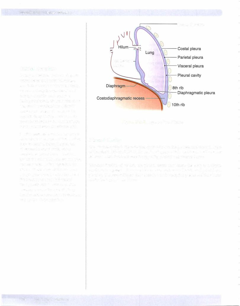
The pleura is the serous membrane that invests the lungs in the lateral compart ments of the thoracic cavity (Figure III-2-5). The external parietal pleura lines and attaches to the inner surfaces ofthe chest wall, diaphragm, and mediastinum. The innermost visceral layer reflects from the parietal layer at the hilum of the lungs and is firmly attached to and follows the contours ofthe lung. Visceral and parietal pleura are continuous at the root ofthe lung.
The parietal pleura is regionally named by its relationship to the thoracic wall and mediastinum (Figure III-2-5):
•Costal parietal pleura is lateral and lines the inner surfaces of the ribs and intercostal spaces
•Diaphragmatic parietalpleura lines the thoracic surface of the dia phragm
•Mediastinal parietalpleura is medial and lines the mediastinum. The mediastinal pleura reflects and becomes continuous with the visceral pleura at the hilum.
•Cervicalparietalpleura extends into the neck above the first rib where it covers the apex of the lung.
The visceral pleura tightly invests the surface of the lungs, following all of the fissures and lobes of the lung.
innervation of Pleura
The parietal pleura has extensive somatic sensory innervation provided by nerves closely related to different aspects of the pleura.
•The intercostal nerves supply the costal and peripheral portions of the diaphragmatic pleura.
•The phrenicnerve supplies the central portion of the diaphragmatic pleura and the mediastinal pleura.
The visceral pleura is supplied by visceral sensory nerves that course with the autonomic nerves.
Chapter 2 • Thorax
Clinical Correlate
Respiratory distress syndrome is caused by a deficiency of surfactant (type II pneumocytes). This condition is associated with premature infants, infants of diabetic mothers, and prolonged intrauterine asphyxia. Thyroxine and cortisol treatment increase the production of surfactant.
Surfactant deficiency may lead to hyaline membrane disease, whereby repeated gasping inhalations damage the alveolar lining. Hyaline membrane disease is characterized histologically by collapsed alveoli (atelectasis) and eosinophilic (pink) fluid covering the alveoli.
Clinical Correlate
Inflammation ofthe parietal pleural layers (pleurisy) produces sharp pain upon respiration. Costal inflammation produces local dermatome pain ofthe chestwallvia the intercostal nerves; whereby mediastinal irritation produces referred pain via the phrenic nerve to the shoulder dermatomes of C3-5.
MEDICAL 189
Section Ill • Gross Anatomy
Clinical Correlate
Open pneumothroax occurs when air enters the pleural cavity following a penetrating wound ofthe chest cavity. Air moves freely through the wound during inspiration and expiration. During inspiration, air enters the chest wall and the mediastinum will shift toward other side and compress the opposite lung. During expiration, air exits the wound and the mediastinum moves back toward the affected side.
Tension pneumothorax occurs when a piece oftissue covers and forms a flap over the wound. During inspiration, air enters the chest cavity, which results in a shift of the mediastinum toward the other side, compressing the opposite lung. During expiration, the piece oftissue prevents the air from escaping the wound, which increases the pressure and the shift toward
the opposite side is enhanced. This severely reduces the opposite lung function and venous return to the heart and can be life-threatening.
Cervical pleura
Mediastinal pleura
Figure 111-2-5. Layers of the Pleura
Pleural Cavity
The pleural cavity is the potential space between the parietal and visceral layers of the pleura (Figure IIl-2-5). It is a closed space which contains a small amount of serous fluid that lubricates the opposing parietal and visceral layers.
The introduction of air into the pleural cavity may cause the lung to collapse, resulting in a pneumothorax which causes shortness of breath and painful res piration. The lung collapses due to the loss ofthe negative pressure ofthe pleural cavity during a pneumothroax.
190 MEDICAL


Chapter 2 • Thorax
Fetal Circulation
Venoussystems associated with thefetal heart
There are 3 major venous systems that flow into the sinus venosus end of the heart tube.
• Viteline (omphalomesenteric) veins drain deoxygenated blood from the yolk stalk; they will coalesce and form the veins of the liver (sinusoids, hepatic portal vein, hepatic vein) and part of the inferior vena cava.
•Umbilicalvein carries oxygenated blood from the placenta.
•Cardinalveins carry deoxygenated blood from the body of the embryo;
they will coalesce and contribute to some of the major veins of the body (brachiocephalic, superior vena cave, inferior vena cava, azygos, renal).
Arterialsystems associated with thefetal heart
During fetal circulation, oxygenated blood flood from the placenta to the fetus passes through the umbilical vein. Three vascular shunts develop in the fetal circulation to bypass blood flow around the liver and l gs (Figure IIl-2-12).
1 . The ductus venosus allows oxygenated blood in the umbilical vein to bypass the sinusoids of the liver into the inferior vena cava and to the right atrium. From the right atrium, oxygenated blood flows mostly through the foramen ovale into the left atrium then left ventricle and into the systemic circulation.
2. The foramen ovale develops during atrial septation to allow oxygenated blood to bypass the pulmonary circulation. Note that this is a right-to left shunting of blood during fetal life.
3. During fetal circulation, the superior vena cava drains deoxygenated blood from the upper limbs and head into the right atrium. Most of this blood flow is directed into the right ventricle and into the pulmonary trunk. The opens into the underside of the aorta just distal to the origin of the left subclavian artery and shunts this deoxy genated blood from the pulmonary trunk to the aorta to bypass the pul monary circulation.
The shunting of blood through the foramen ovale and through the ductus arteriosus (right to left) during fetal life occurs because of a
sure gradient.
MEDICAL 197


Chapter 2 • Thorax
Postnatal circulation
Following birth, these 3 shunts will close because of changes in the pressure gra dients and in oxygen tensions. The umbilical vein closes and reduces blood flow into the right atrium. The ductus venosus also closes. Lung expansion reduces pulmonary resistence and results in increased flow to the lungs and increased venous return to the left atrium.
•Closure of the foramen ovale occurs as a result of the increase in left atrial pressure and reduction in right atrial pressure.
•Closure of the ductus venosus and ductus arteriosus occurs over the next several hours as a result of the contraction of smooth muscles in its wall and increased oxygen tension.
•The release of bradykinin and the immediate drop of prostaglandin E at birth also facilitate the closure of the ductus arteriosus.
The changes which occur between preand postnatal circulation are summarized in Table ill-2-2.
Table 111-2-2. AdultVestiges Derived from the Fetal Circulatory System
Changes After Birth |
Remnant in Adult |
Closure of right and left umbilical |
Medial umbilical ligaments |
arteries |
|
Closure ofthe umbilical vein |
Ligamentum teres of liver |
Closure of ductus venosus |
Ligamentum venosum |
Closure of foramen ovale |
Fossa ovalis |
Closure of ductus arteriosus |
Ligamentum arteriosum |
SEPTATION OF THE HEART TUBE
Except for the sinus venosus of the embryonic heart tube that initially develops into right and left horns, the ventricular, atrial, and truncus parts ofthe heart tube, which are originally a common chamber, willundergo septation into a right and left heart structure. The septation of the atria and ventricles occurs simultane ously beginning in week 4 and is mostly finished in week 8. Most ofthe common congenital cardiac anomalies result from defects in the formation ofthese septa.
Atrial Septation
During fetal life, blood is shunted from the right to the left atrium via the fora men ovale (FO). Note that during fetal circulation, right atrial pressure is higher than left due to the large bolus of blood directed into the right atrium from the placenta and to high pulmonary resistance.
The FO has to remain open and functional during the entire fetal life to shunt oxygenated blood from the right atrium into the left atrium.
MEDICAL 199


Section Ill • Gross Anatomy
Truncus arteriosus defects
Three classic cyanotic congenital heart abnormalities occur with defects in the development of the aorticopulmonary septum and are related to the failure of neural crest cells to migrate into the truncus arteriosus:
1 . TetralogyofFallot (Figure III-2- 1 9) is the mostcommon cyanotic congenital heart defect. Tetralogy occurs when the AP septum fails to align properly and shifts anteriorly to the right. This causes right-to-left shunting of blood with resultant cyanosis that is usually present sometime after birth. Imaging typi cally shows a boot-shaped heart due to the enlarged right ventricule.
•There are 4 major defects in Tetralogy of Fallot:
-Pulmonary stenosis (most important)
Overriding aorta (receives blood from both ventricles) Membranous interventricular septal defect
-Right ventricular hypertrophy (develops secondarily)
Aorta
1 . Pulmonary stenosis
2. Ventricular septa! defect
3. Hypertrophied right ventricle
4. Overriding aorta
Figure 111-2-19. Tetralogy of Fallot
2.Transposition ofthe great vessels (Figure Ill-2-20) occurs when the AP sep tum fails to develop in a spiral fashion and results in the aorta arising from the right ventricle and the pulmonary trunk arising from the left ventricle. This causes right-to-left shunting of blood with resultant cyanosis.
•Transposition is the most common cause of severe cyanosis that per sists immediately at birth. Transposition results in producing 2 closed circulation loops.
•Infants born alive with this defect usually have other defects (PDA, VSD, ASD) that allow mixing of oxygenated and deoxygenated blood to sustain life.
204 MEDICAL


Chapter 2 • Thorax
Anterior Mediastinum
The anterior mediastinum is the small interval between the sternum and the anterior surface of the pericardium. It contains fat and areolar tissue and the infe rior part ofthe thymus gland. A tumor ofthe thymus (thymoma) can develop in the anterior or superior mediastina
Posterior Mediastinum
Theposteriormediastinumis located between the posterior surface ofthe pericar dium and the T5-Tl2 thoracic vertebrae. Inferiorly, it is closed by the diaphragm.
There are 4 vertically oriented structures coursing within the posterior medias tinum:
•Thoracic (descending) aorta
-Important branches are the bronchial, esophageal, and posterior inter costal arteries
-Passes through the aortic hiatus (with the thoracic duct) at the T12 vertebral level to become the abdominal aorta
•Esophagus
-Lies immediately posterior to the left primary bronchus and the left atrium, forming an important radiological relationship
- |
Covered by the anterior and posterior esophagealplexuseswhich are |
- |
Passes through the esophageal hiatus (with the vagal nerve trunks) at |
|
derived from the left and right vagus nerves, respectively |
|
the Tl0 vertebral level |
-Is constricted (1) at its origin from the pharynx, (2) posterior to the arch of the aorta, (3) posterior to the left primary bronchus, and (4) at the esophageal hiatus of the diaphragm.
•Thoracic duct
-Lies posterior to the esophagus and between the thoracic aorta and azygos vein
-Ascends the posterior and superior mediastina and drains into the junction of the left subclavian and internal jugular veins
-Arises from the cisterna chyli in the abdomen (at vertebral level L1) and enters the mediastinum through the aortic hiatus of the diaphragm
•Azygos system ofveins
-Drains the posterior and thoracic lateral wall
-Communicates with the inferior vena cava in the abdomen and ter- minates by arching over the root of the right lung to empty into the superiorvena cava above the pericardium
-Forms a collateralvenous circulation between the inferior and superior vena cava
Middle Mediastinum
The middle mediastinum contains the heart and great vessels and pericardium and willbe discussed later.
MEDICAL 207


Chapter 2 • Thorax
The relationships ofthese structures in the superior mediastinum are best visual ized in a ventral to dorsal orientation between the sternum anteriorly and the vertebrae posteriorly:
• Thymus: Located posterior to the manubrium, usually atrophies in the adult and remains as fatty tissue
• Rightandleftbrachiocephalicveins: Rightvein descends almostvertically and the left vein obliquely crosses the superiormediastinumposterior to the thyrnic remnants.
The 2 veins join to form the superiorvenacava posterior to the right first costalcartilage.
The superiorvena cava descends and drains into the right atrium deep to the right third costal cartilage.
• Aortic arch and its 3 branches: Aortic arch begins and ends at the plane ofthe sternal angle and is located just inferior to the left brachiocephalic vein. As a very important radiological landmark, the origins ofthe
3 branches ofthe aortic arch (brachiocephalic,left common carotid,and left subclavian) are directly posterior to the left brachiocephalic vein.
• Trachea: Lies posterior to the aortic arch and bifurcates at the level of
T4 vertebra to form the right and left primary bronchi. The carina is an internal projection of cartilage at the bifurcation.
• Esophagus: Lies posterior to the trachea and courses posterior to the left primary bronchus to enter the posterior mediastinum
ln addition to these structures, the superior mediastinum also contains the right and leftvagus and phrenic nerves and the superior end ofthe thoracic duct.
Vogus nerves
•Right and left vagus nerves contribute to the pulmonary and cardiac plexuses.
•In the neck, the right vagus nerve gives rise to the right recurrent laryn geal nerve, which passes under the right sublcavian artery to ascend in the groove between the esophagus and the trachea to reach the larynx. Note: The right recurrent laryngeal nerve is not in the mediastinum.
•The left vagus nerve gives rise to the left recurrent laryngealnervein
the superior mediastinum, which passes under the aortic arch and liga mentum arteriosum to ascend to the larynx (Figure
Thoracic duct
The thoracic duct is the largest lymphatic channel in the body. It returns lymph to the venous circulation at the junction ofthe left internal jugular vein and the left subclavian vein.
Phrenicnerves
Phrenic nerves arise from the ventral rami of cervical nerves 3, 4, and 5. The nerves are the sole motor supply of the diaphragm and convey sensory infor mation from the central portion of both the superior and inferior portions of the diaphragm and parietal pleura. Both phrenic nerves pass through the middle mediatstinum lateral between the fibrous pericardium and pleura, and anterior to the root ofthe lung.
Clinical Correlate
The left recurrent laryngeal nerve
(Figure 111-2-23) curves under the aortic arch distal to the ligamentum arteriosum where it may be damaged by pathology (e.g., malignancy or aneurysm of the aortic arch), resulting in paralysis ofthe leftvocal folds. The right laryngeal nerve is not affected because it arises from the right vagus
nerve in the root ofthe neck and passes under the subclavian artery.
Either the right or the left recurrent laryngeal nerve may be lesioned with thyroid gland surgery.
MEDICAL 209


Section Ill • Gross Anatomy
•The circumflex artery courses around the left border of the heart in the coronary sulcus and supplies ( 1) the left border ofthe heart via the marginal branch and (2) ends on the posterior aspect of the left ven tricle and supplies the posterior-inferior left ventricular wall.
Venous Drainage ofthe Heart
The major cardiac veins (Figure IIl-2-34) draining the heart course in the sulci and accompany the arteries but do not carry the same names. The major veins are the following:
•Coronarysinus
The coronary sinus is the main vein of the coronary circulation; it lies in the posterior coronary sulcus. It drains to an opening in the right atrium (Figure III-2-34). It develops from the left sinus venosus.
•Great cardiacvein
The great cardiac vein lies in the anterior interventricular sulcus with the LAD artery. It is the main tributary of the coronary sinus.
•Middle cardiac vein
The middle cardiac vein lies in the posterior interventricular sulcus with the posterior interventricular artery. It joins the coronary sinus.
•Venae cordis minimae (thebesian veins) and anterior cardiac veins
The venae cordis minimae and anterior cardiac veins open directly to the chambers of the heart.
Coronary
sinus
Great
vein
Anterior |
Posterior |
Middle |
interventricular |
interventricular |
cardiac |
sulcus |
sulcus |
vein |
Anterior |
Posterior |
|
Figure 111-2-34. Venous Drainage of the Heart
220 MEDICAL
Chapter 2 • Thorax
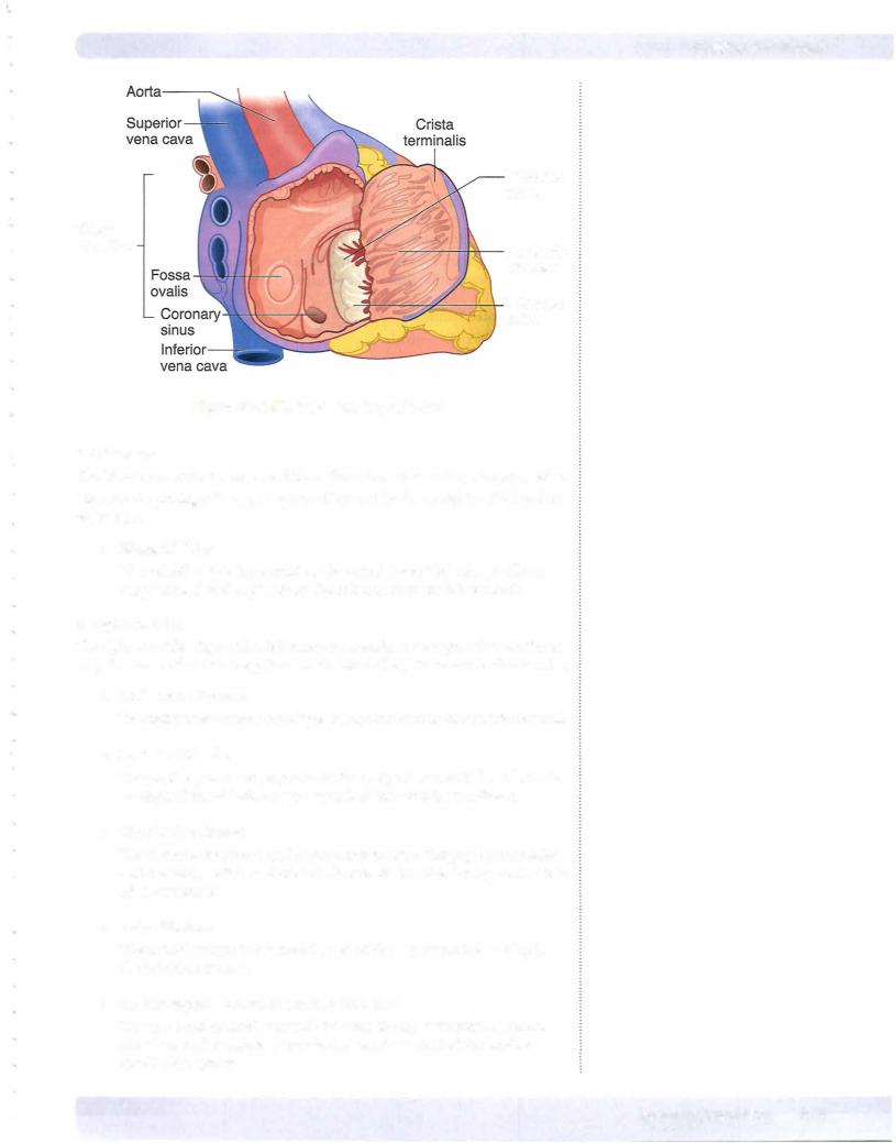
Conducting System ofthe Heart
SA node
The SA node initiates the impulse for contraction ofheart muscle (and is therefore termed the "pacemaker" of the heart). It is located at the superior end of the crista terminalis, where the superior vena cava enters the right atrium (Figure IIl-2-35).
The SA node is supplied bythe SA nodal branch ofthe right coronary artery.
Impulse production is speeded up by sympathetic nervous stimulation; it is slowed by parasympathetic (vagal) stimulation.
AVnode
The AV node receives impulses from the SA node. The AV node is located in the interatrial septum near the opening ofthe coronary sinus. The AV node slows the impulse so that it reaches the ventricles after it has reached the atria.
The AV node is supplied by the rightcoronaryartery.
Bundle ofHis
The bundle ofHis originates in the AV node. It conducts impulses to the right and left ventricles. It is supplied by the LAD artery.
In the right ventricle, the moderator band (septomarginal trabecula) contains the right bundle branch.
Impulses pass from the right and left bundle branches to the papillary muscles and ventricular myocardium.
Innervation
The cardiac plexus is a combination of sympathetic and parasympathetic (vagal) fibers.
•Sympathetic stimulation increases the heart rate. Nerves that sense pain associated with coronary artery ischemia (angina) follow the sympa thetic pathways back into spinal cord segments Tl-TS.
•Parasympathetic stimulation slows the heart rate. Sensory nerves that carry the afferent limb of cardiac reflexes travel with the vagus nerve.
MEDICAL 221


Section Ill • Gross Anatomy
Superior |
Ascending |
Bifurcation of |
Descending |
Vena Cava |
Aorta |
Trachea |
Aorta |
Ribs |
T4 Vertebra |
Scapula |
||
|
Figure 111-2-41 . Chest: CT, T4 |
|
||
Right |
Superior |
Body of |
AscendingPulmonary |
|
Pulmonary |
||||
Artery |
Vena Cava |
Sternum |
Aorta |
Trunk |
Descending Aorta T5 Vertebra |
Spinal Cord |
Figure 111-2-42. Chest: CT, T5
226 MEDICAL
Chapter 2 • Thorax
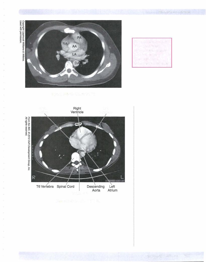
PA= pulmonary artery
RA = right atrium
AA= ascending aorta
LA= left atrium
E = esophagus
DA= descending aorta
|
Figure 111-2-43. Chest: CT, T5 |
Right |
Left |
Atrium |
Ventricle |
Esophagus
Figure 111-2-44. Chest: CT, T6
MEDICAL 227

Section Ill • Gross Anatomy
ChapterSummary
•The chest wall is formed by 1 2 thoracic vertebrae, 1 2 pairs of ribs, and the sternum. An important landmark on the anterior chest wall is the sternal angle found where the 2nd rib articulates with the sternum.
•The respiratory system develops as an endodermal outgrowth ofthe foregut. The tracheoesophageal septum separates the lung buds from the foregut. Improper development ofthis septum will produce an abnormal communication between the trachea and esophagus, a tracheoesophageal fistula.
•The lungs are surrounded by the pleura, which is divided into the parietal
pleura lining, the inner surface ofthe thoracic cavity, and the visceral pleura that is attached to the surface ofthe lung. Between these 2 layers is the pleural cavity containing a small amount of serous fluid. The lungs demonstrate costal, mediastinal, and diaphragmatic surfaces and an apex
that projects through the thoracic inlet into the root ofthe neck. Oblique and horizontal fissures divide the lungs into lobes.
•Heart development begins with the formation of a primitive heart tube, which develops from the lateral plate mesoderm in the third week. The arterial end of the heart tube is called the truncus arteriosus and will develop into the aorta and pulmonary trunk. The sinus venosus at the venous end ofthe heart tube will develop into the coronary sinus and the smooth part ofthe right atrium. The primitive atrial and ventricle chambers divide into right and left chambers following development of interatrial and interventricular septae. Ventricular septal defects result from failure of the membranous septum
to develop. Failure ofthe foramen ovale to close at birth results in atrial septal defects. Fetal circulation involves 3 shunts: ductus venosus, ductus arteriosum, and the foramen ovale. After birth these shunts shut down following changes in the circulatory system.
•The thoracic cavity is divided into the superior mediastinum above the plane of the sternal angle and the inferior mediastinum (anterior, middle, and posterior mediastina) below that sternal plane. The superior mediastinum contains the superiorvena cava, aortic arch and its branches, trachea, esophagus, thoracic duct, and the vagus and phrenic nerves. The anterior mediastinum is anterior to the heart and contains remnants ofthe thymus. The middle mediastinum contains the heart and great vessels and the posterior mediastinum containing the thoracic aorta, esophagus, thoracic duct, azygos veins, and vagus nerve. The inferiorvena cava passes through the diaphragm atthe caval hiatus at the level ofthe 8th thoracic vertebra, the esophagus through the esophageal hiatus at the 10th thoracic vertebra, and the aorta course through the aortic hiatus at the level ofthe 12th thoracic vertebra.
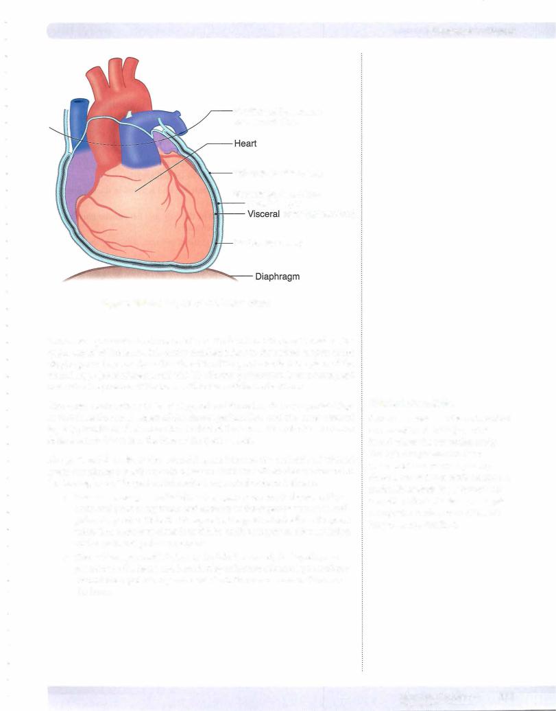
•Covering the heart is the pericardium formed by an outer, tough fibrous layer and a doubled-layered serous membrane divided into parietal and visceral layers. The pericardia! cavity is located between these 2 serous layers and includes the transverse and oblique pericardiaI sinuses.
(Continued)
228 MEDICAL

Chapter 2 • Thorax
ChapterSummary
•The external surface of the heart consists of several borders: the right border formed by the rightatrium, the left border formed by the leftventricle, the base formed bythe 2 atria, and the apex at the tip ofthe leftventricle. The anterior surface is formed bythe rightventricle, the posterior surface formed mainly by the left atrium, and a diaphragmatic surface is formed primarily by the left ventricle.
•Arterial supply to the heart muscle is provided by the right and left coronary arteries, which are branches ofthe ascending aorta. The right coronary artery supplies the right atrium, the right ventricle, the sinoatrial and atrioventricular nodes, and parts ofthe left atrium and left ventricle. The distal branch ofthe right coronary artery is the posterior interventricular artery that supplies, in part, the posterior aspect of the interventricular septum.
•The left coronary artery supplies most ofthe leftventricle, the left atrium, and the anterior part ofthe interventricular septum. The 2 main branches ofthe left coronaryartery are the anterior interventricular artery and the circumflex artery.
•Venous drainage ofthe heart is provided primarily by the great cardiac and middle cardiac veins and the coronary sinus, which drains into the right atrium.
•Sympathetic innervation increases the heart rate while the parasympathetics slows the heart rate. These autonomics fibers fire upon the conducting system ofthe heart.
-The sinoatrial node initiates the impulse for cardiac contraction.
-The atrioventricular node receives the impulse from the sinoatrial node and transmits that impulse to the ventricles through the bundle of His. The bundle divides into the right and left bundle branches and Purkinje fibers to the 2 ventricles.
MEDICAL 229

Abdomen, Pelvis, and Perineum |
3 |
ANTERIOR ABDOMINAL WALL
Surface Anatomy
Linea Alba
The linea alba is a shallow groove that runs vertically in the median plane from the xiphoid to the pubis. It separates the right and left rectus abdominis muscles. The components of the rectus sheath intersect at the linea alba.
Linea Semilunaris
The linea semilunaris is a curved line defining the lateral border of the rectus abdorninis, a bilateral feature.
Planes and Regions
The anterior abdominal wall is divided into 9 regions separated by several planes and lines (Figure III-3- 1) .
Subcostalplane
The subcostal plane (horizontal) passes through the inferior margins of the 10th costal cartilages at the level of the third lumbar vertebra.
Transpyloricplane
The transpyloric plane passes through the Ll vertebra, being half the distance be tween the pubis and the jugular notch. The plane passes through several important abdominal landmarks useful for radiology: pylorus of the stomach (variable), fun dus of gallbladder, neck and body of the pancreas, hila of kidneys, first part of the duodenum, and origin ofthe superior mesenteric artery
Midclavicularlines
The midclavicular lines (vertical) are the 2 planes that pass from the midpoint of the clavicle to the midpoint of the inguinal ligament on each side.
MEDICAL 231

Section Ill • Gross Anatomy
Internal abdominal oblique muscle and aponeurosis: This middle layer of the 3 flat muscles originates, in part, from the lateral two-thirds ofthe inguinal ligament. The internal oblique fibers course medially and arch over the inguinal canal in parallel with the arching fibers of the transversus abdominis muscle. The contributions of the internal abdominal oblique to the abdominal wall and inguinal region are the following:
Conjoint tendon (falxinguinalis) is formed by the combined arching fibers of the internal oblique and the transversus abdominis muscles that insert on the pubic crest posterior to the superficial inguinal ring.
Rectus sheath: The internal aponeuroses contribute to the layers of the rectus sheath.
Cremasteric muscle and fascia represent the middle layer ofthe sper matic fascia covering the spermatic cord and testis in the male. It forms in the inguinal canal.
Transversus abdominis muscle and aponeurosis: This is the deepest ofthe flat muscles. The transversus muscle originates, in part, from the lateral one-third of the inguinal ligament and arches over the inguinal canal with the internal oblique fibers to contribute to the conjoint tendon. The aponeuroses ofthe transversus muscle also contribute to the layers of the rectus sheath. Note that it does not contribute to any ofthe layers ofthe spermatic fasciae.
Abdominopelvic Fasciae and Peritoneum
Transversalis fascia: This fascia forms a continuous lining ofthe entire abdomi nopelvic cavity. Its contributions to the inguinal region include the following (Figure III-3-2):
• Deep inguinal ring is formed by an outpouching ofthe transversalis fascia immediately above the midpoint ofthe inguinal ligament and rep resents the lateral and deep opening ofthe inguinal canal. The inferior epigastric vessels are medial to the deep ring.
• Internal spermatic fascia is the deepest of the coverings of the sper rnatic cord formed at the deep ring in the male.
• Femoral sheath is an inferior extension of the transversalis fascia deep to the inguinal ligament into the thigh containing the femoral artery and vein and the femoral canal (site of femoral hernia).
• Rectus sheath: The transversalis fascia contributes to the posterior layer of the rectus sheath.
Extraperitoneal connective tissue: This is a thin layer ofloose connective tissue and fat surrounding the abdominopelvic cavity, being most prominent around the kidneys. The gonads develop from the urogenital ridge within this layer.
Parietal peritoneum: This is the outer serous membrane that lines the abdomi nopelvic cavity.
234 M EDICAL


Section Ill • Gross Anatomy
Clinical Correlate
A varicocele develops when blood collects in the pampiniform venous plexus and causes dilated and tortuous veins. This may result in swelling
and enlargement ofthe scrotum or enlargement ofthe spermatic cord above the scrotum. Varicoceles are more prominent when standing because of the blood pooling into the scrotum. A varicocele will reduce in size when the individual is horizontal.
Clinical Correlate
Cancers of the penis and scrotum will metastasize to the superficial inguinal lymph nodes, and testicular cancer will metastasize to the aortic (lumbar) nodes.
Clinical Correlate
In males, a cremasteric reflex can be demonstrated by lightly touching the skin of the upper medial thigh, resulting in a slight elevation of the testis. The sensory fibers of the reflex are carried by the Ll fibers of the ilioinguinal nerve and the motor response is a function ofthe genital
branch of the genitofemoral nerve that innervates the cremasteric muscle.
The entrance into the canal is the deep inguinal ring, located just lateral to the inferior epigastric vessels and immediately superior to the midpoint of the ingui nal ligament.
The superficial inguinal ring is the medial opening of the canal superior to the pubic tubercle.
Contents ofthe Inguinal Canal
1. Female Inguinal Canal
Round ligament ofthe uterus: The round ligament extends between the uterus and the labia majora and is a remnant of the gubernaculum that forms during descent of the ovary.
Ilioinguinal nerve (Ll ) is a branch of the lumbar plexus that exits the superficial ring to supply the skin of the anterior part of the mons pubis and labia majora.
2.Male Inguinal Canal
Ilioinguinalnerve (LI) is a branch of the lumbar plexus that exits the superficial ring to supply the skin of the lateral and anterior scrotum.
The spermatic cord is formed during descent of the testis and contains struc tures that are related to the testis. The cord begins at the deep ring and courses through the inguinal canal and exits the superficial ring to enter the scrotum. The spermatic cord is covered by 3 layers of spermatic fascia: external, middle, and internal. The cord contains the following:
•Testicular artery: A branch of the abdominal aorta that supplies the testis.
•Pampiniform venous plexus: An extensive network of veins draining the testis located within the scrotum and spermatic cord. The veins of the plexus coalesce to form the testicular vein at the deep ring. The venous plexus assists in the regulation of the temperature of the testis.
•Vas deferens (ductus deferens) and its artery
•Autonomic nerves
•Lymphatics: Lymphatic drainage of the testis will drain into the lumbar (aortic) nodes of the lumbar region and not to the superficial inguinal nodes which drain the rest of the male perineum.
Fascial Layers ofSpermatic Cord
There are 3 fascial components derived from the layers ofthe abdominal that sur round the spermatic cord (Figure III-3-2):
l.External spermatic fascia is formed by the aponeuroses of the external ab dominal oblique muscle at the superficial ring.
2.Middle or cremasteric muscle and fasciaare formed by fibers ofthe internal abdominal oblique within the inguinal canal The cremasteric muscle elevates the testis and helps regulate the thermal environment ofthe testis.
3.Internalspermaticfasciais formed by the transversalisfascia at the deep ring.
236 MEDICAL

Chapter 3 • Abdomen, Pelvis, and Perineum
Boundaries of the Inguinal Canal
Roof
Formed by fibers ofthe internal abdominal oblique and the transverse abdominis muscles arching over the spermatic cord (Figure IIl-3-2)
AnteriorWall
Formed by aponeurosis ofthe external abdominal oblique throughout the ingui nal canal and the internal abdominal oblique muscle laterally
Floor
Formed by inguinal ligament throughout the entire inguinal canal and the la cunar ligament at the medial end
Posterior Wall:The posterior wall is divided into lateral and medial areas:
•Medialarea is formed and reinforced by the fused aponeurotic fibers of the internal abdominal oblique and transversus abdominis muscles
(conjoint tendon).
•Lateral area is formed bythe transversalis fascia and represents the weak area of the posterior wall.
•Inferior epigastric arteryandvein ascend the posterior wall just lateral to the weak area and just medial to the deep ring.
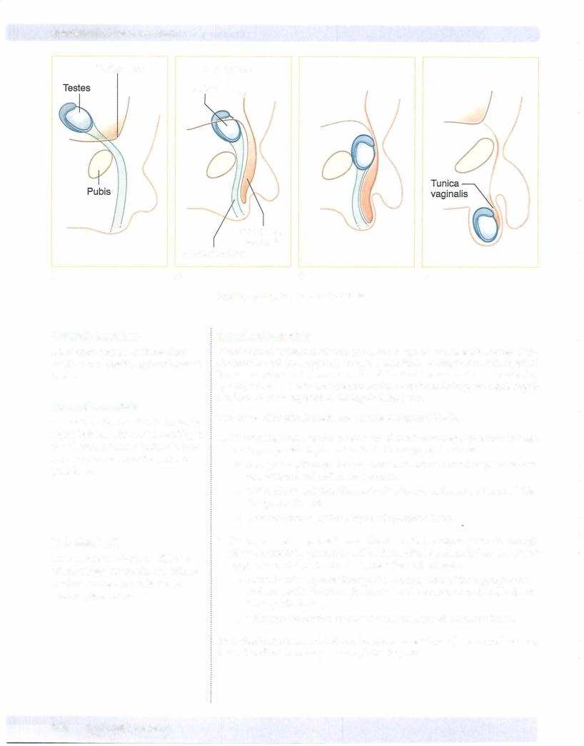
Descent ofthe Testes
The testis develops from the mesoderm of the urogenital ridge within the extra peritoneal connective tissue layer.
•During the last trimester, the testis descends the posterior abdominal wall inferiorly toward the deep inguinal ring guided by the fibrous gubernaculums.
•An evagination of the parietal peritoneum and the peritoneal cavity extends into the inguinal canal called the processus vaginalis (Figure IIl-3-3). The open connection of the processus vaginalis with the perito neal cavity closes before birth.
•A portion of the processus vaginalis remains patent in the scrotum and surrounds the testis as the tunicavaginalis.
Clinical Correlate
Failure of one or both ofthe testes to descend completely into the scrotum results in cryptorchidism, which may lead to sterility if bilateral.
MEDICAL 237

Chapter 3 • Abdomen, Pelvis, and Perineum
Clinical Correlate
Direct inguinal hernias usually pass through the inguinal (Hasselbach's) triangle:
•Lateral border: inferior epigastric vessels
•Medial border: rectus abdominis muscle
•Inferior border: inguinal ligament
Inferior epigastric artery & vein
Indirect
Inguinal
triangle
Direct
Superficial inguinal ring
Figure 111-3-4. Inguinal Hernia
MEDICAL 239


Chapter 3 • Abdomen, Pelvis, and Perineum
Table 111-3-1. Adult Structures Derived from Each of the 3 Divisions of the Primitive Gut Tube
Foregut Midgut Hindgut
Artery: celiac |
Artery: superior mesenteric |
Parasympathetic innervation: vagus |
Parasympathetic innervation: vagus |
nerves |
nerves |
Sympathetic innervation: |
Sympathetic innervation: |
• Preganglionics: thoracic |
• Preganglionics: thoracic |
splanchnic nerves, T5-T9 |
splanchnic nerves, T9-Tl2 |
•Postganglionic cell bodies: celiac ganglion
Referred Pain: Epigastrium
Foregut Derivatives
Esophagus
Stomach
Duodenum (first and second parts) Liver
Pancreas
Biliary apparatus Gallbladder
Amniotic cavity (AM)
•Postganglionic cell bodies: superior mesenteric ganglion
Referred Pain: Umbilical
Midgut Derivatives
Duodenum (second, third, and fourth parts) Jejunum
Ileum
Cecum
Appendix Ascending colon
Transverse colon (proximal two-thirds)
Pharyngeal
pouches
1
Stomach
Hepatic diverticulum
Gallbladder
Artery: inferior mesenteric
Parasympathetic innervation: pelvic splanchnic nerves
Sympathetic innervation:
•Preganglionics: lumbar splanchnic nerves, L1-L2
•Postganglionic cell bodies: inferior mesenteric ganglion
Referred Pain: Hypogastrium
Hindgut Derivatives
Transverse colon (distal third splenic flexure)
Descending colon Sigmoid colon Rectum
Anal canal (above pectinate line)
Esophagus
Lung bud
Foregut 90° rotation to right along
longitudinal axis
Vitelline Vitelline
duct duct
Cloaca
Superior mesenteric artery
270° rotation
counterclockwise Midgut and herniation
(6-10th week)
Hindgut Septation
Celiac artery
Dorsal pancreatic bud
Ventral pancreatic bud
Figure 111-3-6. Development of Gastrointestinal Tract
MEDICAL 241
Section Ill • Gross Anatomy
In A Nutshell
The lower respiratory tract, liver and biliary system, and pancreas all develop from an endodermal outgrowth of the foregut.
Development and Rotation of Foregut
After body foldings and the formation of the gut tube, the foregut is suspended from the dorsal body wall by the dorsalembryonicmesenteryand from the ven tral body wall by the ventral embryonic mesentery (Figure III-3-7A). Note that the liver develops in the ventral embryonic mesentery, and the spleen and dorsal pancreatic bud develop in the dorsal embryonic mesentery.
•The abdominal foregut rotates 90° (clockwise) around its longitudinal axis. The original left side of the stomach before rotation becomes the ventral surface after rotation and its anterior and posterior borders before rotation will become the lesser and greater curvatures, respectively.
•Foregut rotation results in the liver, lesser omentum (ventral embry onic mesentery), pylorus of the stomach, and duodenum moving to the right; and the spleen, pancreas, and greater omentum (dorsal embryonic mesentery) moving to the left (Figure III-3-7A) .
•The ventral embryonic mesentery will contribute to the lesser omentum
and the falciform ligament, both of which attach to the liver.
•The dorsal embryonic mesentery will contribute to the greater omen tum and the gastro-splenic and splenorenal ligaments, all of which attach to the spleen or the greater curvature of the stomach.
242 MEDICAL


Section Ill • Gross Anatomy
Epiploic Foramen (ofWinslow)
The epiploic foramen is the opening between omental bursa and greater peri toneal sac (Figures III-3-7, III-3-8, and III-3-9). The boundaries of the epiploic foramen are the following:
Anteriorly: Hepatoduodenal ligament and the hepatic portal vein Posteriorly: Inferior vena cava
Superiorly: Caudate lobe of the liver Inferiorly: First part of the duodenum
Falciform ligament (contains ligamentum teres of liver)
Epiploic foramen
Descending colon
Figure 111-3-8. Peritoneal Membranes
246 MEDICAL


Chapter 3 • Abdomen, Pelvis, and Perineum
Extrahepatic BiliaryAtresia
Occurs when the lumen ofthe biliary ducts is occluded owing to incomplete re canalization.This condition is associated with jaundice, white-colored stool, and dark-colored urine.
Annular Pancreas
Occurs when the ventral and dorsal pancreatic buds form a ring around the duo denum, thereby causing an obstruction ofthe duodenum and polyhydrarnnios
DuodenalAtresia
Occurs when the lumen ofthe duodenum is occluded owing to failed recanaliza tion. This condition is associated with polyhydramnios, bile-containing vomitus, and a distended stomach.
Omphalocele
An omphalocele occurs when the midgut loop fails to return to the abdominal cavity and remains in the umbilical stalk.
•The viscera herniate through the umbilical ring and are contained in a shinysac of amnion at the base of the umbilical cord.
•Omphalocele is often associated with multiple anomalies of the heart and nervous system with a high mortality rate (25%).
Gastroschisis
Gastroschisis occurs when the abdominal viscera herniate through the body wall directly into the amniotic cavity, usually to the right of the umbilicus.
•This is a defect in the closure of the lateral body folds and a weakness of the anterior wall at the site of absorption of the right umbilical vein.
•Note that the viscera do not protrude through the umbilical ring and are not enclosed in a sac of amnion.
lleal (Meckel) Diverticulum
Occurs when a remnant of the vitelline duct persists, thereby forming a blind pouch on the antimesenteric border of the ileum. This condition is often asymp tomatic but occasionally becomes inflamed ifit contains ectopic gastric, pancre atic, or endometrial tissue, which may produce ulceration. They are found 2 feet from the ileocecal junction, are 2 inches long, and appear in 2% ofthe population.
Vitelline Fistula
Occurs when the vitelline duct persists, thereby forming a direct connection be tween the intestinal lumen and the outside ofthebodyatthe umbilicus. This condi tion is associated with drainage ofmeconium from the umbilicus.
MEDICAL 249

Section Ill • Gross Anatomy
Malrotation of Midgut
Occurs when the midgut undergoes only partial rotation and results in abnormal position of abdominal viscera. This condition may be associated with volvulus (twisting of intestines) .
Colonic Aganglionosis (Hirschsprung Disease)
Results from the failure ofneural crest cells to form the myenteric plexus in the sigmoid colon and rectum. This condition is associated with loss of peristalsis and immobility of the hindgut, fecal retention and abdominal distention of the transverse colon (megacolon).
ABDOMINALVISCERA
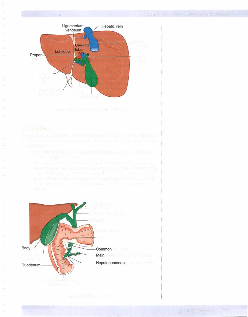
Liver
The liver has 2 surfaces: a superior or a visceral surface (Figure III-3- 1 abdominal cavity and is protected by peritoneum:
or diaphragmatic surface and an inferior 1) . lt lies mostly in the right aspect of the the rib cage. The liver is invested by visceral
•The reflection of visceral peritoneum between the diaphragmatic surface of the liver and the diaphragm forms the falciform ligament, which continues onto the liver as the coronary ligament and the right and left triangular ligaments.
•The extension of visceral peritoneum between the visceral surface of the liver and the first part of the duodenum and the lesser curvature of the stomach forms the hepatoduodenal and hepatogastric ligaments of the lesser omentum, respectively.
The liver is divided into 2 lobes of unequal size as described below (Figure Ill-
3- 1 1).
•Fissures for the ligamentum teres and the ligamentum venosum, the porta hepatis, and the fossa for the gallbladder further subdivide the right lobe into the right lobe proper, the quadrate lobe, and the caudate lobe.
•The quadrate and caudate lobes are anatomically part of the right lobe but functionally part of the left. They receive their blood supply from the left branches of the portal vein and hepatic artery and secrete bile to the left hepatic duct.
The liver has a central hilus, or porta hepatis, which receives venous blood from the portal vein and arterial blood from the hepatic artery.
•The central hilus also transmits the common bile duct, which collects bile produced by the liver.
•These structures, known collectively as the portal triad, are located in the hepatoduodenal ligament, which is the right free border of the lesser omentum.
The hepatic veins drain the liver by collecting blood from the liver sinusoids and returning it to the inferior vena cava.
250 MEDICAL

Chapter 3 • Abdomen, Pelvis, and Perineum
Spleen
The spleen is a peritoneal organ in the upper left quadrant that is deep to the left 9th, 10th, and 11th ribs The visceral surface ofthe spleen is in contact with the left colic flexure, stomach, and left kidney. Inasmuch as the spleen lies above the costal margin, a normal-sized spleen is not palpable.
The splenic artery and vein reach the hilus ofthe spleen by traversing the spleno renalligament.
Stomach
The stomachhas a right lesser curvature, which is connected to the porta hepatis of the liver by the lesser omentum (hepatogastric ligament), and a left greater curvature from which the greater omentum is suspended (Figure IIl-3-8).
The cardiac region receives the esophagus; and the dome-shaped upper portion ofthe stomach, which is normally filledwithair, is the fundus. The main central part of the stomach is the body. The pyloric portion of the stomach has a thick muscular wall and narrow lumen that empties into the duodenum approximately in the transpyloric plane (Ll vertebra).
Duodenum
The duodenum is C-shaped, has 4 parts, and is located retroperitoneal except for the first part.
•The first part is referred to as the duodenal cap (bulb). The gastroduode nal artery and the common bile duct descend posterior to the first part.
•The second part (descending) receives the common bile duct and main pancreatic duct (Figures III-3-10 and III-3-12) at the hepatopancreatic ampulla (ofVater). Smooth muscle in the wall ofthe duodenal papilla is known as the sphincter of Oddi.
•Note that the foregut terminates at the point of entry of the common bile duct; the remainder ofthe duodenum is part of the midgut.
Jejunum and Ileum
The jejunum begins at the duodenojejunal junction and comprises 2/5 ofthe re maining small intestine. The beginning ofthe ileum is not dearly demarcated; it consists ofthe distal 3/5 ofthe small bowel.
The jejunoileum is suspended from the posterior body wall by the mesentery proper. Although the root ofthe mesentery is only 6 inches long, the mobile part ofthe small intestine is approximately 22 feet in length.
Colon
Cecum
The cecum is the firstpart ofthe colon, or large intestine, andbegins at the ileoce caljunction. It is a blind pouch, which often has a mesentery and gives rise to the vermiform appendix. The appendixhas its own mesentery, the mesoappendix.
Clinical Correlate
The spleen may be lacerated with a fracture ofthe 9th, 10th, or 11th rib on the left side.
Clinical Correlate
A sliding hiatal hernia occurs when the cardia ofthe stomach herniates through the esophageal hiatus ofthe diaphragm. This can damage the vagal trunks as they pass through the hiatus.
MEDICAL 253

Section Ill • Gross Anatomy
Ascending colon
The ascending colon lies retroperitoneally and lacks a mesentery. It is continuous with the transverse colon at the right (hepatic) flexure of colon.
Transverse colon
The transverse colon has its own mesentery called the transverse mesocolon. It becomes continuous with the descending colon at the left (splenic) flexure of co lon. The midgut terminates at the junction of the proximal two-thirds and distal one-third of the transverse colon.
Descending colon
The descending colon lacks a mesentery. It joins the sigmoid colon where the large bowel crosses the pelvic brim.
Sigmoid colon
The sigmoid colon is suspended by the sigmoid mesocolon. It is the terminal portion of the large intestine and enters the pelvis to continue as the rectum.
Rectum
The superior one-third of the rectum is covered by peritoneum anteriorly and laterally. It is the fixed, terminal, straight portion of the hindgut.
Anal Canal
•The anal canal is about 1 .5 inches long and opens distally at the anus. The anal canal is continuous with the rectum at the pelvic diaphragm where it makes a 90-degree posterior bend (anorectal flexure) below the rectum.
•The puborectalis component of the pelvic diaphragm pulls the flexure forward, helping to maintain fecal continence.
•The internal anal sphincter is circular smooth muscle that surrounds the anal canal. The sympathetics (lumbar splanchnics) increase the tone of the muscle and the parasympathetics (pelvic splanchnics) relax the muscle during defecation.
•The external anal sphincter is circular voluntary skeletal muscle sur rounding the canal that is voluntarily controlled by the inferior rectal branch of the pudenda!nerve and relaxes during defecation.
•The anal canal is divided in an upper and lower parts separated by the pectinate line, an elevation of the mucous membrane at the distal ends of the anal columns. A comparison of the features of the anal canal above and below the pectinate line is shown in Table III-3-3.
254 MEDICAL


Section Ill • Gross Anatomy
Clinical Correlate
The most common site for an abdominal aneurysm is in the area between the renal arteries and the bifurcation of the abdominal aorta. Signs include decreased circulation to the lower limbs and pain radiating down the back ofthe lower limbs. The most common site of atherosclerotic plaques is at the bifurcation of the abdominal aorta.
Clinical Correlate
•The splenic artery may be subject to erosion by a penetrating ulcer of the posteriorwall of the stomach into the lesser sac.
•The left gastric artery may be subject to erosion by a penetrating ulcer ofthe lesser curvature ofthe stomach.
•The gastroduodenal artery may be subject to erosion by a penetrating ulcer ofthe posterior wall ofthe first part of the duodenum.
Three Unpaired Visceral Arteries
CeliacArtery(Trunk)
The celiac artery (Figure IIl-3- 15) is the blood supply to the structures derived from the foregut. The artery arises from the anterior surface of the aorta just in ferior to the aortic hiatus at the level ofT12-Ll vertebra. The celiac artery passes above the superior border of the pancreas and then divides into 3 retroperitoneal branches.
The left gastric artery courses superiorly and upward to the left to reach the lesser curvature of the stomach. The artery enters the lesser omentum and follows the lesser curvature distally to the pylorus. The distribution of the left gastric includes the following:
•Esophageal branch to the distal one inch of the esophagus in the abdo men
•Most ofthe lesser curvature
The splenic artery is the longest branch of the celiac trunk and runs a very tortu ous course along the superior border of the pancreas. The artery is retroperitone al until it reaches the tail of the pancreas, where it enters the splenorenal ligament to enter the hilum of the spleen. The distributions of the splenic artery include:
•Direct branches to the spleen
•Direct branches to the neck, body, and tail ofpancreas
•Left gastroepiploic artery that supplies the left side of the greater cur vature of the stomach
•Short gastric branches that supply to the fundus of the stomach
The common hepatic artery passes to the right to reach the superior surface of the first part of the duodenwn, where it divides into its 2 terminal branches:
•Properhepatic artery ascends within the hepatoduodenal ligament of the lesser omentum to reach the porta hepatis, where it divides into the right and left hepatic arteries. The right and left arteries enter the
2 lobes of the liver, with the right hepatic artery first giving rise to the cystic artery to the gallbladder.
•Gastroduodenal artery descends posterior to the first part of the duo denum and divides into the right gastroepiploic artery (supplies the pyloric end of the greater curvature of the stomach) and the superior pancreaticoduodenal arteries (supplies the head of the pancreas, where it anastomoses the inferior pancreaticoduodenal branches of the supe rior mesenteric artery) .
256 MEDICAL


Chapter 3 • Abdomen, Pelvis, and Perineum
POSTERIOR ABDOMINAL BODYWALL
Embryology of Kidneys and Ureter
Renal development is characterized by 3 successive, slightly overlapping kidney systems: pronephros, mesonephros, and metanephros (Figure IIl-3-21).
Stomach
Midgut
Cecum
Pronephros
Mesonephros
Hindgut
Figure 111-3-21 . Pronephros, Mesonephros, and Metanephros
Pronephros
During week 4, segmented nephrotomes appear in the cervical intermediate me soderm ofthe embryo. These structures grow laterally and canalize to form neph ric tubules. The first tubules formed regress before the last ones are formed. By the end ofweek 4, the pronephros disappears and does not function.
Mesonephros
In week 5, the mesonephros appears as S-shaped tubules in the intermediate mesoderm of the thoracic and lumbar regions of the embryo.
•The medial end of each tubule enlarges to form a Bowman's capsule into which a tuft of capillaries, or glomerulus, invaginates.
•The lateral end of each tubule opens into the mesonephric (Wolffian) duct, an intermediate mesoderm derivative. The duct drains into the hindgut.
•Mesonephric tubules function temporarily and degenerate by the begin ning of month 3. The mesonephric duct persists in the male as the ductus epididyrnidis, ductus deferens, and the ejaculatory duct. It disap pears in the female.
Metanephros
During week 5, the metanephros, or permanent kidney, develops from 2 sources: the uretericbud, a diverticulum of the mesonephric duct, and the metanephric mass (blastema), from intermediate mesoderm of the lumbar and sacral regions (Figure IIl-3-22).
MEDICAL 263


Section Ill • Gross Anatomy
Clinical Correlate
Blockage by Renal Calculi
The most common sites of ureteral constriction susceptible to blockage by renal calculi are:
•Where the renal pelvis joins the ureter
•Where the ureter crosses the pelvic inlet
•Where the ureter enters the wall of the urinary bladder
Congenital Abnormalities ofthe Renal System
•Renal agenesis
Renal agenesis results from failure of one or both kidneys to develop because of early degeneration of the ureteric bud. Unilateral genesis is fairly common; bilateral agenesis is fatal (associated with oligohydram nios, and the fetus may have Pottersequence: clubbed feet, pulmonary hypoplasia, and craniofacial anomalies) .
•Pelvic and horseshoekidney
Pelvic kidney is caused by a failure of one kidney to ascend. Horseshoe kidney (usually normalrenalfunction, predisposition to calculi) is a fusion of both kidneys at their ends and failure of the fused kidney to ascend. The horseshoe kidney hooks under the origin of the inferior mesenteric artery.
•Double ureter
Caused by the early splitting of the ureteric bud or the development of 2 separate buds
•Patent urachus
Failure ofthe allantois to be obliterated results in urachal fistulas or sinuses. In male children with congenital valvular obstruction of the pros tatic urethra or in older men with enlarged prostates, a patent urachus maycause drainage ofurine through the umbilicus.
Posterior Abdominal Wall and Pelvic Viscera
Kidneys
The kidneys are a pair ofbean-shaped organs approximately 1 2 cm long. They ex tend from vertebral level Tl2 to L3 when the body is in the erect position (Figure III-3-24A and -24B). The right kidney is positioned slightly lower than the left because of the mass of the liver.
•Kidney's Relation to the PosteriorAbdominalWall
Both kidneys are in contact with the diaphragm, psoas major, and qua dratus lumborum (Figure III-3-24) .
-Right kidney-contacts the above structures and the 1 2th rib
-Left kidney-contacts the above structures and the 1 1th and 12th ribs
Ureters
Ureters are fibromuscular tubes that connect the kidneys to the urinary bladder in the pelvis. They run posterior to the ductus deferens in males and posterior to the uterine artery in females. They begin as continuations of the renal pelves and run retroperitoneally, crossing the external iliac arteries as they pass over the pelvic brim.
Ureter's Relation to the PosteriorAbdominalWall
The ureter lies on the anterior surface of the psoas major muscle.
266 MEDICAL

Section Ill • Gross Anatomy
Clinical Correlate
Spastic bladder results from lesions of the spinal cord above the sacral spinal cord levels. There is a loss of inhibition ofthe parasympathetic nerve fibers that innervate the detrusor muscle during the filling stage. Thus, the detrusor muscle responds to a minimum amount of stretch, causing urge incontinence.
Clinical Correlate
Atonic bladder results from lesions to the sacral spinal cord segments or the sacral spinal nerve roots. Loss of pelvic splanchnic motor innervation
with loss of contraction of the detrusor muscle results in a full bladder with a continuous dribble of urine from the bladder.
Clinical Correlate
Weakness of the puborectalis part of the levator ani muscle may result in rectal incontinence.
Weakness of the sphincter urethrae part of the urogenital diaphragm may result in urinary incontinence.
268 MEDICAL
• Muscles
The detrusor muscle forms most of the smooth-muscle walls of the bladder and contracts during emptying of the bladder (micturition). The contraction of these muscles is under control of the parasympa thetic fibers of the pelvic splanchnics (52, 3, 4)
The internal urethral sphincter (sphincter vesicae) is smooth-muscle fibers that enclose the origin of the urethra at the neck ofthe bladder. These muscles are under control of the sympathetic fibers of the lower thoracic and lumbar splanchnics (Tl l -L2) and are activated during the filling phase of the bladder to prevent urinary leakage.
-The external urethralsphincter (sphincter urethrae) is the voluntary skeletal muscle component of the urogenital diaphragm that encloses the urethra and is relaxed during micturition (voluntary muscle
of micturition). The external sphincter is innervated by perineal branches of the pudendal nerve.
Urethra
The male urethra is a muscular tube approximately 20 cm in length. The urethra in men extends from the neck of the bladder through the prostate gland (prostat ic urethra) to the urogenital diaphragm ofthe perineum (membranous urethra), and then to the external opening of the glans (penile or spongy urethra) (Figure
III-3-26).
The male urethra is anatomically divided into 3 portions: prostatic, membra nous, and spongy (penile).
•The distal spongyurethra of the male is derived from the ectodermal cells of the glans penis.
The female urethra is approximately 4 cm in length and extends from the neck of the bladder to the external urethral orifice ofthe vulva (Figure III-3-27).
PELVIS
Pelvic and Urogenital Diaphragms
The pelvic andurogenital diaphragms (Figure III-3-25) are 2 important skeletal muscle diaphragms that provide support of the pelvic and perinea! structures. They are each innervated by branches of the pudenda! nerve.
•The pelvic diaphragm forms the muscular floor of the pelvis and separates the pelvic cavity from the perineum. The pelvic diaphragm is a strong support for the pelvic organs and transmits the distal parts of the genitourinary system and GI tract from the pelvis to the perineum.
-The diaphragm is formed by 2 layers of fascia and the 2 muscles: the levator ani and coccygeus.
-The puborectalis component of the levator ani muscle forms a muscu
lar sling around the anorectal junction, marks the boundary between the rectum and anal canal and is important in fecal continence.


Section Ill • Gross Anatomy
Clinical Correlate
Support for pelvic viscera is provided by the pelvic and urogenital diaphragms, perinea! membrane, perinea! body, and the transverse (cardinal) cervical and uterosacral ligaments. Weakness of support structures may result in prolapse of the uterus into the vagina or herniation of the bladder or rectum into the vagina.
Uterus and BroadLigament
Figure III-3-28 illustrates a posteriorview of the female reproductive tract. |
|||
|
d |
{ |
|
Broa |
|
Mesosalpinx |
Round |
ligament |
Mesovarium |
ligaments |
|
|
|
Mesometrium |
of uterus |
Ovarian
artery
Clinical Correlate
The ureter passes inferior to the uterine artery 1 to 2 centimeters from the cervix ("water under the bridge") and must be avoided during surgical procedures.
Clinical Correlate
A pudenda( nerve block to anesthetize the perineum is performed as the pudenda! nerve crosses posterior to the ischial spine (Figure 111-3-248).
Uterine artery |
|
Transverse (cardinal) |
("water under bridge") |
|
|
|
Ureter |
cervical ligament |
|
Uterosacral ligament |
|
|
|
|
|
Figure 111-3-28. Broad Ligament |
|
PERINEUM
The perineum is the diamond-shaped outlet of the pelvis located below the pelvic diaphragm. The perineum is divided by a transverse line between the ischial tuber osities into the anal and urogenital triangles (Figure III-3-29).
•The sensory and motor innervation to the perineum is provided by the pudenda! nerve (S2, 3, 4) of the sacral plexus.
•The blood supply is provided by the internal pudenda! artery, a branch of the internal iliac artery.
•The pudendal nerve and vessels cross the ischial spine posteriorly to enter the perineum (Figure III-3-24 B).
AnalTriangle
The anal triangle is posterior and contains the anal canal surrounded by the fat-filled ischioanal fossa.
•The anal canal is guarded by a smooth-muscle internal anal sphincter innervated by the ANS and an externalanal sphincter of skeletal mus cle innervated by the pudendal nerve.
•The pudenda! canal transmitting the pudenda! nerve and internal pudenda! vessels is found on the lateral aspect of the ischioanal fossa (Figure III-3-29) .
272 MEDICAL


Chapter 3 • Abdomen, Pelvis, and Perineum
Complete androgen insensitivity (CAIS, ortesticularfeminization syndrome)
•Occurs when a fetus with a 46,)CY genotype develops testes and female external genitalia with a rudimentary vagina; the uterus and uterine tubes are generally absent
•Testes may be found in the labia majora and are surgically removed to circumvent malignant tumor formation.
•Individuals present as normal-appearing females, and their psychosocial(AR) orientation is female despite their genotype.
Most common cause is a mutation in the androgen receptor gene AR inactive.• that renders the
Abnormalities ofthe Penis and Testis
Hypospadias
•Occurs when the urethral folds fail to fuse completely, resulting in the external urethral orifice opening onto the ventral surface of the penis.
•Generally associated with a poorly developed penis that curves ventrally, known as chordee.
Epispadias
•Occurs when the external urethral orifice opens onto the dorsal surface of the penis.
•Generally associated with exstrophy of the bladder.
Undescendedtestes (cryptorchidism)
Occurs when the testes fail to descend into the scrotum. Normally occurs within 3 months after birth.
•Bilateral cryptorchidism results in sterility.
•The undescended testes may be found in the abdominal cavity or in the inguinal canal.
Hydrocele ofthe testes
Occurs when a small patency of the processus vaginalis remains so that perito neal fluid can flow into the processus vaginalis. Results in a fluid-filled cyst near the testes.
MEDICAL 277

Section Ill • Gross Anatomy
RADIOLOGY OF THE ABDOMEN AND PELVIS
Duodenum Pylorus Stomach Jejunum
Ileum
Figure 111-3-31 . Abdomen: Upper GI, Small Bowel
Hepatic |
Transverse |
Splenic |
Flexure |
Colon |
Flexure |
Descending
Colon
Sigmoid
Colon
Figure 111-3-32. Abdomen: Barium Enema
278 MEDICAL

Chapter 3 • Abdomen, Pelvis, and Perineum
|
Inferior |
Liver |
Vena Cava Aorta Diaphragm Stomach |
Figure 111-3-33. Abdomen: CT, T1 1
Liver |
Portal Vein |
Descending Colon |
Inferior Vena Cava Diaphragm Aorta Stomach Spleen
Figure 111-3-34. Abdomen: CT, T12
MEDICAL 279

Section Ill • Gross Anatomy
Liver |
Ascending |
Descending |
Colon |
Aorta Stomach Colon |
Spleen
Inferior Vena Cava |
Diaphragm |
Left Kidney |
|
Figure 111-3-35. Abdomen: CT, T12 |
|||
|
Superior |
|
|
Mesenteric |
Splenic |
Spleen |
|
Liver Pancreas |
Artery |
Vein |
|
Left Adrenal Gland
Figure 111-3-36. Abdomen: CT, L1
280 MEDICAL

Chapter 3 • Abdomen, Pelvis, and Perineum
Ascending |
Superior |
Superior |
Mesenteric |
Mesenteric |
|
Colon Duodenum |
Vein |
Artery Jejunum |
Right |
Renal |
Inferior |
Aorta |
Descending |
|
Kidney |
Pelvis |
Vena Cava |
Colon |
||
Figure 111-3-37. Abdomen: CT, L2 |
|||||
|
Inferior |
Superior |
|
||
|
Mesenteric |
|
|||
Duodenum |
Vena Cava |
|
Artery |
Aorta |
|
Right Kidney Right Ureter Left Psoas Major
Figure 111-3-38. Abdomen: CT, L3
MEDICAL 281
Section Ill • Gross Anatomy
Inferior |
Left Common |
Ureter Vena Cava |
Iliac Artery |
Psoas |
Right Common |
Ureter |
Major |
Iliac Artery |
|
Figure 111-3-39. Abdomen: CT, L4
ChapterSummary
•The abdominal wall consists primarily of 3 flat muscles (external oblique, internal oblique, and transversus abdominis muscles), rectus abdominis muscle, and the transversalis fascia.
•The inguinal canal contains the round ligament in the female and the spermatic cord in the male. The inguinal canal is an oblique canal through the lower abdominal wall beginning with the deep inguinal ring laterally and the superficial inguinal ring medially. Weakness ofthe walls ofthe canal can result in 2 types of inguinal hernias: direct and indirect.
•A direct hernia emerges through the posterior wall ofthe inguinal canal medial to the inferior epigastric vessels.
•An indirect hernia passes through the deep inguinal ring lateral to the inferior epigastria vessels and courses through the inguinal canal to reach the superficial inguinal ring.
•A persistent processus vaginalis often results in a congenital indirect inguinal hernia.
•The gastrointestinal (GI) system develops from the primitive gut tube formed by the incorporation ofthe yolk sac into the embryo during body foldings.
The gut tube is divided in the foregut, midgut, and hindgut.
•Defects in the development of the GI tract include annular pancreas, duodenal atresia, Meckel diverticulum, and Hirschsprung disease.
(Continued)
282 MEDICAL

Chapter 3 • Abdomen, Pelvis, and Perineum
ChapterSummary (Cont'd)
•The foregut, midgut, and hindgut are supplied by the celiac trunk, superior mesenteric artery, and inferior mesenteric artery, respectively. These arteries and their branches reach the viscera mainly by coursing in different parts of the visceral peritoneum. Venous return from the abdomen is provided by the tributaries of the inferior vena cava, exceptfor the GI tract. Blood flow from the GI tract is carried by the hepatic portal system to the liver before returning to the inferior vena cava by the hepatic veins.
•Diseases ofthe liver result in obstruction offlow in the portal system and portal hypertension. Four collateral portal-caval anastomoses develop to provide retrograde venous flow back to the heart: esophageal, rectal, umbilical, and retroperitoneal.
•The viscera ofthe GI system are covered by the peritoneum, which is divided into the parietal layer lining the body wall and the visceral layer extending from the bodywall and covering the surface ofthe viscera. Between these layers is the potential space called the peritoneal cavity.
•The peritoneal cavity is divided into the greater peritoneal sac and the lesser peritoneal sac (omental bursa). Entrance into the omental bursa from the greater sac is the epiploic foramen that is bound anteriorly by the lesser omentum and posteriorly by the inferior vena cava.
•The kidneys develop from intermediate mesoderm by 3 successive renal systems: pronephros, mesonephros, and metanephros. The mesonephric kidney is the first functional kidney that develops during the first trimester.
The final or metanephric kidney develops from 2 sources: the ureteric bud that forms the drainage part of the kidney and the metanephric mass thatforms the nephron ofthe adult kidney.
•The urinary bladder develops from the urogenital sinus, which is formed after division of the cloaca by the urorectal septum.
•The kidneys are located against the posteriorabdominal wall between the T12 and L3 vertebrae. Posterior to the kidneys lie the diaphragm and the psoas major and quadratus lumborum muscles. The superior pole of the kidney
lies against the parietal pleura posteriorly. The ureters descend the posterior abdominal wall on the ventral surface ofthe psoas major muscle and cross the
pelvic brim to enterthe pelvic cavity. |
(Continued) |
|
MEDICAL 283

Section Ill • Gross Anatomy
ChapterSummary(Cont,d)
Pelvis
•The pelvic cavity contains the inferior portions ofthe GI and urinary systems along with the reproductive viscera. The pelvic viscera and their relationships are shown for the male and female pelvis in Figures 111-3-26 and 111-3-27, respectively.
•There are 2 important muscular diaphragms related to the floor of the pelvis and the perineum: the pelvic diaphragm and the urogenital diaphragm, respectively. Both of these consist of 2 skeletal muscle components under voluntary control and are innervated by somatic fibers ofthe lumbosacral plexus.
•The pelvic diaphragm forms the floor of the pelvis where it supports the weight ofthe pelvic viscera and forms a sphincter for the anal canal. The urogenital diaphragm is located in the perineum (deep perineal space) and forms a sphincter for the urethra. Both diaphragms are affected by an epidural injection.
•The broad ligament ofthe female is formed by 3 parts: the mesosalpinx, which is attached to the uterine tube; the mesovarium attached to the ovary; and the largest component, the mesometrium, attached to the lateral surface ofthe uterus. In the base of the broad ligament, the ureter passes inferior to the uterine artery just lateral to the cervix.
•The suspensory ligament of the ovary is a lateral extension of the broad ligament extending upward to the lateral pelvic wall. This ligament contains , the ovarian vessels, lymphatics, and autonomic nerves.
Perineum
•The perineum is the area between the thighs bounded by the pubic symphysis, ischial tuberosity, and coccyx. The area is divided into 2 triangles. Posteriorly, the anal triangle contains the anal canal, external anal sphincter, and the pudenda! canal that contains the pudenda! nerve and internal pudenda! vessels. Anteriorly is the urogenital triangle, containing the external and deep structures ofthe external genitalia.
•The urogenital triangle is divided into 2 spaces. The superficial perinea[ space contains the root structures ofthe penis and clitoris, associated muscles, and the greatervestibular gland in the female. The deep perineal space is formed by the urogenital diaphragm and contains the bulbourethral gland in the male.
284 MEDICAL


Section Ill • Gross Anatomy
MUSCLE INNERVATION
Terminal Nerves of Upper Limbs
The motor innervation by the 5 terminal nerves of the arm muscles is summa rized in Table III-4-1 .
Table 111-4-1. Major Motor Innervations by the 5 Terminal Nerves
Terminal Nerve
Musculocutaneous nerve CS-6
Median nerve
CS-Tl
Ulnar nerve
C8-Tl
Axillary nerve
CS-6
Radial nerve
CS-Tl
Muscles Innervated
All the muscles ofthe anterior compartment ofthe arm
A. forearm
Anterior compartment except 1.S muscles by ulnar nerve (flexor e:arpi ulnaris and the ulnar half ofthe flexor digitorum profundus)
B. Hand
Thenar compartment
Centralcompartment
Lumbricals: Digits 2 and 3
A.Forearm
Anterior Compartment:
1 [1/2] muscles not innervated by the median nerve
B.Hand
•Hypothenar compartment
•Central compartment
-lnterossei muscles: Palmar and Dorsal
•Lumbricals: Digits 4 & 5
•Adductor pollicis
Deltoid
feres minor
Posterior compartment muscles ofthe arm and forearm
PrimaryActions
Flex elbow
Supination (biceps brachii)
Flexwrist and all digits
Pronation
Opposition ofthumb
Flex metacarpophalangeal (MP) and extend interphalangeal ( IP and DIP) joints of digits 2 and 3
Flex wrist (weak) and digits 4 and 5
Dorsal - Abduct digits 2-5 (DAB)
Palmar - Adduct digits 2-5 (PAD)
Assist Lumbricals in MP flexion and IP extension digits 2-5
Flex MP and extend PIP & DIP joints of digits 4 and 5
Adduct the thumb
Abduct shoulder-15°-110°
Lateral rotation of shoulder
Extend MP, wrist, and elbow
Supination (supinator muscle)
286 MEDICAL

ChapterIf • Upper Limb
Collateral Nerves
In addition to the S terminal nerves, there are several collateral nerves that arise from the brachia! plexus proximal to the terminal nerves (i.e., from the rami, trunks, or cords). These nerves innervate proximal limb muscles (shoulder girdle muscles). Table III-4-2 summarizes the collateral nerves.
Table 111-4-2. The Collateral Nerves ofthe Brachial Plexus
Collateral Nerve
Dorsal scapular nerve
Longthoracic nerve
Muscles or Skin Innervated
Rhomboids
Serratus anterior-protracts and rotates scapular superiorly
Suprascapular nerve |
Supraspinatus-abduct shoulder 0-1 5° |
(5-6 |
lnfraspinatus-laterally rotate shoulder |
Lateral pectoral nerve |
Pectoralis major |
Medial pectoral nerve |
Pectoralis major and minor |
Upper subscapular nerve |
Subscapularis |
Middle subscapular |
Latissimus dorsi |
(thoracodorsal) nerve |
|
Lower subscapular nerve |
Subscapularis and teres major |
Medial brachia! |
Skin of medial arm |
cutaneous nerve |
|
Medial antebrachial |
Skin of medial forearm |
cutaneous nerve |
|
Segmental Innervation to Muscles of Upper Limbs
The segmental innervation to the muscles ofthe upper limbs has a proximal distal gradient, i.e., the more proximal muscles are innervated by the higher segments (CS and C6) and the more distal muscles are innervated by the lower
segmendtsprtwimal(C8 and Tl)forearm. Therefore,musclestheareintrinsicinnervatedshoulderby C6musclesand C7,areandinnervatedthe more by CS and C6, the intrinsic hand muscles are rvated by C8 and Tl, distal
:ll'm
distal forearm muscles are innervated by C7 and C8.
SENSORY INNERVATION
The skin of the palm is supplied by the median and ulnar nerves. The median supplies the later.,µ 3¥2 digits and the adjacent area of the lateral palm and the thenar eminence. The ulnar supplies the medial 1Yz digits and skin of the hypo thenar eminence. The radial nerve supplies skin ofthe dorsum ofthehand in the area of the first dorsal web space, including the skin over the anatomic snuffbox.
The sensory innervation of the hand is summarized in Figure III-4-2.
MEDICAL 287


Chapter 4 • Upper Limb
UPPER AND LOWER BRACHIAL PLEXUS LESIONS
Upper(CS and C6) Brachial Plexus Lesion: Erb-Duchenne
Palsy (Waiter'sTip Syndrome)
•Usually occurs when the head and shoulder are forcibly separated (e.g., accident or birth injury or herniation of disk)
•Trauma will damage CS and C6 spinal nerves (roots) of the upper trunk.
•Primarily affects the axillary, suprascapular, and musculocutaneous
nerveswith the loss of intrinsic muscles of the shoulder and muscles of the anterior arm(Figure III-4- 1).
•Arm is medially rotated and adducted at the shoulder: Loss of axillary and suprascapular nerves. The unopposed latissimus dorsi and pecto ralis major muscles pull the limb into adduction and medial rotation at the shoulder.
•The forearm is extended and pronated: loss of musculocutaneous nerve.
•Sign is "waiter's tip:'
•Sensory loss on lateral forearm to base of thumb: loss of musculocuta neous nerve
Lower (CS and Tl) Brachial Plexus Lesion: Klumpke's
Paralysis
•Usually occurs when the upper limb is forcefully abducted above the head (e.g., grabbing an object when falling, thoracic outlet syndrome or birth injury)
•Trauma will injure the CS and Tl spinal nerve roots of inferior trunk.
•Primarily affects the ulnarnerve and the intrinsic muscles of the hand with
a weakness of the median innervated muscles of the hand (Figure III-4-1 )
•Sign is combination of"dawhand" and "ape hand" (median nerve).
•May include a Horner syndrome.
•Sensory loss on medial forearm and medial 11/2 digits
Table 111-4-3. Lesions of Roots of Brachia( Plexus
Lesioned Root |
CS |
C6 |
CS |
T1 |
Dermatome |
Lateral border of |
Lateral forearm |
Medial forearm |
Medial arm |
paresthesia |
upper arm |
to thumb |
to little finger |
to elbow |
Muscles |
Deltoid |
Biceps |
Finger flexors |
Hand muscles |
affected |
Rotator cuff |
Brachioradialis |
Wrist flexors |
|
|
Serratus |
Brachialis |
Hand muscles |
|
|
anterior |
Supinator |
|
|
|
Biceps |
|
|
|
|
Brachioradialis |
|
|
|
Reflextest |
|
Biceps tendon |
|
|
Causes of |
Upper trunk |
Uppertrunk |
Lowertrunk |
Lower trunk |
lesions |
compression |
compression |
compression |
compression |
MEDICAL 289

Section Ill • Gross Anatomy
LESIONS OF BRANCHES OF THE BRACHIAL PLEXUS
•Sensory deficits precede motor weakness
•Proximal lesions: more signs
Radial Nerve
Axilla: (Saturdaynightpalsy orusing crutches)
•Loss of extension at the elbow, wrist and MP joints
•Weakened supination
•Sensory loss on posterior arm, forearm, and dorsum of thumb
•Distal sign is "wrist drop."
Mid-shaffofhumerus atradialgroove orlateral elbow (lateral epicondyle orradial head dislocation)
•Loss of forearm extensors of the wrist and MP joints
•Weakened supination
•Sensory loss on the posterior forearm and dorsum of thumb
•Distal sign is "wrist drop!'
Note: Lesions ofradial nerve distal to axilla, elbow extension are spared.
Wrist: laceration
•No motor loss
•Sensory loss only on dorsal aspect of thumb (first dorsal web space)
Median Nerve
Elbow: {Supracondylarfracture ofhumerus)
•Weakened wrist flexion (with ulnar deviation)
•Loss of pronation
• Loss of digital flexion of lateral 3 digits resulting in the inability to make a complete fist; sign is "hand of_benediction"
•Loss of thumb opposition (opponens pollicis muscle); sign is ape (simian) hand
•Loss of first 2 lumbricals
•Thenar atrophy (flattening of thenar eminence)
•Sensory loss on palmar surface of the lateral hand and the palmar sur faces of the lateral 3V2 digits
Note: A lesion of median nerve at elbowresults in the "hand ofbenediction" and "ape hand:'
Wrist: carpal tunnel orlaceration
•Loss of thumb opposition (opponens pollicis muscle); sign is ape or simian hand
•Loss of first 2 lumbricals
290 MEDICAL

Chapter 4 • Upper Limb
•Thenar atrophy (flattening of thenar eminence)
•Sensory loss on the palmar surfaces oflateral 31/2 digits. Note sensory loss on lateral palm may be spared (Figure III-4-2).
Note: Lesions ofmedian nerve at the wrist present without benediction hand andwithnormalwristflexion, digital flexion, and pronation.
Ulnar Nerve
Elbow (medial epicondyle), wrist (lacerations), orfracture ofhookof hamate
•Loss of hypothenar muscles, third and fourth lumbricals, allinterossei and adductor pollicis
•With elbow lesion there is minimal weakening of wrist tlexion with radial deviation
•Loss of abduction and adduction of digits 2-5 (interossei muscles)
•Weakened interphalangeal (IP) extension of digits 2-5 (more pro- nounced in digits 4 and 5)
•Loss of thumb adduction
•Atrophy of the hypothenar eminence
•Sign is "clawhand:'Note that clawing is greater with a wrist lesion.
•Sensory loss on medial 1 Vz digits
Axillary Nerve
Fracture ofthe surgical neck ofthe humerus orinferiordislocation ofthe shoulder
•Loss of abduction of the arm to the horizon
•Sensory lost over the deltoid muscle
Musculocutaneous Nerve
•Loss of elbow tlexion and weakness in supination
•Loss of sensation on lateral aspect of the forearm
LongThoracic Nerve
•Often damaged during a radical mastectomy or a stab wound to the lat eral chest (nerve lies on superficial surface of serratus anterior muscle).
•Loss of abduction of the arm above the horizon to above the head
•Sign of "winged scapula"; patient unable to hold the scapula against the posterior thoracic wall
Suprascapular Nerve
•Loss of shoulder abduction between 0 and 1 5 degrees (supraspinatus muscle)
•Weakness oflateral rotation of shoulder (infraspinatus muscle)
MEDICAL 291
Section Ill • Gross Anatomy
Table 111-4-4. Effects of Lesions to Branches ofthe Brachial Plexus |
|
|
|||
Lesioned |
Axillary |
Musculo- |
Radial |
Median |
Ulnar |
Nerve |
(CS, C6) |
cutaneous |
(CS, C6, C7, CS) |
(C6, C7, CS, Tl) |
(CS, Tl) |
|
|
||||
|
|
(CS, C6, C7) |
|
|
|
Altered |
Lateral arm |
Lateral forearm |
Dorsum of hand |
Lateral 31/i |
Medial 11/i digits; |
sensation |
|
|
over first dorsal |
digits; lateral |
medial palm |
|
|
|
|||
|
|
|
interosseous and |
palm |
|
|
|
|
anatomic snuffbox |
|
|
Motor |
Abduction at |
Flexion of |
Wrist extension |
Wrist flexion |
Wrist flexion |
weakness |
shoulder |
forearm |
Metacarpo- |
Finger flexion |
Finger spreading |
|
|
|
|||
|
|
Supination |
phalangeal |
Pronation |
Thumb adduction |
|
|
|
extension |
||
|
|
|
Thumb |
Finger extension |
|
|
|
|
Supination |
||
|
|
|
opposition |
|
|
|
|
|
|
|
|
Common sign |
|
|
Wrist drop |
Ape hand |
Claw hand |
oflesion |
|
|
|
Hand of |
Radial deviation |
|
|
|
|
||
|
|
|
|
benediction |
at wrist |
|
|
|
|
Ulnar deviation |
|
|
|
|
|
at wrist |
|
Causes of |
Surgical neck |
Rarely lesioned |
Saturday night palsy |
Carpal tunnel |
Fracture of medial |
lesions |
fracture of |
|
Midshaft fracture of |
compression |
epicondyle of |
|
humerus |
|
|
humerus |
|
|
|
humerus |
Supracondylar |
||
|
|
|
|
||
|
Dislocated |
|
Subluxation of |
fracture of |
Fracture of hook |
|
humerus |
|
humerus |
of hamate |
|
|
|
radius |
|||
|
|
|
|
|
|
|
|
|
Dislocated humerus |
Pronator teres |
Fracture of clavicle |
|
|
|
syndrome |
|
|
|
|
|
|
|
|
292 MEDICAL

Chapter If • Upper Limb
ARTERIAL SUPPLYAND MAJOR ANASTOMOSES
Arterial Supply to the Upper Limb (Figure 111-4-3)
Subclavian artery
Branch ofbrachiocephalic trunk on the right and aortic arch on the left.
Axillaryartery
•From the first rib to the posterior edge of the teres major muscle
•Three major branches:
-Lateral thoracic artery-supplies mammary gland; runs with long thoracic nerve
-Subscapular artery-collateral to shoulder with suprascapular branch of subclavian artery
-Posterior humeral circumflex artery-at surgical neck with axillary nerve
Brachial artery
Profunda brachii artery with radial nerve in radial groove-at midshaft of hu merus
Radial artery
Deep palmar arch
Ulnarartery
•Common interosseus artery
•Superficial palmar arch
MEDICAL 293


RADIOLOGY
Glenoid Clavicle Coracoid fossa
Surgical neck of humerus (axillary nerve and posterior circumflex humeral artery)
Mid-shaft of humerus radial groove (radial nerve and profunda brachii artery)
Figure 111-4-6. Upper Extremities: Anteroposterior
View of Shoulder (External Rotation)
Location of median nerve
Lateral epicondyle (location of radial nerve)
Coronoid process of ulna
Ulna
From the IMC, DxR Development
Group, Inc. All rights reserved.
Figure 111-4-7. Upper Extremities: Anteroposterior View of Elbow
Chapter If • Upper Limb
Clinical Correlate
Humeral Surgical Neck Fracture
The axillary nerve accompanies the posterior humeral circumflex artery as it passes around the surgical neck of the humerus.
A fracture in this area could lacerate both the artery and nerve.
Mid-Shaft (Radial Groove) Humeral
Fracture
The radial nerve accompanies the profunda brachii artery.
Both could be damaged as a result of a mid-shaft humeral fracture.
What deficits would result from laceration of the radial nerve?
MEDICAL 297