

- •Contents
- •1. Cell Biology and Epithelia
- •2. Connective Tissue
- •3. Cartilage and bone
- •5. Nervous Tissue
- •6. Immune Tissues
- •7. Respiratory System
- •8. Gastrointestinal System
- •12. Integument
- •1. Gonad Development
- •4. Embryonic Period (Weeks 3-8)
- •1. Back and Autonomic Nervous System
- •2. Thorax
- •5. Lower Limb
- •4. The Spinal Cord
- •5. The Brain Stem
- •7. Basal Ganglia
- •11. Limbic System
- •Index

The Brain Stem |
5 |
The brain stem is divisible into 3 continuous parts: the midbrain, the pons, and the medulla. The midbrain is most rostral and begins just below the diencephalon. The pons is in the middle and is overlain by the cerebellum.
The medulla is caudal to the pons and is continuous with the spinal cord.
The brain stem is the home of the origins or sites of termination of fibers in 9 of the 12 cranial nerves (CNs).
CRANIAL NERVES
Two cranial nerves, the oculomotor and trochlear (CN III and IV), arise from the midbrain (Figure IV-5- 1).
Four cranial nerves-the trigeminal, abducens, facial, and vestibulocochlear nerves (CN V, VI, VII, and VIII)-enter or exit from the pons.
Three cranial nerves-the glossopharyngeal, vagus, and hypoglossal nerves (CN IX, X, and XII)- enter or exit from the medulla. Fibers ofthe accessory nerve arise from the cervical spinal cord.
MEDICAL 383


Section IV • Neuroscience
SENSORYAND MOTOR NEURAL SYSTEMS
Each of the following 5 ascending or descending neural tracts, fibers, or fasciculi courses through the brain stem and will be found at every transverse sectional level.
Medial Lemniscus
The medial lemniscus (ML) contains the axons from cellbodies found in the dor sal column nuclei (gracilis and cuneatus) in the caudal medulla and represents the second neuron in the pathway to the thalamus and cortex for discriminative touch, vibration, pressure, and conscious proprioception. The axons in the ML cross the midline ofthe medulla immediately after emerging from the dorsal col umn nuclei. Lesions in the ML, in any part of the brain stem, result in a loss of discriminative touch, vibration, pressure, and conscious proprioception from the contralateral side of the body.
Spinothalamic Tract (Part ofAnterolateral System)
The spinothalamic tract has its cells of origin in the spinal cord and represents the crossed axons ofthe second neuron in the pathway conveying pain and tempera ture to the thalamus and cortex. Lesions of the spinothalamic tract, in any part of the brain stem, results in a loss of pain and temperature sensations from the contralateral side of the body.
CorticospinalTract
The corticospinal tract controls the activity oflower motoneurons, and interneu ron pools for lower motoneurons course through the brain stem on their way to the spinal cord. Lesions of this tract produce a spastic paresis in skeletal muscles of the body contralateral to the lesion site in the brain stem.
Descending Hypothalamic Fibers
The descending hypothalamic fibers arise in the hypothalamus and course with out crossing through the brain stem to terminate on preganglionic sympathetic neurons in the spinal cord. Lesions ofthis pathway produce an ipsilateral Horner syndrome. Horner syndrome consists of miosis (pupillary constriction), ptosis (drooping eyelid), and anhidrosis (lack of sweating) in the face ipsilateral to the side of the lesion.
Descending hypothalamic fibers course with the spinothalamic fibers in the lat eral part of the brain stem. Therefore, brain stem lesions producing Horner syn drome may also result in a contralateral loss of pain and temperature sensations from the limbs and body.
Medial Longitudinal Fasciculus
The medial longitudinal fasciculus is a fiber bundle interconnecting centers for hor izontal gaze, the vestibular nuclei, and the nerve nuclei of CN III, rv,and VI, which
innervate skeletal muscles that move the eyeball. This fiber bundle courses close to the dorsal midline ofthe brain stem and also contains vestibulospinalfibers, which
course through the medulla to the spinal cord. Lesions of the fasciculus produce internuclear ophthalmoplegia and disrupt the vestibulo-ocular reflex.
390 MEDICAL

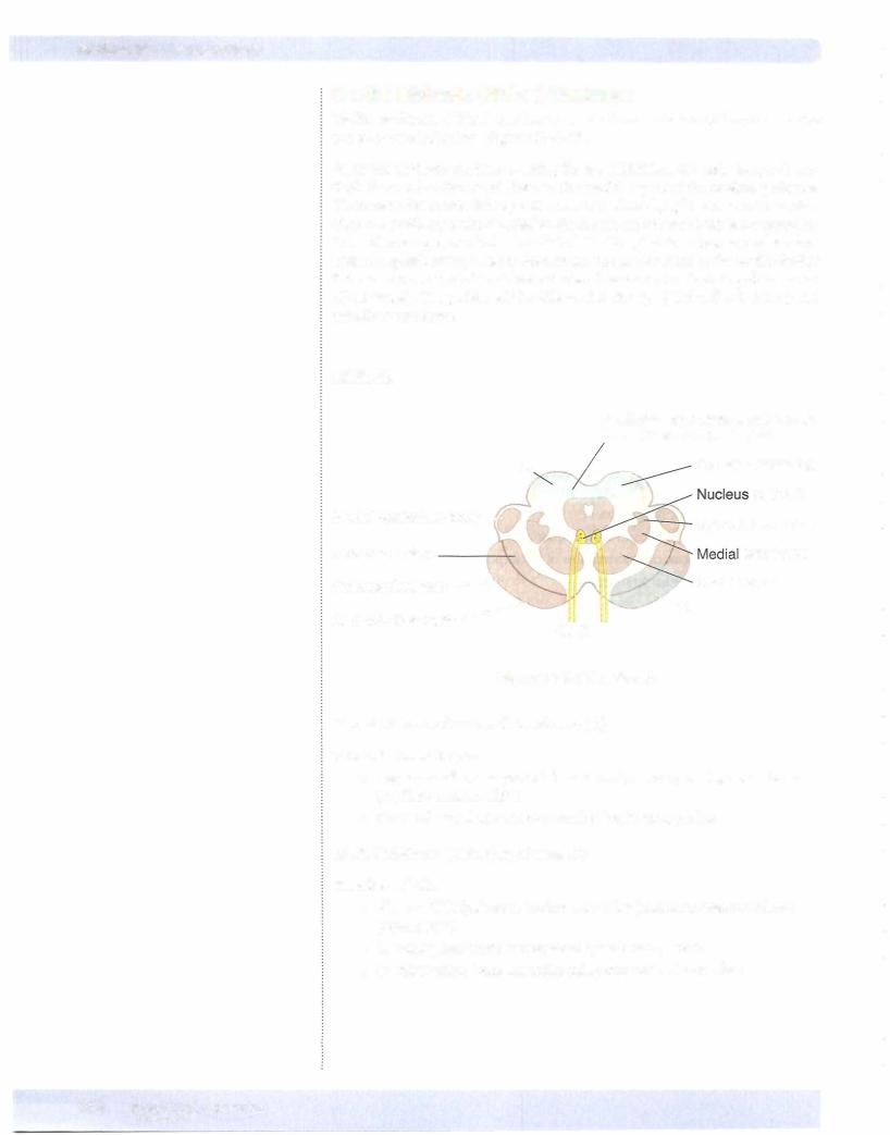
MEDULLA
In the caudal medulla, 2 of the neural systems-the corticospinal and dorsal column-medial lemniscal pathways-send axons across the midline. The nucleus gracilis and nucleus cuneatus give rise to axons that decussate in the caudal me dulla (the crossing axons are the internal arcuate fibers), which then form and ascend in the medial lemniscus.
The corticospinal (pyramidal) tracts, which are contained in the pyramids, course ventromedially through the medulla. Most of these fibers decussate in the caudal medulla just below the crossing of axons from the dorsal column nuclei, and then travel down the spinal cord as the (lateral) corticospinal tract.
The olives are located lateral to the pyramids in the rostral two-thirds ofthe medul la. The olives contain the convoluted inferior olivary nuclei. The olivary nuclei send climbing (olivocerebellar) fibers into the cerebellum through the inferior cerebellar peduncle. The olives are a key distinguishing feature ofthe medulla.
The spinothalamic tract and the descending hypothalamic fibers course together in the lateral part of the medulla below the inferior cerebellar peduncle and near the spinal nucleus and tract of CN V.
Cranial Nerve Nuclei
Spinal nucleus ofV
The spinal nucleus of the trigeminal nerve (CN V) is located in a position analo gous to the dorsal horn of the spinal cord. The spinal tract ofthe trigeminal nerve lies just lateral to this nucleus and extends from the upper cervical cord (C2) to the point of entry of the fifth cranial nerve in the pons. Central processes from cells in the trigeminal ganglion conveying pain and temperature sensations from the face enter the brain stem in the rostral pons but descend in the spinal tract of CN V and synapse on cells in the spinal nucleus (Figure IV-5-3).
Solitary nucleus
The solitary nucleus receives the axons of all general and special visceral afferent fibers carried into the CNS by CN VII, IX, and X. These include taste, cardiore spiratory, and gastrointestinal sensations carried by these cranial nerves. Taste and visceral sensory neurons allhave their cell bodies in ganglia associated with CN VII, IX, and X outside the CNS.
Nucleus ambiguus
The nucleus ambiguus is a column of large motoneurons situated dorsal to the inferior olive. Axons arising from cells in this nucleus course in the ninth and tenth cranial nerves. The component to the ninth nerve is insignificant. In the tenth nerve, these fibers supply muscles of the soft palate, larynx, pharynx, and upper esophagus. A unilateral lesion will produce ipsilateral paralysis of the soft palate causing the uvula to deviate away from the lesioned nerve and nasal regur gitation of liquids, weakness of laryngeal muscles causing hoarseness, and pha ryngeal weakness resulting in difficulty in swallowing.
Chapter 5 • The Brain Stem
MEDICAL 391
Section IV • Neuroscience
Dorsal motornucleus ofCNX
These visceral motoneurons ofCN X arelocated lateral to the hypoglossal nucleus in the floor ofthe fourth ventricle. This is a major parasympathetic nucleus ofthe brain stem, and it supplies preganglionic fibers innervatingterminalganglia in the thorax and the foregut and midgut parts ofthe gastrointestinal tract.
Hypoglossal nucleus
The hypoglossal nucleus is situated near the midline just beneath the central ca nal and fourth ventricle. This nucleus sends axons into the hypoglossal nerve to innervate all ofthe tongue muscles except the palatoglossus.
The accessory nucleus
The accessorynucleus is found in the cervical spinal cord. The axons ofthe spinal accessory nerve arise from the accessory nucleus, pass through the foramen mag num to enter the cranial cavity, and join the fibers ofthe vagus to exit the cranial cavity through the jugular foramen. As a result, intramedullary lesions do not affect fibers ofthe spinal accessory nerve. The spinal accessory nerve supplies the sternocleidomastoid and trapezius muscles.
Therootlets ofthe glossopharyngeal (CN IX) andvagus (CN X) nerves exitbetween the olive and the fibers of the inferior cerebellar peduncle. The hypoglossal nerve (CN XII) exits more mediallybetween the olive and the medullarypyramid.
PONS
The pons is located between the medulla (caudally) and the midbrain (rostrally). The cerebellum overlies the pons. It is connected to the brain stem by 3 pairs of cerebellar peduncles. The fourth ventricle is found between the dorsal surface ofthe pons and the cerebellum. The ventral surface ofthe pons is dominated by fibers, which form a large ventral enlargement that carries fibers from pontine nuclei to the cerebellum in the middle cerebellar peduncle. This ventral enlarge ment is the key distinguishing feature ofthe pons.
The corticospinal tracts are more diffuse in the pons than in the medulla and are embedded in the transversely coursing fibers that enter the cerebellum in the middle cerebellar peduncle.
The medial lemniscus is still situated near the midline but is now separated from the corticospinal tracts by the fibers forming the middle cerebellar peduncle. The mediallemniscus has changed from a dorsoventral orientation in the medulla to a more horizontal orientation in the pons.
The spinothalamic tract and the descending hypothalamic fibers continue to course together in the lateral pons.
The lateral lemniscus, an ascending auditory pathway, is lateral and just dorsal to the medial lemniscus. The lateral lemniscus carries thebulkofascending auditory fibers from both cochlear nuclei to the inferior colliculus ofthe midbrain.
The medial longitudinal fasciculus (MLF) is located near the midlinejustbeneath the fourth ventricle.
392 M E D I CAL

Chapter 5 • The Brain Stem
Cranial Nerve Nuclei
Abducens nucleus
The abducens nucleus is found near the midline in the floor of the fourth ven tricle just lateral to the MLF.
Facial motornucleus
The facial motor nucleus is located ventrolateral to the abducens nucleus. Fibers from the facial nucleus curve around the posterior side of the abducens nucleus (the curve forms the internal genu of the facial nerve), then pass ventrolaterally to exit the brain stem at the pontomedullary junction.
Superiorolivary nucleus
The superior olivary nucleus lies immediately ventral to the nucleus of CN VII and receives auditory impulses from both ears by way ofthe cochlear nuclei. The cochlear nuclei are found at the pontomedullary junction just lateral to the infe rior cerebellar peduncle.
Vestibular nuclei
The vestibular nuclei are located near the posterior surface of the pons lateral to the abducens nucleus, and extend into the medulla.
Cochlearnuclei
The dorsal and ventral cochlear nuclei are found at the pontomedullary junction.
All of the fibers of the cochlear part of CN VIII terminate here.
Trigeminal nuclei
Motor Nucleus-Pons
The motor nucleus of CN V is located in the pons just medial to the main sensory nucleus ofthe trigeminal and adjacent to the point ofexit or entry ofthe trigeminal nerve fibers. These motor fibers supply the muscles ofmastication (masseter, tem poralis, and medial and lateral pterygoid (Figure IV-5-3).
Main Sensory Nucleus-Pons
The main sensory nucleus is located just lateral to the motor nucleus.
The main sensory nucleus receives tactile and pressure sensations from the face, scalp, oral cavity, nasal cavity, and dura.
Spinal Trigeminal Nucleus-Spinal cord to pons
The spinal trigeminal nucleus is a caudal continuation ofthe main sensory nu cleus, extending from the mid pons through the medulla to the cervical cord. Central processes from cells in the trigeminal ganglion conveying pain and tem perature sensations from the face descend in the spinal tract ofV and synapse on cells in the spinal nucleus.
MEDICAL 393


Chapter 5 • The Brain Stem
Cranial NervesV, VI, VII, and VIII
Four cranial nerves emerge from the pons. Cranial nerves VI, VII, and VIII emerge from the pontomedullary junction. The facial nerve is located medial to the vestibu locochlear nerve. The abducens nerve (CN VI) emerges near the midline lateral to the corticospinal tract. The trigeminal nerve (C V) emerges from the middle of the pons.
MIDBRAIN
The midbrain (mesencephalon) is located between the pons and diencephalon. The cerebral aqueduct, a narrow channel that connects the third and fourth ven tricles, passes through the midbrain. The inferior colliculi and superior colliculi are found on the dorsal aspect of the midbrain above the cerebral aqueduct. The inferior colliculus processes auditory information received bilaterally from the co chlear nuclei by axon fibers of the lateral lemniscus. The superior colliculi help direct movements ofboth eyes in gaze. The pretectal region is locatedjust beneath the superior colliculi and in front of the oculomotor complex. This area contains interneurons involved in the pupillary light reflex. The massive cerebral peduncles extend ventrally from the midbrain. 1be cerebral peduncles contain corticospinal and corticobulbar fibers. The interpeduncular fossa is the space between the cere bral peduncles.
The substantia nigra is the largest nucleus of the midbrain. It appears black to dark brown in the freshly cut brain because nigral cells contain melanin pig ments. Neurons in the substantia nigra utilize Dopamine and GABA as neu rotransmitters.
The medial lemniscus and spinothalamic tract and descending hypothalamic fi bers course together ventrolateral to the periaqueductal gray.
The MLF continues to be located near the midline, just beneath the cerebral aq ueduct.
The mesencephalic nuclei ofthe trigeminal nerve are located on either side ofthe central gray.
Cranial Nerve Nuclei
The trochlear nucleus is located just beneath the periaqueductal gray near the midline between the superior and inferior colliculi. The oculomotor nucleus and the nucleus of Edinger-Westphal are found just beneath the periaqueductal gray near the midline at the level of the superior colliculi.
Two cranial nerves emerge from the midbrain: the oculomotor (CN III) and the trochlear (C IV) nerves.
The oculomotor nerve arises from the oculomotor nucleus and exits ventrally from the midbrain in the interpeduncular fossa. CN III also contains preganglion ic parasympathetic axons that arise from the nucleus of Edinger-Westphal, which lies adjacent to the oculomotor nucleus.
Axons of the trochlear nerve decussate in the superior medullary velum and exit the brain stem near the posterior midline just inferior to the inferior colliculi.
MEDICAL 395


Section IV • Neuroscience
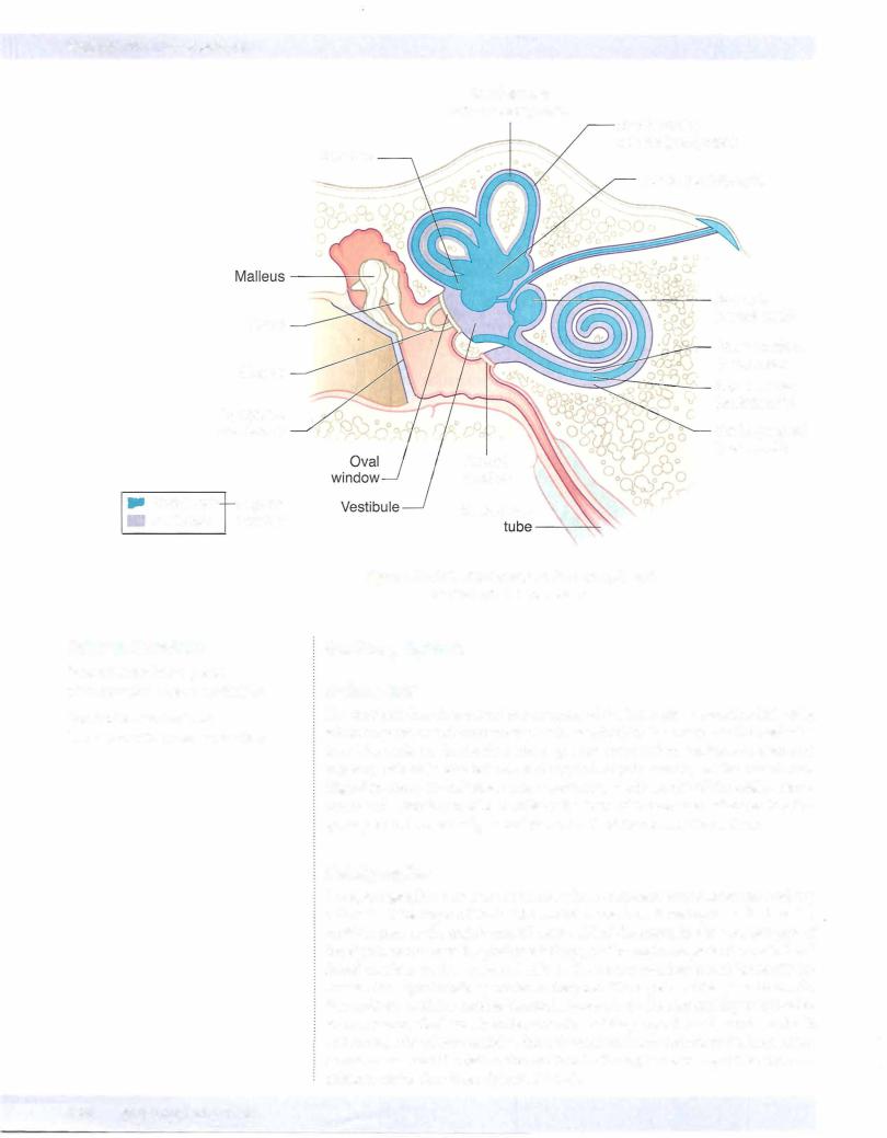
COMPONENTS OF THE EAR, AUDITORY, AND VESTIBULAR SYSTEMS
Each ear consists of 3 components: 2 air-filled spaces, the external ear and the middle ear; andthe fluid-filled spaces ofthe inner ear (Figures IV-5-6 and IV-5-7).
Theexternal ear includes thepinna andthe external auditorymeatus, which extends to the tympanic membrane. Soundwavestravelthrough the external auditory canal and cause the tympanic membrane (eardrum) to vibrate. Movement ofthe eardrum causes vibrations ofthe ossicles in the middle ear (i.e., the malleus, incus, and sta pes). Vibrations ofthe ossicles are transferred through the ovalwindow and into the inner ear.
The middle ear lies in the temporal bone, where the chain of 3 ossicles connect the tympanic membrane to the oval window. These auditory ossicles amplify the vibrations received by the tympanic membrane and transmit them to the fluid of the inner ear with minimal energy loss. The malleus is inserted in the tympanic membrane, and the stapes is inserted into the membrane ofthe ovalwindow. Two small skeletal muscles, the tensor tympani and the stapedius, contract to prevent damage to the inner ear when the ear is exposed to loud sounds. The middle-ear cavity communicates with the nasopharynxvia the eustachian tube,which allows air pressure to be equalized on both sides ofthe tympanic membrane.
The inner ear consists ofa labyrinth (osseous and membranous) ofinterconnected sacs (utricle and saccule) and channels (semicircular ducts and the cochlear duct) that contain patches of receptor or hair cells that respond to airborne vibrations or movements ofthe head. Both the cochlear duct and the sacs and channels of the vestibular labyrinth are filled with endolymph, which bathes the hairs ofthe hair cells. Endolymph is unique because it has the inorganic ionic composition ofan intracellular fluid but it lies in an extracellular space. The intracellular ionic composition ofendolymph is important for the function ofhair cells. Perilymph, ionically like a typical extracellular fluid, lies outside the endolymph-filled laby rinth (Figure IV-5-7).
398 MEDICAL


Section IV • Neuroscience
Auditory Tests
Weber test: place tuning fork on vertex of skull. If unilateral conductive loss vibration is louder in affected ear; if unilateral sensorineural loss vibration is louder in normal ear.
Rinne test: place tuning fork on mastoid process (bone conduction) until vibra tion is not heard, then place fork in front of ear (air conduction). If unilateral conductive loss no air conduction after bone conduction is gone; ifunilateral sensorineural loss air conduction present after bone conduction is gone.
VestibularSystem
Sensoryreceptors
The vestibular system contains 2 kinds of sensory receptors, one kind in the utri cle and the saccule and the other in the semicircular ducts.
The utricle and the saccule are 2 large sacs, each containing a patch of hair cells in a macula. Each macula responds to linear acceleration and detects positional changes in the head relative to gravity. There are 3 semicircular ducts in the inner ear, each lying in a bony semicircular canal. Each semicircular duct con tains an ampullary crest of hair cells that detect changes in angular acceleration resulting from circular movements of the head. The 3 semicircular ducts-an- terior, posterior, and horizontal-are oriented such that they lie in the 3 planes of space. Circular movements of the head in any plane will depolarize hair cells in a semicircular duct in one labyrinth and hyperpolarize hair cells in the cor responding duct in the opposite labyrinth.
Vestibularnuclei
There are 4 vestibular nuclei located in the rostral medulla and caudal pons. The vestibular nuclei receive afferents from the vestibular nerve, which innervates re ceptors located in the semicircular ducts, utricle, and saccule. Primary vestibular fibers terminate in the vestibular nuclei and the flocculonodular lobe of the cer ebellum.
Vestibularfibers
Secondary vestibular fibers, originating in the vestibular nuclei, join the MLF and supply the motor nuclei ofCN III, IV, and VI. These fibers are involved in the pro duction ofconjugate eye movements. These compensatory eye movements repre sent the efferent limb of the vestibulo-ocular reflex, which enables the eye to re main focused on a stationary target during movement of the head or neck. Most of our understanding of the vestibulo-ocular reflex is based on horizontal head turning and a corresponding horizontal movement of the eyes in the direction opposite to that of head turning. For example, when the head turns horizontally to the right, both eyes will move to the left using the following vestibulo-ocular structures. Head turning to the right stimulates hairs cells in the right semicir cular ducts. The right eighth nerve increases its firing rate to the right vestibular nuclei. These nuclei then send axons by way of the MLF to the right oculomotor nucleus and to the left abducens nucleus. The right oculomotor nerve to the right medial rectus adducts the right eye, and the left abducens nerve to the left lateral rectus abducts the left eye. The net effect of stimulating these nuclei is that both eyes will look to the left.
402 M EDICAL


Eye MovementControl Systems
For the eyes to move together (conjugate gaze), the oculomotor nuclei and abducens nuclei are interconnected by the medial longitudinal fasciculus (MLF).
Horizontal gaze is controlled by 2 gaze centers: 1 . Frontal eye field (contralateral gaze)
2. PPRF (paramedian pontine reticular formation, ipsilateral gaze)
Nystagmus
Nystagrnus refers to rhythmic oscillations of the eyes slowly to one side followed by a rapid reflex movement in the opposite direction. Nystagmus is defined by the direction of the rapid reflex movement or the fast phase. It is usually horizontal, although rotatory or vertical nystagmus may also occur.
Unilateral vestibular nerve or vestibular nucleus lesions may result in a vestibular nystagmus. In a pathologic vestibular nystagmus, the initial slow phase is the re sponse to the pathology, and the fast phase is the correction attempt made bythe cortex in response to the pathology. Consider this example: ifthe left vestibular nerve or nuclei are lesioned, because of the loss of balance between the 2 sides, the right vestibular nuclei are unopposed and act as if they have been stimulated, causing both eyes to look slowly to the left. This is the slow phase of a pathologic vestibular nystagmus. Because the head did not move, the cortex responds by moving both eyes quickly back to the right, the direction of the fast phase of the nystagmus.
Tests for Nystagmus
The integrity of the vestibulo-ocular reflex can be an indicator of brain-stem in tegrity in comatose patients. To test this reflex, a vestibular nystagmus is induced byperforming a caloric test in which an examiner introduces warm or cool water into an external auditory meatus. Warm water introduced into the external ear stimulates the horizontal semicircular duct and causes the eyes to move slowly in the opposite direction. Because the head did not turn, the eyes are moved quickly back by the cortex (if intact) toward the same ear where the warm water was introduced, producing a fast phase of nystagmus to the same side. Intro duction of cool water into the external ear mimics a lesion; the horizontal duct activity is inhibited on the cool water side, and the opposite vestibular complex moves the eyes slowly toward the cool-water ear. The corrective or fast phase of the nystagrnus moves the eyes quickly away from the ear where the cool water was introduced. A mnemonic which summarizes the direction of the fast phase of vestibular nystagmus in a caloric test toward the warm-water side and away from the cool-water side is COWS; cool, opposite; warm, same.
HORIZONTAL CONJUGATE GAZE
The eyeballs move together in conjugate gaze. The ocular muscles function to move and position both eyes as a unit so that an image falls on a corresponding spot on the retina of each eye. The slightest weakness in the movements of one eye causes diplopia, the presence of a double image, indicating that the image has been shifted to a different position on the retina of the affected side. Although gaze in allplanes is possible, the muscles and cranial nerves involved in horizon tal conjugate gaze, or abduction and adduction ofboth eyes together, are the most important eye movements (Figure IV-5- 1 1).
Chapter 5 • The Brain Stem
MEDICAL 405
Section IV • Neuroscience
Abduction ofeach eyeballisperformedlargelybythelateral rectus muscle, which is innervated by the abducens nerve (CN VI). Adduction of the eyeball is per formedbythemedialrectusmuscle,whichisinnervatedbythe oculomotornerve (CN III). Therefore, forboth eyes to lookto the right in horizontal gaze, the right abducens nerve and the right lateral rectus muscle must be active to abduct the righteye, and the left oculomotornerve and theleftmedial rectus muscle mustbe active to adduct the left eye. The net effect is that both eyes willlook to the right.
In the brain stem, the abducens nucleus (CN VI) and the oculomotor nucleus (CN III) are situated close to the mid.linejust beneath the fourth ventricle or the cerebral aqueduct, in the pons and rnidbrain. These nuclei are interconnected by thefibers in the MLF. It is the fibers in the MLF thatpermitconjugategaze, either when the target moves or when the head moves, through their interconnections to gaze centers and thevestibularsystem.
Control of Horizontal Gaze
Horizontalgaze is controlledby2 interconnected gaze centers. Onecontrol centeris in the frontal lobe, the frontal eye field (Brodmann area 8). Thisarea acts as a center forcontralateralhorizontal gaze. In the pons is a second gaze center, known as the pontine gaze center or the PPRF, the paramedian pontine reticular formation. This is a center for ipsilateral horizontal gaze. When activatedby neurons in the frontal eye field, the pontine gaze center neurons send axons to synapse with cell bodies in the abducens nucleus, which is actually contained within the pontine gaze center. The pontine gaze center also sends axons that cross immediately and course in the contralateral MLF to reach the contralateral oculomotor nucleus. The net effect of stimulation ofthe left frontal eye field, therefore, is activation ofthe pontine gaze centerontherightanda saccadichorizontal eyemovement ofboth eyes to the right. Horizontal gaze to the right results from activation ofthe right abducens nucleus andthe left oculomotor nucleusbyfibers inthe MLE
Lesions in the MLF result in an internuclear ophthalmoplegia in which there is an inability to adduct one eye on attempted gaze to the opposite side. For example, a lesion in the right MLF results in an inability to adduct the right eye on an at tempted gaze to the left. The left eye abducts normallybut exhibits anystagmus. If the MLF is lesioned bilaterally (as mightbe the case in multiple sclerosis), neither eye adducts on attempted gaze (Figures IV-5-1 1 and IV-5-12), and the abducting eye exhibits a nystagmus.
406 MEDICAL

Right |
Left |
|
Paramedian pontine |
Cerebral cortex frontal |
|
eye fields (Area |
8) |
|
reticular formation (PPRF) |
|
|
Medial longitudinal fasciculus (MLF)
Right lateral |
Left medial |
rectus muscle |
rectus muscle |
Abducts |
|
Right eye |
|
Figure IV-5-1 1 . Voluntary Horizontal Conjugate Gaze
Table IV-5-2. Clinical Correlate
Lesion Examples |
Symptoms |
1 . Right CN VI |
Right eye cannot look right |
2. Right PPRF |
Neither eye can look right |
3. Left MLF |
Internuclear ophthalmoplegia (INO) |
|
Left eye cannot look right; convergence is intact (this is |
|
how to distinguish an INO from an oculomotor lesion); |
|
right eye has nystagmus; seen in multiple sclerosis |
4. Left frontal eye field |
Neither eye can look right; but slow driftto left |
Abbreviations: MLF, medial longitudinal fasciculus; PPRF, paramedian pontine reticular formation
Chapter s • The Brain Stem
Lesion sites are indicated by 1-4.
MEDICAL 407


Chapter 5 • The Brain Stem
BasilarArtery
The basilararteryis formed bythejoining ofthe 2 vertebral arteries at the ponto medullary junction. It ascends along the ventral midline ofthe pons and termi nates near the rostral border ofthepons by dividing into the 2 posterior cerebral arteries. Branches include the anterior inferior cerebellar arteries (AICA) and the paramedian arteries.
Branches ofthe basilar artery include: the labyrinthine artery, which follows the course ofthe eighth cranial nerve and supplies the inner ear; the anterior inferior cerebellar artery, which supplies part of the pons and the anterior and inferior regions of the cerebellum; the superior cerebellar artery, which supplies part of the rostral pons and the superior region ofthe cerebellum; and pontine branches, which supply much ofthe pons via paramedian and circumferential vessels.
At the rostral end ofthe midbrain, the basilar artery divides into a pair ofposte rior cerebral arteries. Paramedian and circumferential branches ofthe posterior cerebral arterysupply the midbrain.
BRAIN-STEM LESIONS
There are 2 keys to localizing brain-stem lesions. First, it is uncommon to injure parts ofthe brain stem without involving one or more cranial nerves. The cranial nerve signs will localize the lesion to the midbrain (CN III or IV), upper pons (CN V), lower pons (CN VI, VII, or VIII), or upper medulla (CN IX, X, or XII). Second, ifthe lesion is in the brain stem, the cranial nerve deficits will be seen with a lesion to one or more of the descending or ascending long tracts (cor ticospinal, medial lemniscus, spinothalamic, descending hypothalamic fibers). Lesions in the brain stem to any ofthe long tracts except forthe descending hy pothalamic fibers will result in a contralateral deficit. A unilateral lesion to the descending hypothalamic fibers that results in Horner syndrome is always seen ipsilateral to the side ofthe lesion.
Medial Medullary Syndrome
Medial medullary syndrome is most frequently the result ofocclusion ofthe ver tebral arteryor the anterior spinal artery (Figure IV-5-15). Medial medullary syn dromepresentswith a lesion ofthe hypoglossal nerve as the cranialnerve sign and lesions to both the medial lemniscus and the corticospinal tract. Corticospinal tract lesions produce contralateral spastic hemiparesis ofboth limbs.
Medial lemniscus lesions produce a contralateral deficit of proprioception and touch, pressure, and vibratory sensations in the limbs and body.
Lesions of the hypoglossal nerve in the medulla produce an ipsilateral paralysis ofhalfthe tongue with atrophy. Upon protrusion, the tongue deviatestoward the side ofthe lesion.
MEDICAL 411

Chapter 5 • The Brain Stem
Lateral Medullary (Wallenberg) Syndrome
Lateralmedullarysyndrome results from occlusionofthe PICA (Figure IV-5-15).
Thecranial nervesornuclei involvedinthe lesion are thevestibularorthe cochle ar parts of CN VIII, the glossopharyngeal and the vagus nerves, and the spinal nucleus or tract ofV The long tracts involved are the spinothalamic tract and the descending hypothalamic fibers.
Spinothalamic tract lesions produce a pain and temperature sensation deficit in the contralaterallimbs andbody.
Lesions of descending hypothalamic fibers produce an ipsilateral Horner syn drome (i.e., miosis, ptosis, and anhidrosis).
Lesions ofthe vestibular nuclei and pathways may produce nystagmus, vertigo, nausea, and vomiting. Ifthere is a vestibular nystagmus, the fast component will be away from the side ofthe lesion.
Lesions of the vagus nerves exiting the medulla may produce dysphagia (diffi cultyin swallowing) orhoarseness. The palate willdroop onthe affected side, and the uvulawill deviate away from the side ofthe lesion.
Lesions ofthe glossopharyngealnerveresultin a diminished orabsentgag reflex.
Lesions ofthe spinal tract and nucleus ofthe trigeminal nerve produce a loss of just pain andtemperaturesensations on the ipsilateral side ofhalfthe face. Touch sensations from the faceandthe cornealblinkreflexwillbe intact. In lateral med ullary syndrome, the pain and temperature losses are alternating; these sensa tionsarelost fromthe face andscalp ipsilateral to the lesionbutarelost fromthe contralateral limbs and trunk.
Taste sensations maybe altered ifthe solitary nucleus is involved.
Medial Pontine Syndrome
Medial pontine syndrome results from occlusion ofparamedian branches ofthe basilar artery (Figure IV-5-16).
At a minimum, this lesion affects the exiting fibers ofthe abducens nerve and the corticospinal tract. The medial lemniscus may be affected ifthe lesion is deeper into the pons, and the facial nerve maybe affected ifthe lesion extends laterally.
The long tract signs will be the same as in medial medullary syndrome, involving the corticospinal and medial lemniscus, but the abducens nerve and the facial nerve lesions localize the lesion to the caudal pons.
Corticospinaltractlesionsproduce contralateralspastichemiparesisofbothlimbs.
Medial lemniscus lesions produce a contralateral deficit of proprioception and touch, pressure, and vibratory sensations in the limbs and body.
Lesions of the abducens nerve exiting the caudal pons produce aninternal stra bismus ofthe ipsilateral eye (from paralysis ofthe lateral rectus). This results in diplopia on attempted lateral gaze to the affected side.
MEDICAL 413

Section IV • Neuroscience
Lesions ofthe facial nerve exiting the caudal pons produce complete weakness of the muscles offacial expression on the side ofthe lesion.
Lesions ofthe facial nerve may also include an alteration oftaste from the anterior two-thirds ofthe tongue, loss oflacrimation (eye dryand red), and loss ofthe motor limb ofthe corneal blink reflex.
If a lesion extends dorsally to include the abducens nucleus (which includes the horizontal gaze center in the PPRF), there may be a lateral gaze paralysis in which both eyes are forcefully directed to the side contralateral to the lesion.
Lateral Pontine Syndrome
Lesions ofthe dorsolateral pons usually result from occlusion ofthe anterior in ferior cerebellar artery (caudal pons) or superior cerebellar artery (rostral pons). The long tracts involved will be the same as in lateral medullary syndrome, the spinothalamic tract and the descending hypothalamic fibers. The cranial nerves involved willbe the facial and vestibulocochlear in the caudal pons, the trigemi nal nerve in the rostral pons, and the spinal nucleus and tract ofV in both lesions (Figure IV-5-17).
Spinothalamic tract lesions produce a pain and temperature sensation deficit in the contralateral limbs and body.
Lesions of descending hypothalamic fibers produce an ipsilateral Horner syn drome (i.e., miosis, ptosis, and anhidrosis).
Lesions ofthe vestibular nuclei and pathways (caudal pons) produce nystagmus, vertigo, nausea, andvomiting. Again, the fastphase ofthe nystagmus willbe away from the side ofthe lesion. Lesions ofthe cochlear nucleus or auditorynerve pro duce an ipsilateral sensorineuralhearing loss.
Lesions ofthe spinal tract andnucleus ofthe trigeminal nerve result onlyin a loss ofpain and temperature sensations on the ipsilateral side ofhalfthe face.
Lesions ofthe facial nerve and associated structures produce ipsilateral facial pa ralysis, loss oftaste from the anterior two-thirds ofthe tongue, loss oflacrimation and salivation, and loss ofthe corneal reflex.
Lesions of the trigeminal nerve (rostral pons) result in complete anesthesia ofthe face on the side ofthe lesion, weakness ofmuscles ofmastication, and deviation of thejawtoward the lesioned side.
414 MEDICAL


Section IV • Neuroscience
Clinical Correlate
Neurons in both the raphe and locus caeruleus degenerate in Alzheimer disease.
Parinaud Syndrome
Parinaud syndrome usually occurs as a result of a pineal tumor compressing the superior colliculi. The most common sign is paralysis of upward or vertical gaze, combined with bilateral pupillary abnormalities (e.g., slightlydilated pupils, which may show an impaired light or accommodation reaction) and signs of el evated intracranial pressure. Compression of the cerebral aqueduct can result in noncommunicating hydrocephalus.
RETICULAR FORMATION
The reticular formation is located in the brain stem and functions to coordinate and integrate the actions of different parts of the CNS. It plays an important role in the regulation of muscle and reflex activity and control of respiration, cardio vascular responses, behavioral arousal, and sleep.
Reticular Nuclei
Raphe nuclei
The raphe nuclei are a narrow column of cells in the midline ofthe brain stem, ex tending from the medulla to the midbrain. Cells in some of the raphe nuclei (e.g., the dorsal raphe nucleus) synthesize serotonin (5-hydroxytryptamine [5-HT]) from L-tryptophan and project to vast areas ofthe CNS. Theyplay a role in mood, aggression, and the induction ofnon-rapid eye movement (non-REM) sleep.
Locuscaeruleus
Cells in the locus caeruleus synthesize norepinephrine and send projections to most brain areas involved in the control of cortical activation (arousal). De creased levels ofnorepinephrine are evident in REM (paradoxic) sleep.
Periaqueductal gray
The periaqueductal (central) gray is a collection of nuclei surrounding the ce rebral aqueduct in the midbrain. Opioid receptors are present on many periaq ueductal gray cells, the projections from which descend to modulate pain at the level ofthe dorsal horn of the spinal cord.
418 MEDICAL

Chapter 5 • The Brain Stem
ChapterSummary
•The brain stem is divided into 3 major subdivisions-the medulla oblongata, pons, and midbrain. The brain stem contains many descending and ascending tracts, the reticular formation, and the sensory and motor cranial nuclei ofCNs Ill-XII. Cranial nerve nuclei forCN Ill and IV are located in the midbrain; cranial nerve nuclei for CN V-Vlll are located in the pons; and cranial nerve nuclei of IX-XII in the medulla. Most ofthe motor nuclei are located medially and the sensory nuclei are located more laterally in the brain stem. Normal functions ofthe cranial nerves and the clinical deficits resulting from lesions ofthe brain stem are listed in Table IV-5-1. Lesions affecting the 3 long tracts to and from the spinal cord will produce contralateral deficits, but lesions ofthe motor or sensory cranial nuclei result in ipsilateral findings.
•The motor nuclei of CNs Ill-VII and IX-XII are lower motor neurons that innervate most ofthe skeletal muscles ofthe head. These lower motor neurons are innervated by upper motor neurons (corticobulbar fibers). The cell bodies of the corticobulbar fibers are found primarily in the motor cortex of the frontal lobe. Corticobulbar innervation of lower motor neurons is primarily bilateral from both the right and left cerebral cortex, except for the innervation ofthe lower facial muscles around the mouth, which are derived only from the contralateral motor cortex. Generally, no cranial deficits will be seen with unilateral corticobulbar lesions, except for drooping ofthe corner of the mouth contralateral to the side of the lesion.
•CN VIII provides sensory pathways for auditory and vestibular systems.
Auditory input depends on the stimulation of hair cells on the organ of Corti due to movement of endolymph within the membranous labyrinth of the inner ear. Axons from the organ of Corti enterthe pons via CN VIII and
synapse in the cochlear nuclei. From the cochlear nuclei, auditory projections bilaterally ascend the brain stem to the superior olivary nuclei, then via the lateral lemniscus to the inferior colliculus, and then to the medial geniculate body of the thalamus. Final auditory projections connect the thalamus with the primary auditory cortex of both temporal lobes. Thus, each auditory cortex receives input from both ears; however, input from the contralateral ear predominates. Lesions of the inner ear or ofthe cochlear nuclei in the
pons will produce total deafness, whereas other lesions central to the cochlear nuclei will primarily affect the ability to localize sound directi(Continued).
MEDICAL 419


Section IV • Neuroscience
From medialto lateral, the deep cerebellar nuclei in the internal white matter are the fastigial nucleus, interposed nuclei, and dentate nucleus.
Two kinds ofexcitatoryinput enter the cerebellum in the form ofclimbingfibers and mossy fibers. Both types influence the firingofdeep cerebellar nucleibyaxon collaterals.
Climbingfibers originate exclusively from the inferiorolivarycomplexofnuclei on the contralateral side ofthe medulla. Climbing fibers provide a direct power ful monosynaptic excitatoryinput to Purkinje cells.
Mossyfibersrepresentthe axonsfromallothersourcesofcerebellarinput. Mossy
fibers provide an indirect, morediffuseexcitatory input to Purkinje cells.
Allmossyfibers exertan excitatory effect on granule cells. Each granule cell sends its axon into the molecular layer, where it gives offcollaterals at a 90-degree angle thatrun parallelto the cortical surface (i.e.,parallelfibers). These granule cell axons stimulate the apical dendrites of the Purkinje cells. Golgi cells receive excitatory input from mossy fibers and from the parallel fibers ofthe granule cells. The Golgi cell in turn inhibits the granule cell, which activatedit in the firstplace.
The basket and stellate cells, which also receive excitatory input from parallel fibers ofgranule cells, inhibit Purkinje cells.
CIRCUITRY
Thebasic cerebellar circuits beginwith Purkinje cells that receiveexcitatoryinput directly from climbing fibers and from parallel fibers ofgranule cells.
Purkinje cell axons project to and inhibit the deep cerebellar nuclei or the ves tibular nuclei in an orderlyfashion (Figure IV-6-3).
•Purkinje cells in the flocculonodular lobe project to the lateral vestibular nucleus.
•Purkinje cells in the vermis project to the fastigial nuclei.
•Purkinje cells in the intermediate hemisphere primarily project to the interposed (globose and emboliform) nuclei.
•Purkinje cells in the lateral cerebellar hemisphere project to the dentate nucleus.
Dysfunction
•Hemisphere lesions -7 ipsilateral symptoms: intention tremor, dysmet ria, dysdiadochokinesia, scanning dysarthria, nystagmus, hypotonia
•Vermal lesions -7 truncal ataxia
MajorPathway
Purkinje cells -7 deep cerebellar nucleus; dentate nucleus -7 contralateral VL -7 first-degree motor cortex -7 pontine nuclei -7 contralateral cerebellarcortex
424 MEDICAL


Section IV • Neuroscience
Clinical Correlate
Anterior vermis lesions are usually the result of degeneration from alcohol abuse and are present with gait ataxia. Posterior vermis lesions result from medulloblastomas or ependymomas and present with truncal ataxia.
Efferents from the deep cerebellar nuclei leave mainlythrough the SCP and influ ence allupper motoneurons. In particular, axons from the dentate and interposed nuclei leave through the SCP, cross the midline, and terminate in the ventrolat eral (VL) nucleus ofthe thalamus.
The VL nucleus ofthe thalamus projects to primary motor cortex and influences the firing ofcorticospinal and corticobulbar neurons.
Axons from other deep cerebellar nuclei influence uppermotoneurons in the red nucleus and in the reticular formation and vestibular nuclei.
Cerebellar Lesions
The hallmark of cerebellar dysfunction is a tremor with intended movement without paralysis or paresis. Symptoms associated with cerebellar lesions are ex pressed ipsilaterally because the major outflow ofthe cerebellum projects to the contralateral motor cortex, and then the corticospinal fibers cross on their wayto the spinal cord. Thus, unilateral lesions ofthe cerebellum willresult in a patient falling toward the side ofthe lesion.
Lesions that include the hemisphere
Lesions that include the hemisphere produce a number of dysfunctions, mostly involving distal musculature.
An intention tremor is seen whenvoluntarymovements are performed. Forexam ple, ifa patient with a cerebellar lesion is asked to pick up a penny, a slight tremor of the fingers is evident and increases as the penny is approached. The tremor is barelynoticeable or is absent at rest.
Dysmetria (past pointing) is the inabilityto stop a movement at the proper place. The patient has difficulty performing the finger-to-nose test.
Dysdiadochokinesia (adiadochokinesia) is the reduced ability to perform alter nating movements, such as pronation and supination of the forearm, at a mod erately quickpace.
Scanning dysarthria is causedby asynergy ofthe muscles responsible for speech. In scanning dysarthria, patients divide words into syllables, thereby disrupting the melody ofspeech.
Gaze dysfunction occurs when the eyes try to fix on a point: They may pass it or stop too soon and then oscillate a few times before they settle on the target. A nystagmus may be present, particularly with acute cerebellar damage. The nys tagmus is often coarse, with the fast component usually directed toward the in volved cerebellar hemisphere.
Hypotonia usually occurs with an acute cerebellar insult that includes the deep cerebellar nuclei. The muscles feel flabby on palpation, and deep tendon reflexes are usually diminished.
Lesions to the vermal region
Verma! lesions result in difficultymaintaining posture, gait, or balance (an ataxic gait). Patients with vermal damage maybe differentiated from those with a lesion of the dorsal columns by the Romberg sign. In cerebellar lesions, patients will swayor lose their balancewith their eyes open; in dorsal column lesions, patients sway with their eyes closed.
426 MEDICAL

Chapter 6 • The Cerebellum
ChapterSummary
•The cerebellum controls posture, muscle tone, and learning of repeated motor functions, and coordinates voluntary motor activity. Diseases ofthe cerebellum result in disturbances of gait, balance, and coordinated motor actions, but there is no paralysis or inability to start or stop movement.
•The cerebellum is functionally divided into (1) the vermis and intermediate
zone, (2) the hemisphere, and (3) the flocculonodular lobe. Each ofthese 3 areas receives afferent inputs mainly from the spinal cord, cortex and inferior olivary nucleus, and vestibular nuclei, respectively. These afferent fibers (mossy and climbing) reach the cerebellum via the inferior and middle cerebellar peduncles, which connect the cerebellum with the brain stem. The afferent fibers are excitatory and project directly or indirectlyvia granule cells to the Purkinje cells ofthe cerebellar cortex. The axons of the Purkinje cells are inhibitory and are the only outflow from the cerebellar cortex. They project to and inhibit the deep cerebellar nuclei (dentate, interposed, and fastigial nuclei) in the medulla. From the deep nuclei, efferents project mainly through the superior cerebellar peduncle and drive the upper motor neurons ofthe motor cortex. The efferents from the hemisphere project through the dentate nucleus, to the contralateral ventral lateral/ventral anterior nuclei of the thalamus, to reach the contralateral precentral gyrus.
These influence contralateral lower motor neurons via the corticospinal tract.
•Symptoms associated with cerebellar lesions are expressed ipsilaterally. Unilateral lesions ofthe cerebellum will result in a patient falling toward the side of the lesion. Hallmarks of cerebellar dysfunction include ataxia, intention tremor, dysmetria, and dysdiadochokinesia.
MEDICAL 427
