

- •Contents
- •1. Cell Biology and Epithelia
- •2. Connective Tissue
- •3. Cartilage and bone
- •5. Nervous Tissue
- •6. Immune Tissues
- •7. Respiratory System
- •8. Gastrointestinal System
- •12. Integument
- •1. Gonad Development
- •4. Embryonic Period (Weeks 3-8)
- •1. Back and Autonomic Nervous System
- •2. Thorax
- •5. Lower Limb
- •4. The Spinal Cord
- •5. The Brain Stem
- •7. Basal Ganglia
- •11. Limbic System
- •Index

The Spinal Cord |
4 |
GENERAL FEATURES
The spinal cord is housedin the vertebral canal. It is continuous with the medulla below the pyramidal decussation and terminates as the conus medullaris at the second lumbar vertebra of the adult. The roots of 31 pairs of spinal nerves arise segmentally from the spinal cord.
There are 8 cervicalpairsof spinal nerves (Cl through CS). The cervical enlarge ment (CS through T l ) gives rise to the rootlets that form the brachia! plexus, which innervates the upper limbs.
There are 12 thoracic pairs of spinal nerves (Tl through T l2). Spinal nerves emanating from thoracic levels innervate most of the trunk.
There are 5 lumbarpairs of spinal nerves (Ll through LS). The lumbar enlarge ment (Ll through S2) gives rise to rootlets that form the lumbar and sacral plex uses, which innervate the lower limbs.
There are 5 sacral pairs of spinal nerves (Sl through SS). Spinal nerves at the sacral level innervate part of the lower limbs and the pelvis.
There is one coccygeal pair of spinal nerves. The cauda equina consists of the dorsal and ventral roots ofthe lumbar, sacral, and coccygeal spinal nerves.
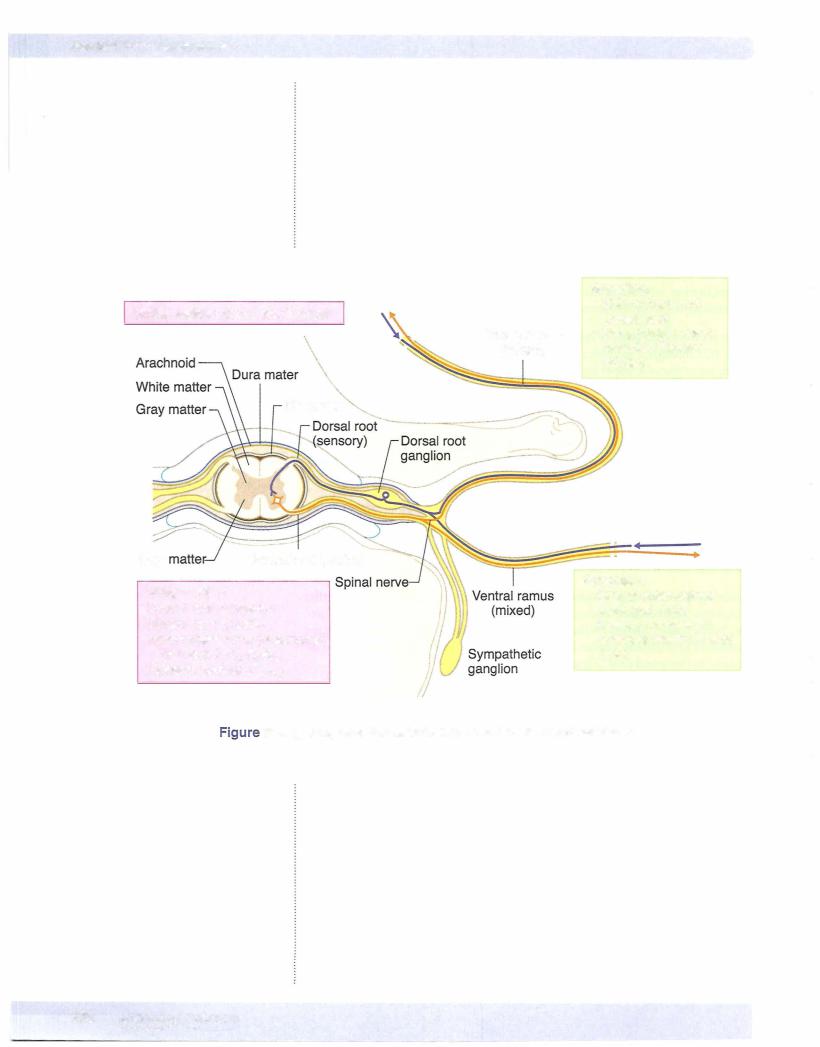
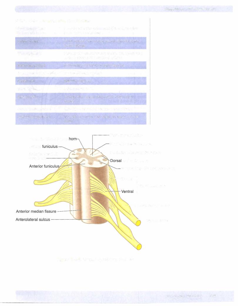
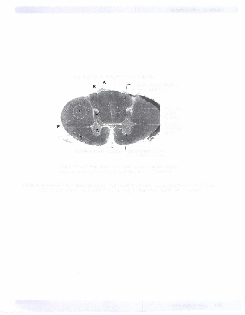
Inside the spinal cord, gray matter is centrally located and shaped like a butterfly. It contains neuronal cell bodies, their dendrites, and the proximal parts of ax ons. White matter surrounds the gray matter on all sides. White matter contains bundles of functionally similar axons called tracts or fasciculi, which ascend or descend in the spinal cord (Figures IV-4- 1 and IV-4-2).
MEDICAL 357
Section IV • Neuroscience
In A Nutshell
Motor end plate of skeletal muscles
UMN, upper motoneuron; LMN, lower motoneuron
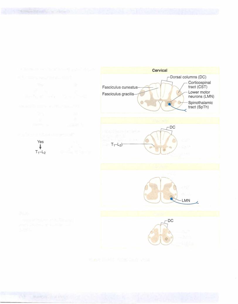
Intermediate Zone
Theintermediate zone ofthe spinal cord from Tl to L2 contains preganglionic sym pathetic neuron cell bodies and Clarke nucleus, which sends unconscious proprio ception to the cerebellum.
NEURAL SYSTEMS
There are 3 major neural systems in the spinal cord that use neurons in the gray matter and tracts or fasciculi of axons in the white matter. These neural systems have components that can be found at all levels of the CNS from the cerebral cortex to the tip ofthe spinal cord. An understanding of these 3 neural systems is essential to understanding the effects oflesions in the spinal cord and brain stem, and at higher levels of the CNS.
Motor System
Voluntaryinnervation ofskeletal muscle
Upper and Lower Motoneurons
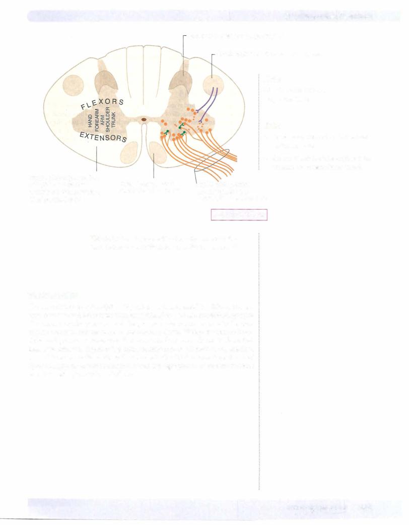
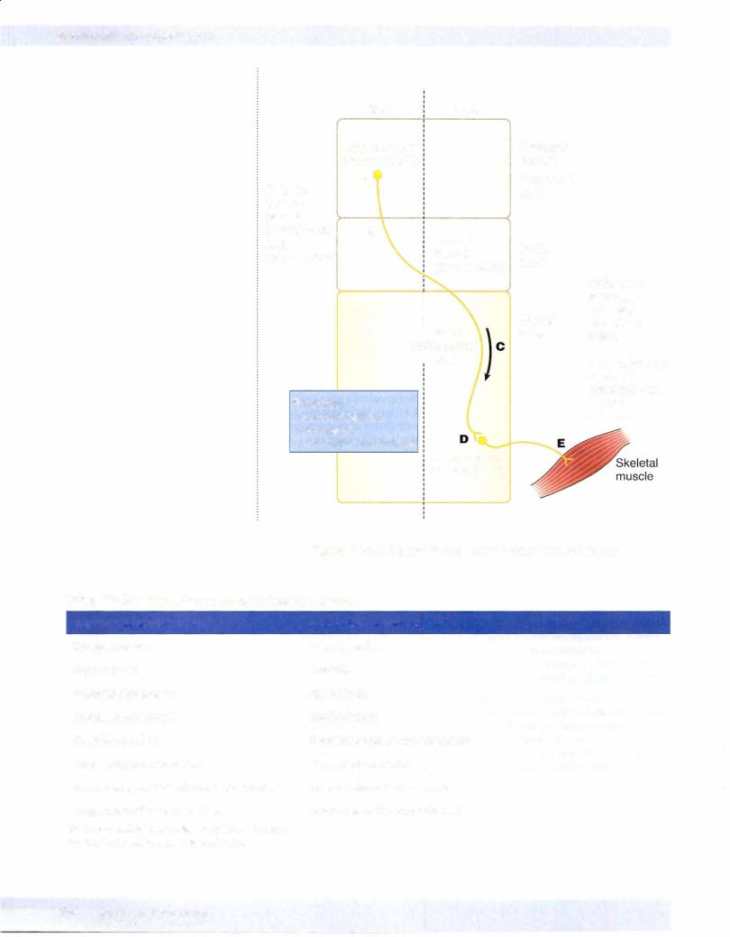
Two motoneurons, an upper motoneuron and a lower motoneuron, together form the basic neural circuit involved in the voluntary contraction of skeletal muscle everywhere in the body. The lower motoneurons are found in the ven tral horn ofthe spinal cord and in cranial nervenuclei in thebrain stem. Ax ons of lower motoneurons of spinal nerves exit in a ventral root, then join the spinal nerve to course in one of its branches to reach and synapse directly at a neuromuscular junction in skeletal muscle. Axons of lower motoneurons in the brain stem exit in a cranial nerve.
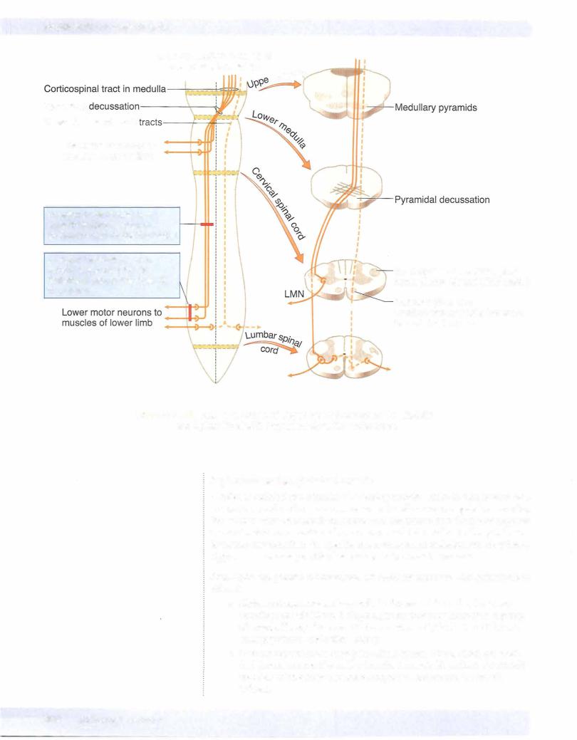
To initiate a voluntary contraction of skeletal muscle, a lower motoneuron must be innervated by an upper motoneuron (Figure IV-4-3). The cell bodies ofupper motoneurons are found in the brain stem and cerebral cortex, and their axons descend into the spinal cord in a tract to reach and synapse on lower motoneurons, or on interneurons, which then synapse on lower motoneurons. At a minimum, therefore, to initiate a voluntary contraction of skeletal muscle, 2 motoneurons, an upper and a lower, must be involved. The upper motoneuron innervates the lower motoneuron, and the lower motoneuron innervates the skeletal muscle.
The cell bodies of upper motoneurons are found in the red nucleus, reticular for mation, and lateral vestibular nuclei of the brain stem, but the most important
location ofupper motoneurons is in thecerebralcortex. Axons of these cortical neurons course in the corticospinal tract.
362 MEDICAL
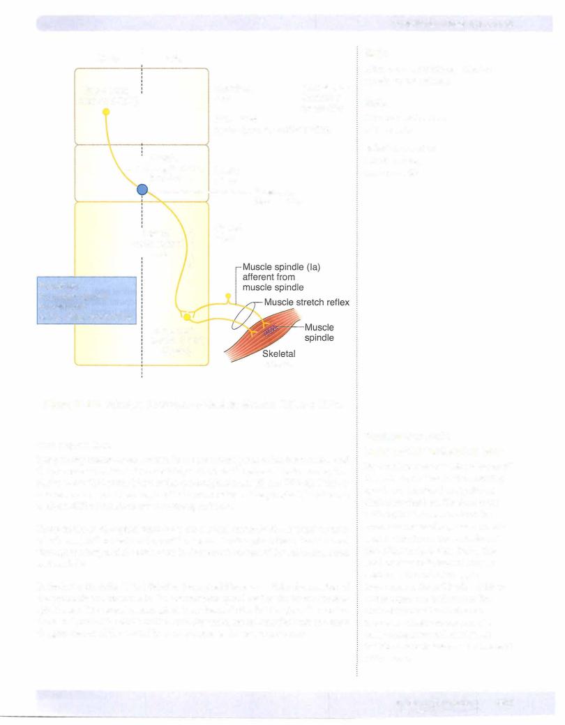
Both ends ofthe muscle spindle are connected in parallel with the extrafusal fibers, so these receptors monitor the length and rate of change in length of extrafusal fibers. Muscles involved with fine movements contain a greater density of spindles than those used in coarse movements.
Commonlytested muscle stretch reflex.es
The deep tendon (stretch, myotatic) reflex is monosynaptic and ipsilateral. The afferent limb consists of a muscle spindle, Ia sensory neuron, and efferent limb
(lower motor neuron). These reflexes are useful in the clinical exam.
Reflex |
Cord Segment Involved |
Muscle Tested |
|
Knee (patellar) |
L2-L4 (femoral n.) |
Quadriceps |
|
Ankle |
51 (tibial n.) |
Gastrocnemius |
|
Elbow |
C5-C6 |
(musculocutaneous n.) |
Biceps |
Elbow |
C7-C8 |
(radial n.) |
Triceps |
Forearm |
C5-C6 |
(radial n.) |
Brachioradialis |
Muscle stretch (myotatic) reflex
1be muscle stretch (myotatic) reflex is the stereotyped contraction of a muscle in response to stretch of that muscle. The stretch reflex is a basic reflexthat oc curs in allmuscles and is the primary mechanism for regulating muscle tone.
Muscle tone is the tension present in all resting muscle. Tension is controlled by the stretch reflexes.
The best example of a muscle stretch or deep tendon reflex is the knee-jerk re flex. Tapping the patellar ligament stretches the quadriceps muscle and its muscle spindles. Stretch of the spindles activates sensory endings (Ia afferents), and af ferent impulses are transmitted to the cord. Some impulses from stretch recep tors carried by Ia fibers monosynaptically stimulate the alpha motoneurons that supply the quadriceps. This causes contraction ofthe muscle and a sudden exten sion of the leg at the knee. Afferent impulses simultaneously inhibit antagonist muscles through interneurons (in this case, hamstrings).
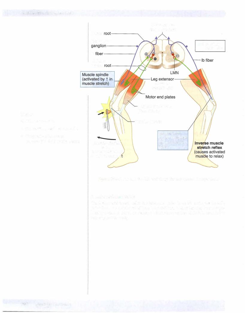
Inverse muscle stretch reflex
The inverse muscle stretch reflex monitors muscle tension. This reflex uses Golgi tendon organs (GTOs). These are encapsulated groups of nerve endings that ter minate between collagenous tendon fibers at the junction of muscle and tendon. GTOs are oriented in series with the extrafusal fibers and respond to increases in force or tension generated in that muscle. Increases in force in a muscle increase the firing rate of lb afferent neurons that innervate the GTOs, which, in turn, polysynaptically facilitate antagonists and inhibit agonist muscles.
Muscle tone and reflex activity can be influenced by gamma motoneurons and by upper motoneurons. Gamma motoneurons directly innervate the muscle spindles and regulate their sensitivity to stretch. Upper motoneurons innervate gamma motoneurons and also influence the sensitivity of muscle spindles to stretch. Stimulation of gamma motoneurons causes intrafusal muscle fibers lo cated at the pole of each muscle spindle to contract, which activates alpha moto neurons, causing an increase in muscle tone.
Chapter It • The Spinal Cord
MEDICAL 365
Chapter If • The Spinal Cord
Clinical Correlate
Upper Motoneuron Versus Lower Motoneuron Muscle Lesions
A fundamental requirement of interpreting the cause of motor weakness in neuroscience cases is the ability to distinguish between a lesion of an upper versus a lower motoneuron. Because a lesion to either an upper or a lower motoneuron produces a weakness in the ability to voluntarily contract skeletal muscles, the key to distinguishing an upper from a lower motoneuron lesion will be the condition of reflexes ofthe affected muscles (Figure IV-4-5).
A lesion of any part of a lower motoneuron will result in hypoactive muscle stretch reflexes and a reduction in muscle tone (hypotonicity) because lower motoneurons form the motor component of the reflex. Therefore, lower motoneuron lesions result in a paresis combined with suppressed or absent muscle stretch reflexes. An early sign of a lower motoneuron lesion is muscle fasciculations, which are twitches or contractions of groups of muscle fibers, that may produce a twitch visible on the skin. Later, lower motoneuron lesions produce fibrillations, which are invisible 1- to 5-ms potentials, detected with electromyography. Muscles denervated by a lower motoneuron lesion undergo pronounced wasting or atrophy. The constellation of lower motoneuron lesion signs combining paresis with suppressed or absent reflexes, fasciculations, and atrophy is known as a flaccid paralysis. With few exceptions, lower motoneuron lesions produce a flaccid paralysis ipsilateral and at the level of the lesion.
Neurologically, upper motoneurons includingthe corticospinal tract have a net overall inhibitory effect on muscle stretch reflexes. As a result, upper motoneuron lesions combine paresis ofskeletal muscles with muscle stretch or deep tendon reflexes that are hyperactive or hypertonic. The hypertonia may be seen as decorticate rigidity (i.e., postural flexion ofthe arm and extension ofthe leg) or decerebrate rigidity (i.e., postural extension ofthe arm and leg) depending on the location ofthe lesion. Lesions above the midbrain produce decorticate rigidity; lesions below the midbrain produce decerebrate rigidity. Upper motoneuron lesions result in atrophy ofweakened muscles only as a result ofdisuse, because these muscles can still be contracted by stimulating muscle stretch reflexes.
Upper motoneuron lesions are also accompanied by reversal of cutaneous reflexes, which normally yield a flexor motor response. The best known of the altered flexor reflexes is the Babinski sign. The test for the Babinski reflex is performed by stroking the lateral surface of the sole ofthe foot with a slightly painful stimulus. Normally, there is plantar flexion of the big toe. With a lesion ofthe corticospinal tract, the Babinski reflex is present, which is characterized by extension of the great toe and fanning of the other toes. Two other flexor reflexes, the abdominal and cremasteric, are also lost in upper motoneuron lesions. The constellation of upper motoneuron lesion signs combining paresis with increases or hyperactive reflexes, disuse atrophy of skeletal muscles, and altered cutaneous reflexes is known as a spastic paresis.
In contrastto lower motoneuron lesions, lesions of upper motoneurons result in a spastic paresis that is ipsilateral or contralateral and below the site of the lesion. Upper motoneuron lesions anywhere in the spinal cord will result in an ipsilateral spastic paresis below the level of the lesion. Upper motoneuron lesions between the cerebral cortex and the medulla above the decussation ofthe pyramids will result in a contralateral spastic paresis below the level of the lesion.
M EDICAL 367

Section IV • Neuroscience
Clinical Correlate
Tabes patients present with paresthesias (pins-and-needles sensations), pain, polyuria, Romberg sign.
Clinical Correlate
Spastic bladder results from lesions of the spinal cord above the sacral spinal cord levels. There is a loss of inhibition of the parasympathetic nerve fibers that innervate the detrusor muscle during the filling stage. Thus, the detrusor muscle responds to a minimum amount of stretch, causing urge incontinence.
Clinical Correlate
Atonic bladder results from lesions to the sacral spinal cord segments or the sacral spinal nerve roots. Loss of pelvic splanchnic motor innervation
with loss of contraction of the detrusor muscle results in a full bladder with a continuous dribble of urine from the bladder.
378 MEDICAL
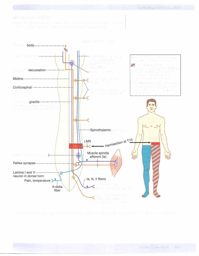
Brown-Sequardsyndrome
Hemisection of the cord results in a lesion of each of the 3 main neural systems: the principal upper motoneuron pathway of the corticospinal tract, one or both dorsal columns, and the spinothalamic tract. The hallmark of a lesion to these 3 long tracts is that the patient presents with 2 ipsilateral signs and one contralat eral sign. Lesion of the corticospinal tract results in an ipsilateral spastic paresis below the level of the injury. Lesion to the fasciculus gracilis or cuneatus results in an ipsilateral loss ofjoint position sense, tactile discrimination, and vibratory sensations below the lesion. Lesion ofthe spinothalamic tract results in a contra lateral loss of pain and temperature sensation starting one or 2 segments below the level of the lesion. At the level of the lesion, there willbe an ipsilateral loss of allsensation, including touch modalities as well as pain and temperature, and an ipsilateral flaccid paralysis in muscles supplied by the injured spinal cord seg ments (Figure IV-4-15).
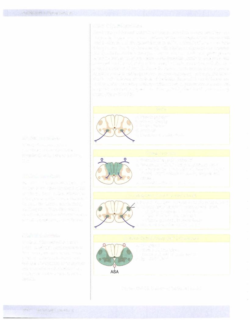
Polio
a.Flaccid paralysis
b.Muscle atrophy
c.Fasciculations
d.Areflexia
e.Common at lumbar levels
Tabes Dorsalis
a."Paresthesias, pain, polyuria"
b.Associated with late-stage syphilis, sensory ataxia, positive Romberg sign: sways with eyes closed, Argyll Robertson pupils, suppressed reflexes
c.Common at lumbar cord levels
Amyotrophic Lateral Sclerosis (ALS)
a.Progressive spinal muscular atrophy (ventral horn)
b.Primary lateral sclerosis (corticospinal tract)
•Spastic paralysis in lower limbs
•Increased tone and reflexes
•Flaccid paralysis in upper limbs
c.Common in ceNical enlargement
Anterior Spinal Artery (ASA) Occlusion
a.DC spared
b.All else bilateral signs
c.Common at mid thoracic levels
d.Spastic bladder
Figure IV-4-1 8. Lesions of the Spinal Cord I
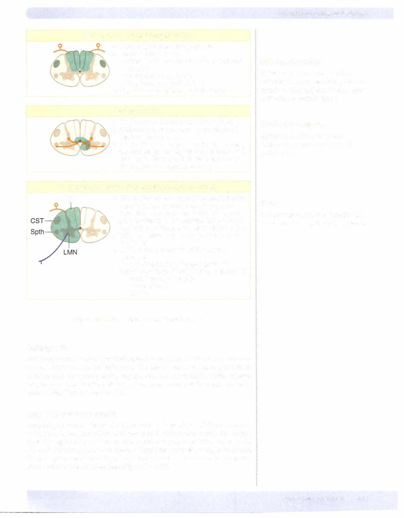
Subacute Combined Degeneration
a.Vitamin 812, pernicious anemia
b.Demyelination of the:
•Dorsal columns (central and peripheral myelin)
•Spinocerebellar tracts
•Corticospinal tracts (CST)
c.Upper thoracic or lower cervical cord
Syringomyelia
a. Cavitation of the cord (usually cervical) b. Bilateral loss of pain and temperature at
the level of the lesion
c.As the disease progresses, there is muscle weakness; eventuallyflaccid paralysis and atrophy of the upper limb muscles due to destruction of ventral horn cells
Hemisection: Brown-Sequard Syndrome (cervical)
DC |
a. DC: lpsilateral loss of position and vibratory |
|
|
senses at and below level of the lesion |
|
|
b. Spinothalamic tract: Contralateral loss of |
|
|
pain and temp 1-2 segments below lesion |
|
|
and ipsilateral loss atthe level of the lesion |
|
|
c. CST: lpsilateral paresis below the level of |
|
|
the lesion |
|
|
d. LMN: Flaccid paralysis atthe level of |
|
|
the lesion |
|
|
e. Descending hypothalamics: lpsilateral |
|
|
Horner syndrome (if cord lesion is above T1) |
|
|
• |
PtosisFacial hemianhydrosis(slight) |
|
• |
Miosis |
|
• |
|
Figure IV-4-1 9. Lesions of the Spinal Cord II
Poliomyelitis
Poliomyelitis results from a relatively selective destruction oflower motoneurons in the ventral horn by the poliovirus. The disease causes a flaccid paralysis of muscles with the accompanying hyporeflexia and hypotonicity. Some patients may recover most function, whereas others progress to muscle atrophy and per manent disability (Figure IV-4-18).
Amyotrophic lateralsclerosis
Amyotrophic lateral sclerosis (ALS, Lou Gehrig disease) is a relatively pure mo tor system disease that affects both upper and lower motoneurons. The disease typically begins at cervical levels of the cord and progresses either up or down the cord. Patients present with bilateral flaccid weakness of the upper limbs and bilateral spastic weakness of the lower limbs. Lower motoneurons in the brainstem nuclei may be involved later (Figure IV-4- 18).
Chapter 4 • The Spinal Cord
Clinical Correlate
Subacute combined degeneration patients present paresthesias, bilateral spastic weakness, Babinski signs, and antibodies to intrinsic factor.
Clinical Correlate
Syringomyelia may present with hydrocephalus and Arnold-Chiari II malformation.
Note
Syringomyelia results in a "belt-like" or "cape-like" loss of pain and temperature.
MEDICAL 379
Section IV • Neuroscience
Occlusion ofthe anteriorspinal artery
This artery lies in the anterior median sulcus of the spinal cord. Occlusion ofthe anterior spinal artery interrupts blood supplyto the ventrolateral parts ofthe cord, including the corticospinal tracts and spinothalamic tracts. Below the level ofthe lesion, the patient exhibits a bilateral spastic paresis and abilateral loss ofpain and temperature (Figure IV-4-18).
Syringomyelia
Syringomyeliais adisease characterizedbyprogressive cavitation ofthe central canal, usually in the cervical spinal cord but may involve other cord regions or the medulla. Early in the disease, there is a bilateral loss ofpain and temperature sensation in the hands and forearms as a result ofthe destruction ofspinothalamic fibers crossing in the anteriorwhite commissure. When the cavitation expands, lower motoneurons in theventralhorns are compressed, resulting inbilateral flaccid paralysis ofupperlimb muscles. A late manifestation ofcavitation is Homer syndrome, which occurs as a result of involvement ofdescending hypothalamic fibers innervating preganglionic sympathetic neurons inthe Tl throughT4 cord segments. Horner syndrome consists ofrniosis (pupillary constriction), ptosis (drooping eyelids), and anhidrosis (lack of sweating) in the face (Figure IV-4-19).
Tabes dorsalis
Tabes dorsalis is one possible manifestation of neurosyphilis. It is caused by bi lateral degeneration of the dorsal roots and secondary degeneration of the dor sal columns. There maybe impaired vibration and position sense, astereognosis, paroxysmal pains, and ataxia, as well as diminished stretch reflexes or inconti nence. Owing to the loss ofproprioceptive pathways, individuals with tabes dor salis are unsure ofwhere the ground is and walk with a characteristic and almost diagnostic "high-step stride" (Figure IV-4-18). Tabetic patients may also present with abnormal pupillary responses (Argyll Robertson pupils).
Subacute combined degeneration
Subacute combined degeneration is seen most commonlyin cases ofvitamin B12 deficiency, sometimes related to pernicious anemia. The disease is characterized by patchy losses ofmyelin in the dorsal columns and lateral corticospinal tracts, resulting in a bilateral spastic paresis and a bilateral alteration oftouch, vibration, and pressure sensations below the lesion sites (Figure IV-4-19). Myelin in both CNS and PNS is affected.
380 MEDICAL
Chapter 4 • The Spinal Cord
ChapterSummary
•The spinal cord is internally divided into 31 segments that give rise to 31 pairs of spinal nerves: 8 cervical, 12 thoracic, 5 lumbar, 5 sacral, and 1 coccygeal. Each segment is divided into an inner gray matter containing neuron cell bodies. The ventral horn of gray contains alpha and gamma motoneurons, the intermediate horn contains preganglionic neurons and Clarke nucleus, and the dorsal horn contains sensory neurons. The outer partofthe spinal cord is the white matter containing ascending and descending axons that form tracts located within funiculi.
Motor Pathways
•The corticospinal tract is involved in the voluntary contraction of skeletal muscle, especially in the distal extremities. This pathway consists of
2 neurons, an upper motor neuron, and a lower motor neuron. Most of the upper motor neurons have their cell bodies in the primary motor cortex and premotor cortex of the frontal lobe. These axons leave the cerebral
hemispheres through the posterior limb of the internal capsule and descend medially through the midbrain, pons, and medulla. In the medulla, 80-90% of these fibers decussate at the pyramids and then descend in
the spinal cord as the lateral corticospinal tract in the lateral funiculus of the white matter. These enter the ventral horn of gray at each cord segment and synapse upon the lower motor neurons. Axons ofthe lower neurons (final common pathway) leave via the ventral root ofthe spinal nerves and innervate the skeletal muscles. Lesions above the decussation (in the brain stem or cortex) produce contralateral deficits, and lesions below the decussation (in the spinal cord) produce ipsilateral findings. Patients with upper motor neuron lesions present with spastic paralysis, hyperreflexia, a clasp-knife reflex, and a positive Babinski. Lower motor neuron lesions present with flaccid paralysis, areflexia, atonia, muscle atrophy, and fasciculations.
Sensory Pathways
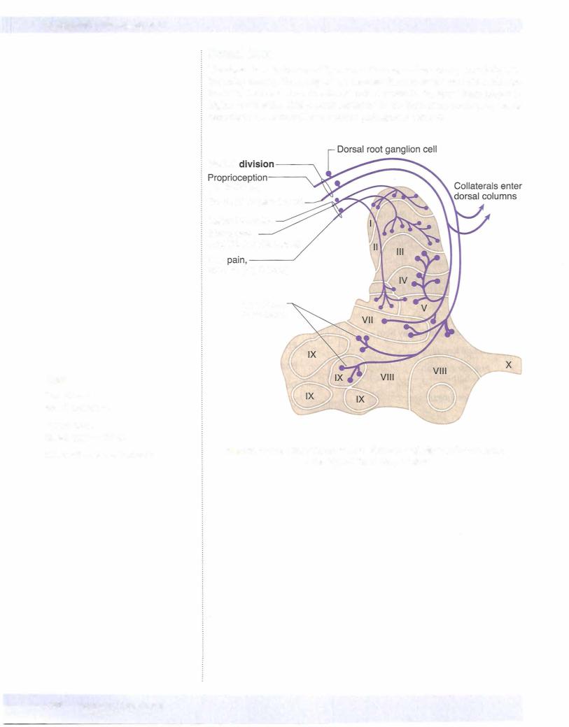
•Most sensory systems use 3 neurons to project sensory modalities to the cerebral cortex. The first neuron (primary afferent neuron) has its cell body in the dorsal root ganglion ofthe spinal nerve. This axon enters the spinal cord and either synapses in the spinal cord or the brain stem. The second neuron will decussate and project to the thalamus. The third neuron then projects from the thalamus to the somatosensory cortex of the parietal lobe.
(Continued)
MEDICAL 381
Section IV • Neuroscience
ChapterSummary(Conrd)
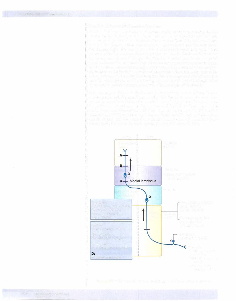
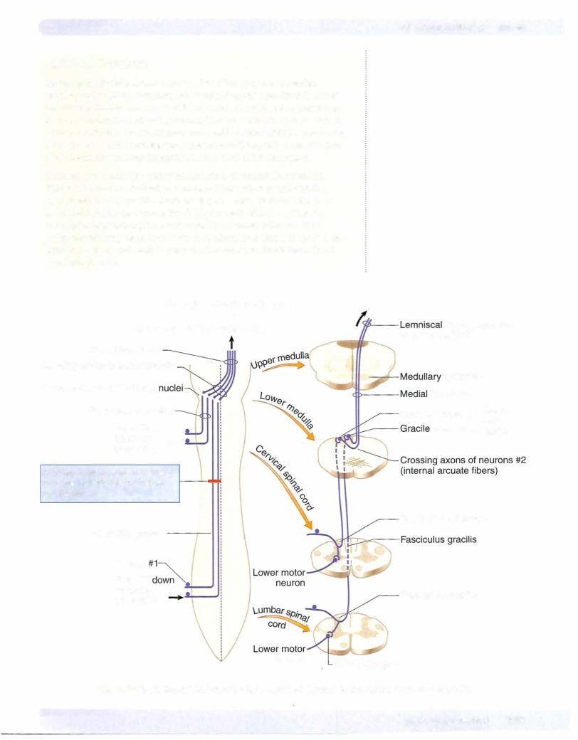
Dorsal Column-Medial Lemniscal System
•This pathway conducts sensory information for touch, proprioception, vibration, and pressure. The primary afferent neurons ofthis pathway have their cell bodies in the dorsal root ganglia. Their axons enter the spinal cord and ascend in the dorsal columns of the white matter as the fasciculus gracilis (from lower limb) or the fasciculus cuneatus (from upper limb). They synapse with the second neuron in the same named nuclei in the lower medulla. Axons ofthe second neuron decussate (internal arcuate fibers) and ascend the midline of the brain stem in the medial lemniscus to reach the ventral posterolateral (VPL) nucleus of the thalamus. The third neuron then projects through the posterior limb ofthe internal capsule to the somatosensory cortex. Lesions above the decussation (in the brain stem or cortex) produce contralateral loss of joint position, vibration, and touch, whereas lesions below decussations (in the spinal cord) produce ipsilateral deficits below the level of the lesion. A positive Romberg test indicates lesions of the sensory input to the cerebellum.
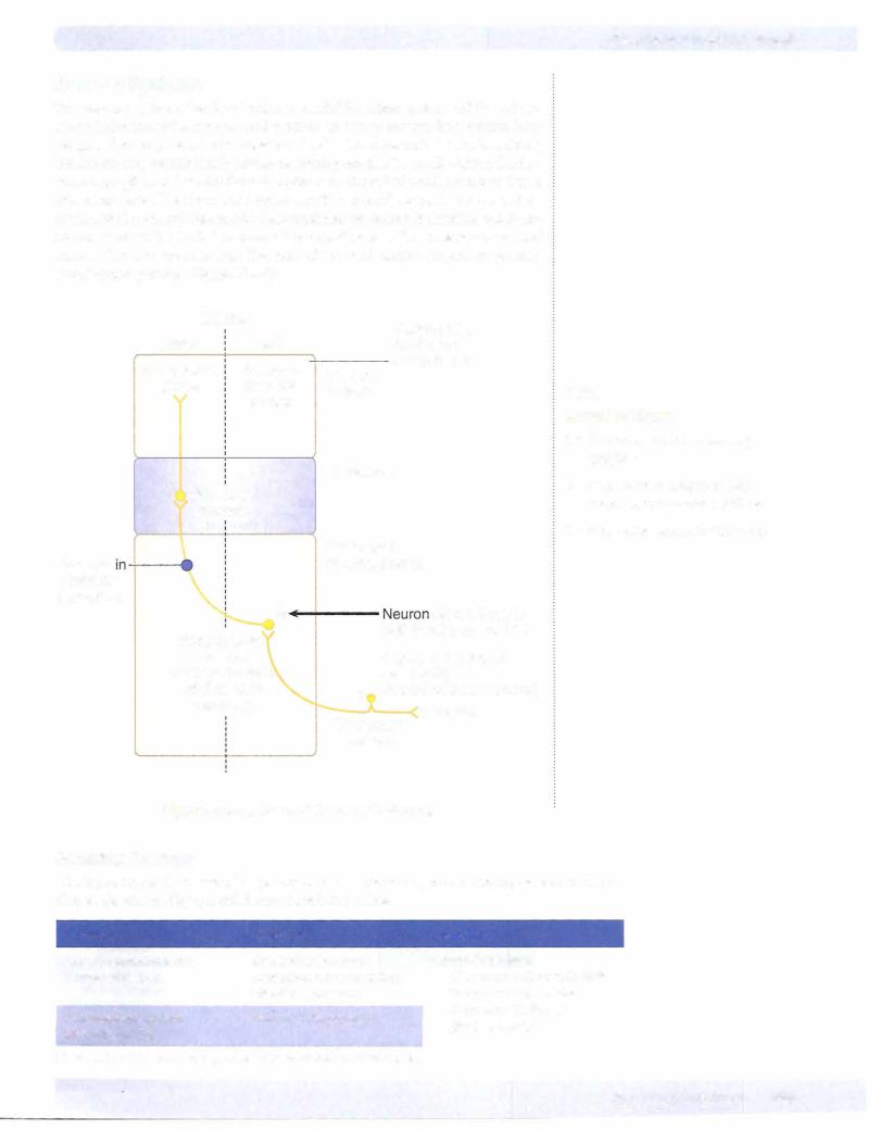
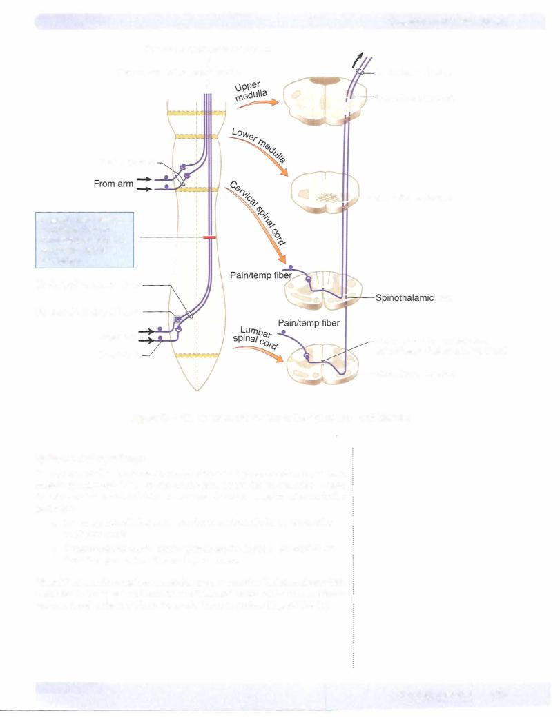
Anterolateral System
•The anterolateral pathway carries pain and temperature sensations. The first neuron fibers enter the spinal cord and synapse in the dorsal horn with the second neurons. The first neuron often ascends or descends one or 2 segments before they synapse. The second neuron axons then
decussate (ventral white commissure) and ascend the spinal cord and form the spinothalamic tract in the lateral funiculus of the white matter. The spinothalamic tract ascends the lateral aspect ofthe brain stem and synapses in the VPL nucleus ofthe thalamus where the third neuron projects to the cortex. All lesions ofthe spinothalamic tract in the spinal cord, brain stem, or cortex produce contralateral loss of pain and temperature below the lesion. Note that a central cord lesion at the spinal canal (syringomyelia) produces bilateral loss of pain and temperature at the level ofthe lesion.
•Lesions ofthe spinal cord that involve the above-mentioned tracts include poliomyelitis, tabes dorsalis, amyotrophic lateral sclerosis, anterior spinal artery occlusion, subacute combined degeneration, syringomyelia, and Brown-Sequard syndrome.
382 MEDICAL