

- •Contents
- •1. Cell Biology and Epithelia
- •2. Connective Tissue
- •3. Cartilage and bone
- •5. Nervous Tissue
- •6. Immune Tissues
- •7. Respiratory System
- •8. Gastrointestinal System
- •12. Integument
- •1. Gonad Development
- •4. Embryonic Period (Weeks 3-8)
- •1. Back and Autonomic Nervous System
- •2. Thorax
- •5. Lower Limb
- •4. The Spinal Cord
- •5. The Brain Stem
- •7. Basal Ganglia
- •11. Limbic System
- •Index

Basal Ganglia |
7 |
GENERAL FEATURES
The basal ganglia initiate and provide gross control over skeletal muscle move ments. The major components of the basal ganglia include:
•Striatum, which consists of the caudate nucleus and the putamen (telen- cephalon)
•External and internal segments of the globus pallidus (telencephalon)
•Substantia nigra (in midbrain)
•Subthalamic nucleus (in diencephalon)
Together with the cerebral cortex and the ventrolateral (VL) nucleus of the thal amus, these structures are interconnected to form 2 parallel but antagonistic circuits known as the direct and indirect basal ganglia pathways (Figures IV-7- 1 and IV-7-2). Both pathways are driven by extensive inputs from large areas of cerebral cortex, and both project back to the motor cortex after a relay in the VL nucleus of the thalamus. Both pathways use a process known as "disinhibition'' to mediate their effects, whereby one population of inhibitory neurons inhibits a second population ofinhibitory neurons.
Direct Basal Ganglia Pathway
In the direct pathway, excitatory input from the cerebral cortex projects to striatal neurons in the caudate nucleus and putamen. Through disinhibition, activated inhibitory neurons in the striatum, which use y-aminobutyric acid (GABA) as their neurotransmitter, project to and inhibit additional GABA neurons in the internal segment ofthe globus pallidus.
The GABA axons ofthe internal segment ofthe globus pallidus project to the thala mus (VL). Because their input to the thalamus is disinhibited, the thalamic input excites the motor cortex. The net effect of the disinhibition in the direct pathway results in an increasedlevel ofcortical excitation and the promotion ofmovement.
Indirect Basal Ganglia Pathway
In the indirect pathway, excitatory input from the cerebral cortex also projects to striatal neurons in the caudate nucleus and putamen. These inhibitory neurons in the striatum, which also use GABA as their neurotransmitter, project to and inhibit additional GABA neurons in the external segment ofthe globus pallidus.
The GABA axons ofthe external segment ofthe globus pallidus project to the sub thalamic nucleus. Through disinhibition, the subthalamic nucleus excites inhibi tory GABA neurons in the internal segment ofthe globus pallidus, which inhibits the thalamus. This decreases the level of cortical excitation, inhibiting movement. The net effect of the disinhibition in the indirect pathway results in a decreased level ofcortical excitation, and a suppression ofunwanted movement.
MEDICAL 429


Dopamine and cholinergic effects
In addition to the GABA neurons, 2 other sources of chemically significant neurons enhance the effects of the direct or indirect pathways.
Dopaminergic neurons in the substantia nigra in the midbrain project to the striatum. The effect of dopamine excites or drives the direct pathway, increasing cortical excitation. Dopamine excites the direct pathway through D1 receptors and inhibits the indirect pathway through D2 receptors.
Cholinergic neurons found within the striatum have the opposite effect. Acetyl choline (Ach)drives the indirect pathway, decreasing cortical excitation.
Figure IV-7-3. MRI of Horizontal Section through
Diencephalon, Basal Ganglia, and Cortex.
(a) Thalamus {b) Head of Caudate Nucleus (c) Genu of Internal Capsule Containing CorticobulbarAxons {d) Posterior Limb of Internal Capsule (e) Primary Visual Cortex (f) Splenium of Corpus Callosum (g) Putamen {h) Broca's Motor Speech Area
(i) Wernicke's Oral Comprehension Area
Chapter 7 • Basal Ganglia
Note
All basal ganglia connections are with ipsilateral cortex.
MEDICAL 431
Section IV • Neuroscience
Figure IV-7-4. Coronal Section through Basal Ganglia and Other Subcortical Structures
(A)caudate nucleus (B) putamen (C) globus pallidus external segment
(D)globus pallidus internal segment (E) septal nuclei (F) fornix (G) lateral ventricle (H) anterior commissure (I) optic chiasm (J) basal nucleus of Meynert (K) preoptic hypothalamus (L} internal capsule, anterior limb
432 MEDI CAL


Section IV • Neuroscience
Clinical Correlate
Lesions or Diseases ofthe Basal Ganglia
Lesions or diseases ofthe basal ganglia generally present with movement disorders, known as dyskinesias, and an involuntary tremor, ortremor at rest.
Most basal ganglia disorders seem to preferentially affect either the direct or the indirect pathways, altering the balance between the two.
Lesions ofthe direct pathway
Lesions of the direct pathway result in an underactive cortex and hypokinetic disturbances in which there is a slowing or absence of spontaneous movements. The best-known disorder of the direct pathway is caused by the degeneration of dopaminergic neurons of the substantia nigra in Parkinson disease. Because the cortex is underactive, Parkinson patients have problems initiating movements, combined with a reduction in the velocity and amplitude of the movements. The tremor at rest is the classic pill-rolling tremor seen in the fingers. Skeletal muscles in the upper limbs exhibit a cogwheel rigidity because of increased muscle tone. Patients also present with a stooped posture, an expressionless face, and a festinating or accelerating gait during which individuals seem to chase their center of gravity. One strategy for Parkinson patients is to give them L-dopa, a dopamine precursor that crosses the blood-brain barrier. Another strategy is to give anticholinergic drugs to inhibit the effects of acetylcholine on the indirect pathway.
Lesions ofthe indirect pathway
Other common disorders of the basal ganglia (chorea, athetosis, dystonia, tics) result from lesions to parts of the indirect pathway, which result in an overactive motor cortex. An overactive cortex produces hyperkinetic disturbances, expressed in numerous spontaneous movements. The involuntary tremors seen in these diseases range from being dancelike in chorea to ballistic with lesions to the subthalamic nucleus.
Chorea produces involuntary movements that are purposeless, quick jerks that may be superimposed on voluntary movements. Huntington chorea exhibits autosomal dominant inheritance (chromosome 4) and is characterized by severe degeneration of GABA neurons in the striatum. In addition to chorea, these patients frequently sufferfrom athetoid movements, progressive dementia, and behavioral disorders. Sydenham chorea is a transient complication in some children with rheumatic fever.
Athetosis refers to slow, worm like, involuntary movements that are most noticeable in the fingers and hands but may involve any muscle group. It is present in Huntington disease and may be observed in many diseases that involve the basal ganglia.
Dystonia refers to a slow, prolonged movement involving predominantly the truncal musculature. Dystonia often occurs with athetosis. Blepharospasm (contraction ofthe orbicularis oculi causing the eyelids to close), spasmodic torticollis (in which the head is pulled toward the shoulder), and writer's cramp (contraction ofarm and hand muscles on attempting to write) are all examples
of dystonic movements.
(Continued)
434 MEDICAL

Chapter 7 • Basal Ganglia
Clinical Correlate (Continued)
Hemiballismus results from a lesion of the subthalamic nucleus usually seen in hypertensive patients. Hemiballismus refers to a violent projectile movement of a limb and is typically observed in the upper limb contralateral to the involved subthalamic nucleus.
Tourette syndrome involves facial and vocaltics that progress to jerking movements ofthe limbs. It is frequently associated with explosive, vulgarspeech.
Wilson disease results from an abnormality of copper metabolism, causing the accumulation of copper in the liver and basal ganglia. Personality changes, tremor, dystonia, and athetoid movements develop. Untreated patients usually succumb because of hepatic cirrhosis. A thin brown ring around the outer cornea, the Kayser-Fleischer ring, may be present and aid in the diagnosis.
ChapterSummary
•The basal ganglia play important motor functions in starting and stopping
voluntary motor functions and inhibiting unwanted movements. The basal ganglia consists of 3 nuclei masses deep in the cerebrum (caudate nucleus, putamen, and globus pallidus), one nucleus in the midbrain (substantia nigra), and the subthalamic nucleus ofthe diencephalon. The striatum combines the caudate nucleus and the putamen while the corpus striatum
consists ofthese 2 nuclei plus the globus pallidus.
•There are 2 parallel circuits (direct and indirect) through the basal ganglia. These circuits receive extensive input from the cerebral cortex that project back to the motor cortex after a relay in the ventrolateral (VL) nucleus ofthe thalamus. Both ofthese pathways demonstrate disinhibition. The direct pathway increases the level of cortical excitation and promotes movement. The indirect pathway decreases the level of cortical excitation and suppresses unwanted movement.
•The striatum is the major input center and the globus pallidus is the major output centerforthe pathways through the basal ganglia. Critical to proper function ofthe striatum is dopamine production by the substantia nigra.
Dopamine excites the direct pathway and inhibits the indirect pathway.
•Lesions of the direct pathway result in an underactive cortex, which produces hypokinetic motor disturbances. The classic disorder caused by degeneration of dopaminergic neurons of the substantia nigra is Parkinson disease. These patients are characterized by tremor at rest (pill-rolling), increased muscle tone, mask face, and hypokinetic movement.
•Hyperkinetic disorders result from lesions ofthe indirect pathway and cause an overactive motor cortex. These movements occur spontaneously at rest and cannot be controlled by the patient. Examples ofthese disorders include chorea (multiple quick movements), athetosis (slow serpentine movements), and hemiballismus (violent flinging movements). Hemiballismus results from hemorrhagic destruction ofthe contralateral subthalamic nucleus.
MEDICAL 435


Section IV • Neuroscience
Open-Angle Glaucoma
A chronic condition (often with increased intraocular pressure [IOP]) due to de creased reabsorption of aqueous humor, leading to progressive (painless) visual loss and, ifleft untreated, blindness. IOP is a balance between fluid formation and its drainage from the globe.
Narrow-Angle Glaucoma
An acute (painful) or chronic (genetic) conditionwith increased IOP due to block ade ofthe canal ofSchlemm. Emergencytreatmentprior to surgeryoften involves cholinomimetics, carbonic anhydrase inhibitors, and/or mannitol.
VISUAL REFLEXES
Pupillary Light Reflex
When light is directed into an eye, it stimulates retinal photoreceptors and results in impulses carried in the optic nerve to the pretectal area. Cells in the pretectal area send axons to the Edinger-Westphal nuclei on both sides.
The Edinger-Westphal nucleus is the parasympathetic nucleus ofthe oculomotor nerve and gives rise to preganglionic parasympathetic fibers thatpass in the third cranial nerve to the ciliary ganglion. Because cells in the pretectal area supply both Edinger-Westphal nuclei, shining light into one eye results in constriction of both the ipsilateral pupil (direct light reflex) and contralateral pupil (consensual light reflex).
Accommodation-Convergence Reaction
This reaction occurs when an individual attempts to focus on a nearbyobject after looking at a distant object. The oculomotor nerve carries the efferent fibers from the accommodation-convergence reaction, which consists of3 components: ac commodation, convergence, and pupillary constriction.
Accommodation refers to the reflex that increasesthe curvature ofthe lens needed for nearvision. Preganglionic parasympathetic fibers arise in the Edinger-Westphal nucleus and pass via the oculomotor nerve to the ciliary ganglion. Postganglionic parasympathetic fibers from the ciliaryganglionsupplythe ciliarymuscle. Contrac tion ofthis muscle relaxes the suspensory ligaments and allows the lens to increase its convexity (become more round). This increases the refractive index ofthe lens, permittingtheimage ofa nearbyobject to focus on the retina.
Convergence results from contraction ofboth medial rectus muscles, which pull the eyes to look toward the nose. This allows the image ofthe near object to focus on the same part ofthe retina in each eye.
Pupillaryconstriction (miosis) results from contraction ofthe constrictor mus cle ofthe iris. A smalleraperturegives the optic apparatus agreater depth offield. With Argyll Robertson pupils, both direct and consensual light reflexes are lost, but the accommodation-convergence reaction remains intact. This type ofpupil is often seen in cases ofneurosyphilis; however, it is sometimes seen in patients with multiple sclerosis, pineal tumors, or tabes dorsalis. The lesion site is believed to occur near the pretectal nuclei just rostral to the superior colliculi.
438 MEDICAL

Chapter 8 • Visual Pathways
Table IV-8-1. Pupillary Light Reflex Pathway
Pretectal area |
Afferent Limb: CN II |
|
|
|
|
|
Light stimulates ganglion retinal cells |
|
Edinger |
impulses travel up CNll, which projects |
|
bilaterally to the pretectal nuclei (midbrain) |
|
|
Westphal |
|
|
nucleus |
The pretectal nucleus projects bilaterally |
|
|
Edinger-Westphal nuclei (CN Ill) |
|
|
Efferent Limb: CN Ill |
|
|
Edinger-Westphal nucleus (preganglionic |
|
|
This is a simplified |
|
|
parasympathetic) ciliary ganglion |
diagram; the ciliary |
|
(postganglionic parasympathetic) pupillary |
ganglion is not shown. |
|
sphincter muscle miosis |
|
|
|
Because cells in the pretectal area supply the Edinger-Westphal nuclei bilaterally, shining light in one eye constriction in the ipsilateral pupil (direct light reflex) and the contralateral pupil (consensual light reflex).
Because this reflex does not involve the visual cortex, a person who is cortically blind can still have this reflex.
The eye is predominantly innervated by the parasympathetic nervous system. Therefore, application of muscarinic antagonists or ganglionic blockers has a large effect by blocking the parasympathetic nervous system.
Table IV-8-2. Pharmacology ofthe Eye
Structure |
Predominant |
Receptor |
Receptor |
|
|
Receptor |
Stimulation |
Blockade |
|
Pupillary sphincter |
M3 receptor (PANS) |
Contraction |
Relaxation |
|
ms. (iris) |
a |
|
miosis |
mydriasis |
Radial dilator |
receptor (SANS) |
Contraction |
Relaxation |
|
ms. (iris) |
|
mydriasis |
miosis |
|
Ciliary ms. |
M3 receptor (PANS) |
Contraction |
Relaxation |
|
|
|
|
accommodation |
focus for far |
|
|
|
for near vision |
vision |
Ciliary body |
receptor (SANS) |
Secretion of |
Decreased |
|
epithelium |
|
|
aqueous humor |
aqueous |
|
|
|
|
humor |
|
|
|
|
production |
Abbreviations: ms., muscle; PANS, parasympathetic nervous system; SANS, sympathetic nervous system
MEDICAL . 439

Section IV • Neuroscience
Table IV-8-3. Accommodation-Convergence Reaction
When an individual focuses on a nearby object after looking at a distant object, 3 events occur:
1.Accommodation
2.Convergence
3.Pupillary constriction (miosis)
In general, stimuli from light visual cortex superior colliculus and pretectal nucleus Edinger-Westphal nucleus (1, 3) and oculomotor nucleus (2).
Accommodation: Parasympathetic fibers contract the ciliary muscle, which relaxes suspensory ligaments, allowing the lens to increase its convexity (become more round). This increases the refractive index ofthe lens, thereby focusing a nearby object on the retina.
Convergence: Both medial rectus muscles contract, adducting both eyes.
Pupillary constriction: Parasympathetic fibers contract the pupillary sphincter muscle miosis.
Table IV-8-4. Clinical Correlates
|
• |
Pupillary Abnormalities |
Argyll Robertson pupil |
No direct or consensual light reflex; accommodation-convergence intact |
|
(pupillary light-near dissociation) |
• Seen in neurosyphilis, diabetes |
|
|
||
Relative afferent (Marcus Gunn) |
|
Lesion of afferent limb of pupillary light reflex; diagnosis made with swinging |
pupil |
|
flashlight |
|
|
Shine light in Marcus Gunn pupil pupils do not constrict fully |
|
• |
Shine light in normal eye pupils constrict fully |
|
Shine light immediately again in affected eye apparent dilation of both pupils |
|
|
because stimulus carried through that CN II is weaker; seen in multiple sclerosis |
|
Horner syndrome |
Caused by a lesion of the oculosympathetic pathway; syndrome consists of miosis, |
|
|
|
ptosis, apparent enophthalmos, and hemianhidrosis |
Adie pupil |
• |
|
Transtentorial (uncal) herniation |
||
|
Dilated pupil that reacts sluggishly to light, but better to accommodation; often seen in women and often associated with loss of knee jerks. Ciliary ganglion lesion
Increased intracranial pressure leads to uncal herniation CN Ill compression fixed and dilated pupil, "down-and-out" eye, ptosis
440 MEDICAL


Chapter 8 • Visual Pathways
Most fibers from the optic tract project to the lateral geniculate body (LGB); some also project to the pretectal area (light reflex), the superior colliculi (reflex gaze), and the suprachiasmatic nuclei (circadian rhythm). The LGB projects to the primary visual cortex (striate cortex, Brodmann area 1 7) of the occipital lobe via the optic radiations.
•Visual information from the lower retina (upper contralateral visual field) temporal lobe (Meyerloop) lingual gyrus
•Visual information from the upper retina (lower contralateral visual field) parietal lobe cuneus gyrus
The lateral geniculate body (LGB) is a laminated structure thatreceivesinputfrom the optic tract and gives rise to axonsthat terminate on cells in the primaryvisual cortex (striate cortex, Brodmann area 17) ofthe occipital lobe. The LGB laminae maintain a segregation ofinputs from the ipsilateral and contralateral retina.
The axons from the LGB thatproject to the striate cortex are known as optic radia tions, visual radiations, or the geniculocalcarine tract. The calcarine sulcus divides the striate cortex (primaryvisual cortex or Brodmann area 17) into the cuneus and the lingual gyri. The cuneus gyrus, which lies on the superior bank ofthe calcarine cortex, receives the medial fibers ofthe visual radiations. The lingual gyrus, which lies on the inferior bank of the calcarine cortex, receives the lateral fibers of the visual radiation. The medial fibers coursing in the visual radiations, which carry input from the upper retina (i.e., the lowercontralateralvisual field), pass from the LGB directlythrough the parietal lobe to reach the cuneus gyrus. Significantly, the lateral fibers coursing in the visual radiations, which carry input from the lower retina (i.e., the upper contralateral visual field), take a circuitous route from the LGB through Meyer loop anteriorly into the temporal lobe. The fibers of Meyer loop then turn posteriorlyand course through the parietal lobe to reach the lingual gyrus in the striate cortex.
-
'
M E D I CAL 443

Section IV • Neuroscience
LESIONS OF THE VISUAL PATHWAYS
Lesions ofthe retina that include destruction of the macula produce a central sco toma. The macula is quite sensitive to intense light, trauma, aging, andneurotoxins.
Lesions of an optic nerve produce blindness (anopsia) in that eye and a loss of the sensory limb ofthe light reflex. The pupil ofthe affected eye constricts when light is shined into the opposite eye (consensual light reflex) but not when light is shinedinto the blinded eye (absence ofdirectlight reflex).
Compression ofthe optic chiasm, often the result ofa pituitarytumor ormeningio ma, results in a loss ofperipheralvision in bothtemporalfields because the crossing fibers from each nasal retina are damaged. The resultingvisual field defect is called a bitemporal heteronymous hemianopia.
Alllesions past the chiasm produce contralateral defects. Lesions of the optic tract result in a loss of visual input from the contralateral visual field. For ex ample, a lesion ofthe right optic tract results in a loss ofinput from the left visual field. This is called a homonymous hemianopia; in this example, a left homony mous hemianopia.
Lesions ofthe visual radiations are more commonthan lesions to the optic tract or lateral geniculate body and produce visual field defects (a contralateral homony mous hemianopia) similar to those ofthe optic tract ifall fibers are involved.
Lesions restricted to the lateral fibers in Meyer loop, usually in the temporal lobe, result in a loss ofvisual input from the contralateral upper quarter of the visual field. For example, a lesion ofthe temporal fibers in the right visual radiation re sults in loss ofvisual input from the upper left quarter ofthe field (a left superior quadrantanopia).
Lesions restricted to the medial fibers in the visual radiation in the parietal lobe result in a loss ofvisual input from the contralateral lower quarter ofthe field (an inferior quadrantanopia).
Lesions inside the primaryvisual cortexare equivalent to those ofthevisualradi ations, resulting in a contralateral homonymous hemianopsia, except that macu lar (central) vision is spared.
Lesions of the cuneus gyrus are equivalent to lesions restricted to the parietal fibers ofthe visual radiation, with macular sparing.
Lesions ofthe lingula aresimilar to lesions ofthe Meyer's loop fibers exceptfor the presence of macular sparing. The pupillary light reflex is spared in lesions of the radiations or insidevisual cortexbecause fibers ofthepupillarylight reflexleavethe optic tracts to terminate in the pretectal area. The combination ofblindness with intact pupillary reflexes is termed cortical blindness.
444 MEDICAL

Chapter 8 • Visual Pathways
ChapterSummary
•The eyeball is formed by 3 layers: the sclera, choroid, and retina. The shape ofthe lens is modified for near and far vision by the ciliary muscle during the accommodation reflex. The sclera is the external layer and continues anteriorly as the cornea, which is transparent and allows light to enter the eye. The intermediate choroid layer is highly vascularized and pigmented. Anteriorly, the choroid layerforms the ciliary body and iris. The retina contains the photoreceptive layer ofrods (for nightvision and dim light) and cones (for colorvision and high visual acuity).
•The axons ofthe ganglionic cells ofthe retina form the optic nerve at the optic disc.
•The visual pathway is a 3-neuron pathway with the first neuron (bipolar neurons) and the second neuron (ganglionic neurons) located in the retina. The ganglionic axons project from the retina through the optic nerve, optic chiasm, and optic tract to synapse with the third neuron located in the lateral geniculate body ofthe thalamus. These thalamic axons then project via the optic radiations (geniculocalcarine tract) in the parietal lobe to reach the primary visual (striate) cortex at the posterior pole of the occipital lobe. Because the lens inverts images like a camera, each nasal retina receives information from the temporal visual fields, and each temporal retina receives information from the nasal visual field. Atthe optic chiasm, the fibers from the nasal half of each retina decussate while the optic fibers from the temporal half of each retina pass through the chiasm without decussating. Thus, central to the chiasm ipsilateral visual fields project through the contralateral visual pathways. Lesions at the lateral aspect of the optic chiasm produce ipsilateral nasal hemianopsia, whereas midline lesions at the chiasm produce bitemporal heteronymous hemianopsia. Any lesion central to the chiasm results in contralateral homonymous hemianopsia.
•In addition, visual pathways carry optic fibers from the superior and inferior quadrants ofthe visual fields through the retina to the lateral geniculate body. The projections of the superior quadrants to the lower retina reach the lateral geniculate body laterally, synapse, and leave through the lateral
course of Meyer loop in the temporal lobe before rejoining the optic radiation to reach the lower (lingual) gyrus of the striate cortex. Thus, lesions ofthe temporal lobe affecting Meyer loop result in contralateral homonymous superior quadrantanopia. Inferior quadrants ofthe visual fields project to the upper retina and then to the medial aspect ofthe lateral geniculate body. After synapsing in the geniculate body, the axons project completely through the optic radiations to reach the upper (cuneus) gyrus of the striate cortex.
Lesions ofthese more medial fibers produce contralateral homonymous inferior quadrantanopia. Vascular lesions ofthe striate cortex due to occlusion of the posterior cerebral artery result in contralateral homonymous hemianopia with macular sparing.
M EDICAL 445


Section IV • Neuroscience
MajorThalamic Nuclei and Their Inputs and Outputs
Anteriornucleargroup (partofthe Papez circuitoflimbicsystem)
Input is from the mammillary bodies via the mammillothalamic tract and from the cingulate gyrus; output is to the cingulate gyrus via the anterior limb ofthe internal capsule.
Clinical Correlate
Thalamic pain syndrome affects the ventral nuclear group. Patients present with burning, aching pain in contralateral limbs or body. Involvement of the dorsal column
medial lemniscal part ofVPL increases the sensitivity to pain and presents as contralateral loss ofvibratory sense and gait ataxia. Thalamic pain syndrome is resistent to analgesic medications.
Medialnucleargroup (partoflimbicsystem)
Input is from the amygdala, prefrontal cortex, and temporal lobe; output is to the prefrontal cortex and cingulate gyrus. The most important nucleus is the dorso medial nucleus.
Ventral nucleargroup
MotorVentralNuclanteriori nucleus (VA): Input to VA is from the globus pallidus, substantia nigraVentral. Outputlateralisnucleusto the premotor(VL): and primary motor cortex.
Input to is mainly from the globus pallidus and
VL
the dentate nucleus of the cerebellum. Output is to the primary motor cortex (Brodmann area 4).
SVentralsoryposterolateralNuclei (VPL) nucleus: Input to VPL conveying somatosensory and nociceptive information ascends in the medial lemniscus and spinothalamic
tract. Output is to primary somatosensory cortex (Brodmann areas 3, 1, and 2) ofVentralthe parieposteromedialal lobe. (VPM) nucleus: Input to VPM is from the ascending tri
geminal and taste pathways. Output is to primary somatosensory cortex (Brod
Medialmann argeniculateas 3, 1 , andbody2)(nucleus):ofthe parietal lobe.
Inputis from auditoryinformation that ascends Lateralfrom thegeniculatei ferior colliculusbody (nucleus):. O tput is to primary auditory cortex.
Input is from the optic tract. Output is in the form ofthe geniculocalcarine or visual radiations that project to the primary vi sual (striate) cortex in the occipital lobe.
Midline and lntralaminar Nuclei
Midline and intralarninar nuclei receive input from the brain-stem reticular for mation, and from the spinothalarnic tract. Intralaminar nuclei send pain informa tion to the cingulate gyrus.
These nuclei appear to be important in mediating desynchronization ofthe elec troencephalogram (EEG) duringbehavioral arousal.
448 MEDICAL

Chapter 9 • Diencephalon
HYPOTHALAMUS
The hypothalamus is composed of numerous nuclei that have afferent and effer ent connections with widespread regions of the nervous system, including the pituitary gland, the autonomic system, and the limbic system (Figure IV-9-1).
Table IV-9-2. Hypothalamus, Epithalamus, Subthalamus
Hypothalamus-helps maintain homeostasis; has roles in the autonomic, endocrine, and limbic systems
Hypothalamic Nuclei |
Functions and Lesions |
|
|
|
||
Lateral hypothalamic |
Feeding center; lesion ---starvationt |
|
|
|
||
Ventromedial |
Satiety center; |
lesion |
---hyperphagia,t |
obesity, savage behavior |
|
|
Suprachiasmatic |
|
|
||||
Regulates circadian rhythms, receives direct retinal input |
|
|||||
Supraoptic and paraventricular |
Synthesizes ADH and oxytocin; regulates water balance |
|
||||
|
Lesion ---diabetesinsipidus, characterized by polydipsia and polyuria |
|
||||
Mamillary body |
Input from hippocampus; damaged in Wernicke encephalopathy |
|
||||
Arcuate |
Produces hypothalamic releasing and inhibiting factors and gives rise to |
|
||||
|
tuberohypophysial tract |
|
|
|
||
|
Has neurons that produce dopamine (prolactin-inhibiting factor) |
|
||||
Anterior region |
Temperature regulation; lesion ---hyperthermia |
|
|
|||
|
Stimulates the parasympathetic nervous system |
|
|
|||
Posterior region |
Temperature regulation; lesion ---poikilothermiat |
(inability to thermoregulate) |
||||
|
Stimulates sympathetic nervous system |
|
|
|||
Preoptic area |
Regulates release of gonotrophic hormones; contains sexually dimorphic |
|
||||
|
nucleus |
|
---t |
|
|
---t |
|
amenorrhea or |
|
|
|
||
|
Lesion before puberty |
arrested sexual development; lesion after puberty |
|
|||
|
|
impotence |
|
|
|
|
Dorsomedial |
Stimulation ---savagetbehavior |
|
|
|
||
Epithalamus-Consists of pineal body and habenular nuclei. The pinealbody secretes melatonin with a circadian rhythm.
Subthalamus-The subthalamic nucleus is involved in basal ganglia circuitry. Lesion ---hemiballismust (contralateral flinging movements of one or both extremities)
Abbreviation: ADH, antidiuretic hormone
MEDICAL 449


Section IV • Neuroscience
Tuberal region
Arcuate Nucleus
Cells in the arcuate nucleus produce releasing hormones and inhibitory factors, which enter capillaries in the tuberoinfundibular tract and pass through the hy pophyseal-portal veins to reach the secondary capillary plexus in the anterior pi tuitary gland. Releasing hormones and inhibitory factors influence the secretory activity of the acidophils and basophils in the anterior pituitary. (See Histology section.)
Ventromedial Nucleus
The ventromedial hypothalamus is a satiety center and regulates food intake. Le sions ofthe ventromedial hypothalamus result in obesity.
Posteriorregion
Mammillary Bodies
The mammillary nuclei are located in the mammillary bodies and are part ofthe limbic system. The mammillothalamic tract originates in the mammillarynuclei and terminates in the anterior nuclear group ofthe thalamus.
Anteriorhypothalamic zone
The anterior hypothalamic zone senses an elevation ofbodytemperature and me diates the response to dissipate heat. Lesions of the anterior hypothalamus lead to hyperthermia.
Posteriorhypothalamic zone
The posterior hypothalamic zone senses a decrease ofbody temperature and me diates the conservation of heat. Lesions of the posterior hypothalamus lead to poikilothermy (i.e., cold-blooded organisms). An individual with a lesion ofthe posterior hypothalamus has a bodytemperature thatvaries with the environmen tal temperature.
Lateral hypothalamiczone
The lateral hypothalamic zone is a feeding center; lesions ofthe lateral hypothala mus produce severe aphagia.
Preoptic area
The preoptic area is sensitive to androgens and estrogens, whereas other areas in fluence the production of sex hormones through their regulation ofthe anterior pituitary. Before puberty, hypothalamic lesions here may arrest sexual develop ment.
After puberty, hypothalamic lesions in this areamayresult in amenorrhea or im potence.
452 MEDICAL

EPITHALAMUS
The epithalamus is the part ofthe diencephalon located in the region ofthe poste rior commissure that consists of the pineal body and the habenular nuclei.
The pineal body is a small, highlyvascularized structure situated above the poste rior commissure and attached by a stalk to the roof ofthe third ventricle.
The pineal body contains pinealocytes and glial cells but no neurons. Pinealo cytes synthesize melatonin, serotonin, and cholecystokinin.
The pineal gland plays a role in growth, development, and the regulation of cir cadian rhythms.
Environmental light regulates the activity ofthe pineal gland through a retinal-su prachiasmaticpineal pathway.
The subthalamus is reviewed with the basal ganglia.
ChapterSummary
•The diencephalon is divided into 4 parts: thalamus, hypothalamus, epithalamus, and subthalamus.
•The thalamus is the major sensory relay for many sensory systems. The long tracks of spinal cord and the trigeminal system synapse in the ventral posterolateral (VPL) and ventral posteromedial (VPM) nuclei, respectively. Auditory input is to the medial geniculate body, and the visual input is to the lateral geniculate body. Motor projections from the basal ganglia and cerebellum synapse in the ventral anterior and ventral lateral nuclei.
•The hypothalamus contains nuclei that have fiber connections with many areas ofthe nervous system, including the pituitary gland in the anterior and tuberal regions ofthe hypothalamus. Other areas control eating, drinking, body temperature, and provide connections with the limbic system.
•The epithalamus consists mainly ofthe pineal gland, which plays a major role in the regulation of circadian rhythms.
•The subthalamic projections are important circuits related to the basal ganglia.
Chapter 9 • Diencephalon
Clinical Correlate
Precocious Puberty
In young males, pineal lesions may cause precocious puberty.
Pineal Tumors
Pineal tumors may cause obstruction of CSF flow and increased intracranial pressure. Compression ofthe upper midbrain and pretectal area by a pineal tumor results in Parinaud syndrome, in which there is impairment of conjugate vertical gaze and pupillary reflex abnormalities.
M EDICAL 453


Section IV • Neuroscience
Note
The internal granular layer is the site oftermination of the thalamocortical projections. In primary visual cortex, these fibers form a distinct Line of Gennari. The internal pyramidal layer gives rise to axons that form the corticospinal and corticobulbar tracts.
Efferent |
Afferent |
cortical fibers |
cortical fibers |
I.Molecular layer
II. External granular layer
Ill. External pyramidal layer
IV. Internal granular layer
V. Internal pyramidal layer
VI. Multiform layer
(layer of polymorphic cells)
t t t
Figure IV-1 0-5. The Six-Layered Neocortex
LANGUAGE AND THE DOMINANT HEMISPHERE
Most people (about 80%) are right-handed, which implies that the left side ofthe brain has morehighlydeveloped hand-controlling circuits. In thevast majorityof right-handed people, speech and language functions are also predominantly or ganized in the left hemisphere. Most left-handed people show language functions bilaterally, although a few, with strong left-handed preferences, show right-sided speech and language functions.
BLOOD SUPPLY
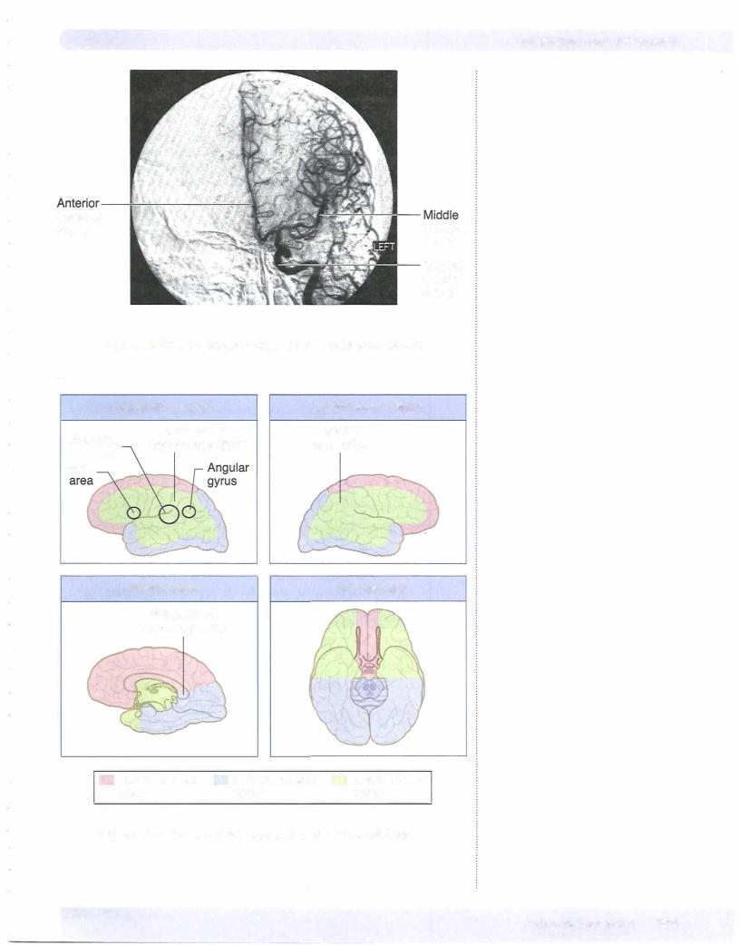
The cortex is supplied by the 2 internal carotid arteries and the 2 vertebral arter ies (Figures IV-10-6 and IV-10-7). On the base (or inferior surface) ofthe brain, branches of the internal carotid arteries and the basilar artery anastomose to form the circle of Willis. The anterior part of the circle lies in front of the optic chiasm, whereas the posterior part is situated just below the mammillary bodies. The circle ofWillis is formedby the terminal part ofthe internal carotid arteries; the proximalparts ofthe anterior and posterior cerebral arteries and the anterior and posterior communicating arteries. The middle, anterior, and posterior cere bralarteries,which arise from the circle ofWillis, supplyall ofthe cerebral cortex, basal ganglia, and diencephalon.
The internal carotid arteryarises from thebifurcation ofthe common carotidand enters the skull through the carotid canal. It enters the subarachnoid space and terminates by dividing into the anterior and middle cerebral arteries.
458 MEDICAL


Section IV • Neuroscience
|
The posterior cerebral artery is formed by the terminal bifurcation of the basilar |
|
|
artery. The posterior communicating artery arises near the termination ofthe in |
|
|
ternal carotid artery and passes posteriorly to join the posterior cerebral artery. |
|
|
The posterior communicating arteries complete the circle of Willis by joining the |
|
|
vertebrobasilar and carotid circulations. The posterior cerebral artery supplies |
|
|
the occipital and temporal cortex on the inferior and lateral surfaces of the hemi |
|
|
sphere, the occipital lobe and posterior 2/3 of the temporal lobe on the medial |
|
|
surface ofthe hemisphere, and the thalamus and subthalamic nucleus. |
|
|
Occlusion ofthe posterior cerebral artery results in a homonymous hemianopia |
|
|
of the contralateral visual field with macular sparing. |
|
Table IV-10-1. Cerebrovascular Disorders |
|
|
Disorder |
Types |
Key Concepts |
Cerebral infarcts |
Thrombotic |
Anemic/pale infarct; usually atherosclerotic complication |
|
Embolic |
Hemorrhagic/red infarct; from heart or atherosclerotic plaques; |
|
|
middle cerebral artery most vulnerable to emboli |
|
Hypotension |
''Watershed" areas and deep cortical layers most affected |
|
Hypertension |
Lacunar infarcts; basal ganglia, internal capsule, and pons most affected |
Hemorrhages |
Epidural hematoma |
Almost always traumatic |
|
|
Rupture of middle meningeal artery after skull fracture |
|
|
Lucid interval before loss of consciousness ("talk and die" syndrome) |
|
Subdural hematoma |
Usually caused by trauma |
|
|
Rupture of bridgingveins (drain brain to dural sinuses) |
|
Subarachnoid hemorrhage |
Ruptured berry aneurysm is most frequent cause |
|
|
Predisposing factors: Marfan syndrome, Ehlers-Dantos |
|
|
type 4, adult polycystic kidney disease, hypertension, smoking |
|
lntracerebral hemorrhage |
Common causes: hypertension, trauma, infarction |
462 MEDI CAL


Section IV • Neuroscience
Clinical Correlate
Lesion of the Frontal Eye Field
The frontal eye field lies in front of the motor cortex in Brodmann area 8. This cortical area is the center for contralateral horizontal gaze. A lesion here results in an inability to make voluntary eye movements toward the contralateral side. Because the activity ofthe intact frontal eye field in the opposite cortexwould also be unopposed after such a lesion, the
result is conjugate slow deviation ofthe eyes toward the side ofthe lesion.
If motor cortex is involved in the lesion, the patient may have a contralateral spastic paresis. The intact frontal
eye field in the opposite hemisphere deviates the eyes away from the paralyzed limbs.
Prefrontal cortex
The prefrontal cortex is located in front ofthe premotor area and represents about a quarter ofthe entire cerebral cortex in the human brain. This area is involved in organizing and planning the intellectual and emotional aspects ofbehavior, much as the adjacent premotor cortex is involved in planning its motor aspects.
Clinical Correlate
Lesions in the Prefrontal Area
Lesions in the prefrontal area produce what is called the frontal lobe syndrome. The patient cannot concentrate and is easily distracted; there is a general lack of initiative, foresight, and perspective. Another common aspect is apathy (i.e., severe emotional indifference). Apathy is usually associated with abulia, a slowing of intellectual faculties, slow speech, and decreased participation in social interactions. Prefrontal lesions also result in the emergence of infantile suckling or grasp reflexes that are suppressed in adults. In the suckling reflex, touching the cheek causes the head to turn toward the side ofthe stimulus as the mouth searches for a nipple to suckle. In the grasp reflex, touching the palm ofthe hand results in a reflex closing ofthe fingers, which allows an infant to grasp anything that touches the hand.
Clinical Correlate
Expressive Aphasia
Broca area is just anterior to the motor cortex region that provides upper motoneuron innervation of cranial nerve motor nuclei. This area in the left or dominant hemisphere is the center for motor speech and corresponds to
Brodmann areas 44 and 45. Damage to Broca area produces a motor, nonfluent, or expressive aphasia that reflects a difficulty in piecing together words to produce expressive speech. Patients with this lesion can understand written and spoken language but normally say almost nothing. When pressed on a question such as "What did you do today?" they might reply "Went town." The ability to write is usually also affected in a similar way (agraphia) in all aphasias, although the hand used for writing can be used normally in all other tasks. Patients are keenly aware of and frustrated by an expressive aphasia, because of their lack ofthe ability to verbalize their thoughts orally or in writing.
Broca area damage often extends posteriorly into the primary motor cortex and might be combined with a contralateral paralysis ofthe muscles ofthe lower face, resulting in a drooping of the corner ofthe mouth. Ifthe lesion is larger, the patient might have a spastic hemiparesis of the contralateral upper limb.
464 MEDICAL

Chapter 10 • Cerebral Cortex
Parietal Lobe
Primary somatosensory cortex
The parietal lobe begins just posterior to the central sulcus with the postcentral gyrus. The postcentral gyrus corresponds to Brodmann areas 3, l, and 2 and con tains primary somatosensorycortex. Likeprimarymotorcortex,thereis a similar somatotopic representation of the body here, with head, neck, upper limb, and trunk represented on the lateral aspect of the hemisphere, and pelvis and lower limb represented medially. These areas are concerned with discriminative touch, vibration, position sense, pain, and temperature. Lesions in somatosensory cor tex result in impairment of all somatic sensations on the opposite side of the body, including the face and scalp.
Posteriorparietal association cortex
Just posterior and ventral to the somatosensory areas is the posterior parietal as sociation cortex, including Brodmannareas 5 and 7.
Clinical Correlate
Lesions, usually in the dominant hemisphere and which include areas 5 and 7 ofthe posterior parietal association areas, often result in apraxia (also seen with lesions to the premotor cortex). Apraxia is a disruption ofthe patterning and execution of learned motor movements. This deficit seems to reflect a lack of understanding how to organize the performance of a pattern of movements (i.e., what should be done first, then next, etc.). The patient may be unable, for example, to draw a simple diagram (constructional apraxia) or describe how to get from his home to his work.
Another deficit, with lesions of areas 5 and 7 is astereognosia (inability to recognize objects by touch). There is no loss oftactile or proprioceptive
sensation; rather, it is the integration ofvisual and somatosensory information that is impaired. Both apraxia and astereognosia are more common after left hemisphere damage than in right hemisphere damage. The astereognosia is usually confined to the contralateral side ofthe body; in contrast, apraxia is usually bilateral. Apraxia is probably a result ofthe loss of input to the premotor cortex (area 6), which is involved in the actual organization of motor movements into a goal-directed pattern.
Wernicke area
The inferior part of the parietal lobe and adjacent part of the temporal lobe in the dominant (left) hemisphere, known as Wernicke area, are cortical regions that function in language comprehension. At a minimum, Wernicke area consists of area 22 in the temporal lobe but may also include areas 39 and 40 in the parietal lobe. Areas 39 (the angulargyrus) and 40 (the supramarginal gyrus) are regions of convergence ofvisual, auditory, and somatosensory information.
Note
Any blockage of the left middle cerebral artery that results in an aphasia (Broca Wernicke, conduction) or Gerstmann syndrome will also result in agraphia.
MEDICAL 465

Section IV • Neuroscience
Clinical Correlate
Receptive Aphasia
Lesions in area 22 in the temporal lobe and area 39 or 40 in the parietal lobe produce a fluent, receptive, or Wernicke aphasia. The patient with Wernicke aphasia cannot comprehend spoken language and may or may not be able to read (alexia) depending on the extent ofthe lesion. The deficit is characterized by fluent verbalization but lacks meaning. Patients are paraphasic, often misusing words as if speaking using a "word salad."
Patients with Wernicke aphasia are generally unaware oftheir deficit and show no distress as a result oftheir condition.
Gerstmann Syndrome
Ifthe lesion is confined to just the angular gyrus (area 39), the result is a loss of ability to comprehend written language (alexia) and to write it (agraphia), but spoken language may be understood. Alexia with agraphia in pure angular gyrus lesions is often seen with 3 other unique symptoms: acalculia (loss of the ability to perform simple arithmetic problems), finger agnosia (inability to recognize one's fingers), and right-left disorientation. This constellation
of deficits constitutes Gerstmann syndrome and underscores the role of this cortical area in the integration of how children begin to count, add, and subtract using their fingers.
Conduction Aphasia
There is a large fiberbundle connecting areas 22, 39, and 40 with Broca area in the frontal lobe, known as the superior longitudinal fasciculus (or the arcuate fasciculus). A lesion affecting this fiber bundle results in a conduction aphasia. In this patient, verbal output is fluent, but there are many paraphrases and word-finding pauses. Both verbal and visual language comprehension are also normal, but if asked to, the patient cannot repeat words or execute verbal commands by an examiner (such as "Count backwards beginning at 100") and also demonstrates poor object naming. This is an example ofa disconnect syndrome in which the deficit represents an inability to send information from one cortical area to another. As with an expressive aphasia, these patients
are aware ofthe deficit and are frustrated by their inability to execute a verbal command that they fully understand.
Transcortical Apraxia
Lesions to the corpus callosum caused by an infarct ofthe anterior cerebralartery may result in anothertype ofdisconnect syndrome known as a transcortical apraxia. As in other cases ofapraxia, there is no motor weakness, but the patient cannot execute a command to move the left arm. They understand the command, which is perceived in the Wernicke area ofthe left hemisphere, butthe callosal lesion disconnects the Wemicke area from the right primary motor cortex so that the command cannot be executed. The patient is still able to execute a command to move the rightarm because Wernicke area in the left hemisphere is able to communicate with the left primary motor cortexwithout using the corpus callosum.
(Continued)
466 MEDICAL

Chapter 10 • Cerebral Cortex
Clinical Correlate (Continued)
Asomatognosia
The integration ofvisual and somatosensory information is important for the formation ofthe "body image" and awareness ofthe body and its position in space. Widespread lesions in areas 7, 39, and 40 in the nondominant right parietal lobe may result in unawareness or neglect of the contralateral half of the body, known as asomatognosia. Although somatic sensation is intact, the patients ignore half oftheir body and may fail to dress, undress, or wash the affected (left) side. Patients will have no visual field deficits, so they can see, but deny the existence ofthings in the left visual field. Asking them to bisect a horizontal line produces a point well to the right of true center. If asked to draw a clock face from memory, they will draw only the numbers on the right side, ignoring those on the left. Patients may deny that the left arm or leg belongs to them when the affected limb is passively brought into their field ofvision. Patients may also deny their deficit, an anosognosia.
Occipital Lobe
The occipital lobe is essential for the reception and recognition of visual stimuli and contains primary visual and visual association cortex.
Visual cortex
The visual cortex is divided into striate (area 17) and extrastriate (areas 18 and 19). Area 17, also referred to as the primary visual cortex, lies on the medial por tion ofthe occipital lobe on either side ofthe calcarine sulcus. Its major thalamic input is from the lateral geniculate nucleus. Some input fibers are gathered in a thick bundle that can be visible on the cut surface of the gross brain, called the line of Gennari. The retinal surface (and therefore the visual field) is represented in an orderly manner on the surface of area 17, such that damage to a discrete part of area 17 willproduce a scotoma (i.e., a blind spot) in the corresponding portion of the visual field. A unilateral lesion inside area 17 results in a contra lateral homonymous hemianopsia with macular sparing, usually caused by an infarct of a branch of the posterior cerebral artery. The area of the macula of the retina containing the fovea is spared because of a dual blood supply from both the posterior and middle cerebral arteries. The actual cortical area serving the macula is represented in the most posterior part of the occipital lobe. Blows to the back of the head or a blockage in occipital branches of the middle cerebral artery that supply this area may produce loss of macular representation of the visual fields. Bilateral visual cortex lesions result in cortical blindness; the patient cannot see, but pupillary reflexes are intact.
Visual association cortex
Anterior to the primary visual or striate cortex are extensive areas of visual as sociation cortex. Visual association cortex is distributed throughout the entire occipital lobe and in the posterior parts of the parietal and temporal lobes. These regions receive fibers from the striate cortex and integrate complex visual input from both hemispheres. From the retina to the visual association cortex, informa tion about form and color, versus motion, depth and spatialinformation are pro cessed separately. Form and color information is processed by the parvocellular blob system. This "cone stream" originates mainly in the central part ofthe retina, relays through separate layers ofthe lateral geniculate, and projects to blob zones
M EDICAL 467

Section IV • Neuroscience
ofprimary visual cortex. Blob zones project to the inferior part ofthe temporal lobe in areas 20 and 21. Unilateral lesions here result in achromatopsia, a com plete loss ofcolorvision inthe contralateral hemifields. Patients see everything in shades ofgray. Additionally, these patients mayalso present with prosopagnosia, an inability to recognize faces.
Motion and depth are processed by the magnocellular system. This "rod stream'' originates in the peripheral part of the retina, relays through separate layers of the lateral geniculate, and projects to thick stripe zones ofprimary visual cortex. Striped areas project through the middle temporal lobe to the parietal lobe in areas 18 and 19. Lesions here result in a deficit in perceiving visual motion; visual fields, color vision, and reading are unaffected.
Clinical Correlate
Visual Agnosia
Damage to parts ofthe temporal lobes involving the cone stream produces a visual agnosia. Visual agnosia is the inability to recognize visual patterns (including objects) in the absence of a visual field deficit. For example, you
might show a patient with an object agnosia a pair of glasses, and the patient would describe them as 2 circles and a bar. Lesions in areas 20 and 21 of the temporal lobe that also include some destruction of adjacent occipital lobe in either hemisphere result in prosopagnosia, a specific inability to recognize faces. The patient can usually read and name objects. The deficiency is an
inability to form associations between faces and identities. On hearing the voice of the same person, the patient can immediately identify the person.
Alexia WithoutAgraphia
A principal "higher-order" deficit associated with occipital lobe damage is alexia without agraphia (or pure word blindness). The patients are unable to read at all and, curiously, often have a color anomia (inability to name colors). However, they are able to write. This is another example of a disconnect syndrome in which information from the occipital lobe is not available to the parietal or frontal lobes to either understand or express what has been seen.
(Recall that alexia with agraphia-inability to read or write-occurs with lesions encompassing the angular gyrus in the dominant parietal lobe. The cause ofthe syndrome is usually an infarction ofthe left posterior cerebral artery that affects not only the anterior part ofthe occipital lobe but the splenium ofthe corpus callosum. Involvement ofthe left occipital cortex results in a right homonymous hemianopsia with macular sparing. Involvement ofthe splenium ofthe corpus callosum prevents visual information from the intact right occipital cortexfrom reaching language comprehension centers in the left hemisphere. Patients can see words in the left visual field but do not understand what the words mean.
468 MEDICAL


Chapter 10 • Cerebral Cortex
Table IV-10-3. CNS Blood Supply and Stroke-related Deficits
System |
Primary Arteries |
Branches |
Supplies |
Deficits after Stroke |
Vertebrobasilar |
Vertebral arteries |
Anterior spinal artery |
Anterior two-thirds |
Dorsal columns spared; all |
(posterior |
|
|
of spinal cord |
else bilateral |
circulation) |
|
Posterior cerebellar |
Dorsolateral |
See Brain-Stem Lesions in |
|
|
|||
|
|
(PICA) |
medulla |
Chapter IV-5. |
|
Basilar artery |
Pontine arteries |
Base of pons |
|
|
|
Anterior inferior |
Inferior cerebellum, |
|
|
|
cerebellar artery (AICA) |
cerebellar nuclei |
|
|
|
Superior cerebellar artery |
Dorsal cerebellar |
|
|
|
|
hemispheres; |
|
|
|
|
superior cerebellar |
|
|
|
|
peduncle |
|
|
|
Labyrinthine artery |
Inner ear |
|
|
(sometimes arises from |
|
|
|
AICA) |
|
|
Posterior cerebral |
|
Midbrain, thalamus, |
|
arteries |
|
occipital lobe |
Internal carotid |
Ophthalmic: artery |
Central artery of retina |
Retina |
(anterior |
Posterior |
|
|
circulation) |
|
|
|
|
communicating |
|
|
|
artery |
|
|
|
Anterior c:erebral |
|
Primary motor and |
|
artery |
|
sensory cortex (leg/ |
|
|
|
foot) |
|
Anterior |
|
|
|
communicating |
|
|
|
artery |
|
|
|
Middle cerebral |
Outer cortical |
Lateral convexity of |
|
artery |
|
hemispheres |
|
|
Lenticulostriate |
Internal capsule, |
|
|
|
caudate, putamen, |
|
|
|
globus pallidus |
*If dominant hemisphere is affected (usually the left) tRight parietal lobe lesion
Contralateral hemianopia with macular sparing
Alexia without agraphia*
Blindness
Second most common aneurysm site (often with CN Ill palsy)
Contralateral spastic paralysis and anesthesia of lower limb
Frontal lobe abnormalities
Most common site of aneurysm
Contralateral spastic paralysis and anesthesia of upper limb/ face
Gaze palsy Aphasia*
Gerstmann syndrome""
Hemi inattention and neglect of contralateral bodyt
MEDICAL 471
Section IV • Neuroscience
Table IV-10-4. Key Features of Lobes
Lobes |
Important Regions |
Frontal |
Primary motor and premotor |
|
cortex |
|
Frontal eye fields |
|
Broca speech area* |
|
(Areas 44, 45) |
Deficit After Lesion
Contralateral spastic paresis (region depends on area of homunculus affected), premotor: apraxia
Eyes deviate to ipsilateral side
Broca aphasia (expressive, nonfluent aphasia): patient can understand written and spoken language, but speech and writing are slow and effortful; patients are aware oftheir problem; often associated with right arm weakness and right lower face weakness.
|
Prefrontal cortex |
Parietal |
Primary somatosensory cortex |
|
Superior parietal lobule |
|
Inferior parietal lobule |
|
(Angular gyrus; Area 39) |
Temporal |
Primary auditory cortex |
Frontal lobe syndrome: symptoms can include poor judgment, difficulty concentrating and problem solving, apathy, inappropriate social behavior
Contralateral hemihypesthesia (region depends on area of homunculus affected)
Contralateral astereognosis/apraxia
Gerstmann syndrome (if dominant hemisphere): right/left confusion, alexia, dyscalculia and dysgraphia, finger agnosia, contralateral hemianopia or lower quadrantanopia; unilateral neglect (nondominant)
Bilateral damage -+ deafness
Unilateral leads to slight hearing loss
|
Wernicke area* |
|
(Area 22) |
|
Hippocampus |
|
Amygdala |
|
Olfactory bulb, tract, primary |
|
cortex |
|
Meyer loop (visual radiations) |
Occipital |
Primary visual cortex |
Wernicke aphasia (receptive, fluent aphasia): patient cannot understand any form of language; speech is fast and fluent, but not comprehensible
Bilateral lesions lead to inability to consolidate short-term to long-term memory
Kliiver-Bucy syndrome: hyperphagia, hypersexuality, visual agnosia
Ipsilateral anosmia
Contralateral upper quadrantanopia ("pie in the sky")
Cortical blindness if bilateral; macular sparing hemianopia
*In the dominant hemisphere. Eighty percent of people are left-hemisphere dominant.
472 MEDICAL

Chapter 10 • Cerebral Cortex
ChapterSummary
•The external layer ofthe gray matter covering the surface ofthe cortex is characterized by numerous convolutions called gyri, separated by grooves called sulci. The cortex is divided into the frontal, parietal, occipital, and temporal lobes by several prominent sulci. Different areas ofthe cortex are concerned with sensory and motor functions. The frontal lobe contains the primary motor and premotor cortex, frontal eye field, and Broca speech area. The primary somatosensory and association cortex is found in the parietal
lobe. The temporal lobe contains the primary auditory cortex and Wernicke area. The primary visual cortex is at the posterior pole ofthe occipital lobe.
•The blood supply ofthe cortex is supplied by branches ofthe 2 internal carotid arteries and 2 vertebral arteries. On the ventral surface ofthe brain, the anterior cerebral and middle cerebral branches ofthe internal carotid arteries connect with the posterior cerebral artery, derived from the basilar arteryform the circle ofWillis. This circle ofvessels is completed by the anterior and posterior communicating arteries. The middle carotid artery mainly supplies the lateral surface ofthe frontal, parietal, and upper aspect ofthe temporal lobe. Deep branches also supply part ofthe basal ganglia and internal capsule. The anterior cerebral artery supplies the medial aspect ofthe frontal and parietal lobes. The entire occipital lobe, lower aspect oftemporal lobe, and the midbrain are supplied by the posterior cerebral artery.
•The homunculus of the motor and sensory cortex indicates that the upper limb and head are demonstrated on the lateral surface of the cortex. The pelvis and the lower limb are represented on the medial surface of the hemispheres. Therefore, the motor and sensory functions of the lower limb are supplied by the anterior cerebral artery while the motor and sensory functions ofthe upper limb and head are supplied by the middle cerebral artery.
•The primary language centers (Broca and Wernicke areas) are functionally located only in the dominant hemisphere, usually the left hemisphere. Both ofthese are supplied by the middle cerebral artery. Lesions ofthe Broca area result in motor or expressive aphasia (intact comprehension). Lesions of the Wernicke area produce receptive aphasia (lack of comprehension). Conduction aphasia results from a lesion ofthe arcuate fasciculus that connects the Broca and Wernicke areas.
•The internal capsule is a large mass ofwhite matter that conducts almost all tracts to and from the cerebral cortex. It is divided into an anterior limb, genu, and posterior limb. The anterior limb is supplied by the anterior cerebral artery, and the genu and posterior limb are supplied by the middle cerebral artery. The primary motor and sensory systems course through the posterior limb and genu.
MEDICAL 473
