


|
108 |
|
Digestive system |
|
|
|
|
|
|
|
|
|
|
|
|
|||||
|
|
|
|
|
|
Apparatus |
digestorius 26 |
Sublingual papilla. Caruncula sublingualis. A |
||||||||||||
1 |
|
1 |
|
DIGESTIVE SYSTEM. |
|
|||||||||||||||
|
|
|
|
(systema alimentarium) |
. |
|
|
|
|
small mucosal eminence on either side of the |
||||||||||
|
|
2 |
ORAL CAVITY. Cavitas oris. |
|
|
|
|
frenulum. It receives the opening of the sub- |
||||||||||||
2 |
|
|
|
|
|
mandibular |
duct and |
the major sublingual |
||||||||||||
|
3 |
Vestibule of mouth. Vestibulum oris. Space be- |
|
|||||||||||||||||
|
|
duct. B |
|
|
|
|
|
|
||||||||||||
|
|
|
|
tween the rows of teeth and the lips or cheeks. |
|
|
|
|
|
|
|
|||||||||
|
|
|
|
27 Sublingual fold. Plica sublingualis. Mucosal |
||||||||||||||||
3 |
|
|
|
B C |
|
|
|
|
|
|
||||||||||
|
|
|
|
|
|
|
|
|
|
fold overlying the sublingual gland and extend- |
||||||||||
|
|
4 |
Oral fissure. Rima oris. Mouth opening be- |
|
||||||||||||||||
|
|
|
ing posterolaterally from the sublingual papilla. |
|||||||||||||||||
|
|
|
|
tween the lips. A |
|
|
|
|
|
|||||||||||
4 |
|
|
|
|
|
|
|
|
B |
|
|
|
|
|
|
|||||
|
5 |
Lips. Labia oris. |
|
|
|
|
|
|
|
|
|
|
|
|
|
|||||
|
|
|
|
|
|
|
|
28 Parotid papilla. Papilla ductus parotidei. Small |
||||||||||||
|
|
|
|
Upper lip. Labium superius. A B C |
|
|
||||||||||||||
|
6 |
|
|
|||||||||||||||||
5 |
|
|
|
mucosal elevation at the opening of the parotid |
||||||||||||||||
|
|
7 Philtrum. Groove extending from nasal septum |
|
duct lateral to the second upper molar tooth. B |
||||||||||||||||
|
|
|
|
|||||||||||||||||
|
|
|
|
|
|
|
|
|
|
|
||||||||||
|
|
|
|
to upper lip. A |
|
|
|
|
|
|
29 |
Transverse palatine folds. Plicae palatinae |
||||||||
6 |
|
|
|
|
|
|
|
|
|
|||||||||||
8 |
Tuberculum. Small eminence on upper lip mark- |
|
transversae. |
Mucosal |
folds |
running |
trans- |
|||||||||||||
|
|
|
|
ing end of philtrum. A |
|
|
|
|
|
versely on the anterior part of the hard palate. |
||||||||||
7 |
|
|
|
|
|
|
|
|
D |
|
|
|
|
|
|
|||||
9 |
Lower lip. Labium inferius. A B C |
|
|
|
|
|
|
|
|
|
|
|||||||||
|
|
|
30 Incisive papilla. Papilla incisiva. Small mucosal |
|||||||||||||||||
|
10 |
Commissure of lips. Commissura |
labiorum. |
|||||||||||||||||
8 |
|
elevation over the incisive foramen at the ante- |
||||||||||||||||||
|
|
|
Transition of upper lip into lower lip at the |
|
||||||||||||||||
|
|
|
|
rior end of the palatine raphe. D |
|
|||||||||||||||
|
|
|
|
angle of the mouth. A B |
|
|
|
|
|
|||||||||||
|
|
|
|
|
|
|
30 a GLANDULAE ORIS. The glands of the mouth. |
|||||||||||||
9 |
11 |
Angle of mouth. Angulus oris. A |
|
|
|
|||||||||||||||
|
|
|
31 Small glands of the oral cavity. Glandulae |
|||||||||||||||||
|
12 |
Cheek. Bucca. |
Lateral |
wall of |
vestibule |
of |
||||||||||||||
|
|
salivariae minores. |
|
|
|
|
||||||||||||||
10 |
|
|
|
mouth. A |
|
|
|
|
|
|
|
|
|
|
|
|||||
|
|
|
|
|
|
|
|
|
32 |
Labial glands. |
Gll. |
labiales. |
Small |
salivary |
||||||
|
13 Buccal fat pad. Corpus adiposum buccae. [[Bi- |
|||||||||||||||||||
|
|
|
glands at the inner aspect of the lips. B |
|||||||||||||||||
|
|
|
|
chat]]. Encapsulated body of fat between the |
|
|||||||||||||||
11 |
|
|
|
33 |
Buccal glands. Gll. buccales. Small mucous |
|||||||||||||||
|
|
|
buccinator and masseter muscles. A |
|
|
|||||||||||||||
|
14 |
Oral cavity proper. Cavitas oris propria. True |
|
salivary glands |
at |
the inner aspect |
of the |
|||||||||||||
|
|
|||||||||||||||||||
12 |
|
cheeks. B |
|
|
|
|
|
|
||||||||||||
|
|
|
oral cavity enclosed anteriorly and laterally by |
|
|
|
|
|
|
|
||||||||||
|
|
|
34 Molar glands. Gll. molares. Salivary glands cor- |
|||||||||||||||||
|
|
|
|
the teeth and extending as far as the isthmus of |
||||||||||||||||
|
|
|
|
|
responding |
to |
the |
buccal |
glands situated |
|||||||||||
13 |
|
|
|
fauces (oropharyngeal isthmus). C |
|
|
|
|||||||||||||
|
|
|
|
|
|
beneath the mucosal at the level of the molar |
||||||||||||||
15 |
Palate. Palatum. Partition between |
oral |
and |
|
||||||||||||||||
|
|
teeth. B |
|
|
|
|
|
|
||||||||||||
|
|
|
|
nasal cavities. |
|
|
|
|
|
|
|
|
|
|
|
|
|
|||
14 |
|
|
|
|
|
|
|
|
|
35 Palatine glands. Gll. palatinae. Salivary glands |
||||||||||
16 |
Hard palate. Palatum durum. Hard, bony part |
|||||||||||||||||||
|
|
situated beneath the |
mucosa of the |
palate. |
||||||||||||||||
|
|
|
|
of the palate. C D |
|
|
|
|
|
|||||||||||
|
|
|
|
|
|
|
|
|
(Two large groups right and left of the midline.) |
|||||||||||
15 |
|
|
|
|
|
|
|
|
||||||||||||
17 |
Soft palate. Palatum molle (velum palatinum). |
|
||||||||||||||||||
|
D |
|
|
|
|
|
|
|||||||||||||
|
|
|
|
Soft, posterior part of the palate. C D |
|
36 |
Lingual glands. Gll. linguales. Numerous |
|||||||||||||
|
|
|
|
|
||||||||||||||||
16 |
18 |
Palatine raphe. Raphe palati. Median mucosal |
||||||||||||||||||
|
mucous, serous and mixed glands primarily in |
|||||||||||||||||||
|
|
|
|
ridge at the junction of the right and left bony |
|
the lateral and posterior areas of the tongue. B |
||||||||||||||
|
|
|
|
|
||||||||||||||||
|
|
|
|
palatal processes. D |
|
|
|
|
37 |
Anterior lingual glands. Gl. lingualis anterior |
||||||||||
17 |
|
|
|
|
|
|
|
|||||||||||||
19 |
Oral mucosa. Tunica mucosa oris. Mucous |
|||||||||||||||||||
|
|
[[gl. apicis linguae, Nuhn’s glands]]. Mixed |
||||||||||||||||||
|
|
|
|
membrane of oral cavity consisting of |
|
glands near the apex of the tongue providing |
||||||||||||||
18 |
|
|
|
|
||||||||||||||||
|
|
|
stratified, |
nonkeratinized |
|
squamous |
|
several drainage ducts on the undersurface of |
||||||||||||
|
|
|
|
epithelium throughout and underlying mixed |
|
the tongue. B |
|
|
|
|
|
|||||||||
|
|
|
|
|
|
|
|
|
|
|||||||||||
19 |
|
|
|
glands. |
|
|
|
|
|
|
|
|
|
|
|
|
|
|
||
|
20 Frenulum of upper lip. Frenulum labii super- |
|
|
|
|
|
|
|
|
|||||||||||
|
|
|
|
|
|
|
|
|
|
|||||||||||
|
|
|
|
ioris. Median mucosal fold between the gums |
|
|
|
|
|
|
|
|
||||||||
20 and upper lip. B
|
21 Frenulum of lower lip. Frenulum labii inferi- |
||
21 |
|
oris. Median mucosal fold between the gums |
|
|
|
and lower lip. B |
|
|
|
||
22 |
22 |
Gums. Gingivae. Mucous membrane united |
|
|
|
firmly with the teeth and jaw bones. B D |
|
|
23 |
Gingival (gum) margin. Margo gingivalis. B D |
|
23 |
|||
24 |
Gingival (interdental) papilla. Papilla ging- |
||
|
|
ivalis (interdentalis). B D |
|
24 |
|
||
25 |
Gingival sulcus. Sulcus gingivalis. Shallow fur- |
||
|
|
row between the gum margin and the tooth. Its |
|
25 |
|
deepening leads to cavity formation. See p. 113. |
|
|
|
A |
|

Digestive system 109
13 10
|
7 |
12 |
|
6 |
8 |
|
|
11 |
|
||
|
4 |
|
|
|
9 |
|
|
|
|
20 |
22 |
6
A Face, anterior view
37 |
|
|
|
32 |
||||||
|
|
|
|
|
|
|
|
|
|
|
36 |
|
|
|
|
|
|
|
|
|
33 |
|
|
|
|
|
|
|
|
|
||
28 |
|
|
|
|
|
|
||||
|
|
|
|
|
|
|
|
|
34 |
|
|
|
|
|
|
|
|
|
|
||
10 |
|
|
|
|
|
|||||
|
|
|
|
|||||||
|
|
|
|
|
|
|
|
|
|
|
|
|
|
|
27 |
|
|
|
|
|
|
26 |
|
|
|
|
|
|
|
|
|
23 |
|
|
|
|
|
|
|
|
|
||
|
|
|
|
|
|
|
|
|||
|
|
|
|
|
|
|
||||
|
9 |
|
|
|
|
|
|
24 |
||
|
|
|
|
|
|
|||||
|
|
|
|
|
|
|
|
|
||
3  21
21 22
22
B Mouth with tongue elevated
|
16 |
|
|
|
|
|
24 |
|
|
|
|
|
|
|
|
|
|||
|
|
|
|
|
|
30 |
|
||
|
|
|
|
|
29 |
|
|
23 |
|
|
|
|
|
|
|
|
|
||
|
|
|
|
|
|
|
|
|
|
6 |
14 |
|
|
|
|
|
|
18 |
|
16 |
16 |
|
|
|
|||||
|
|
|
|
|
|
||||
3 |
|
|
|
17 |
|
|
|
|
|
|
|
|
|
|
|
|
|
||
9 |
|
|
|
|
|
|
|
|
|
|
|
|
|
|
|
|
22 |
||
|
|
|
|
|
35 |
|
|
|
|
|
|
|
17 |
17 |
|
|
|
||
C |
Sagittal section of oral cavity |
D |
Palate, inferior view |
1
2
3
4
5
6
7
8
9
10
11
12
13
14
15
16
17
18
19
20
21
22
23
24
25
|
110 |
Digestive system |
|
|
|
|
|
|||
|
|
|
Major salivary glands. Glandulae salivariae |
|
21 Root of tooth. Radix dentis. Portion of the |
|||||
1 |
|
1 |
|
|||||||
|
||||||||||
|
|
|
majores. |
|
|
|
tooth covered by cementum. E |
|
||
2 |
|
2 |
Sublingual gland. Glandula sublingualis. Pre- |
|
22 |
Apex of root of tooth. Apex radicis dentis. E |
||||
|
|
|
dominantly mucous gland situated on the my- |
|
23 Clinical root. Radix clinica. Portion of the tooth |
|||||
|
|
|
|
lohyoid muscle diaphragma oris. It contains |
|
|
situated below the gum. C |
|
||
3 |
|
|
|
several drainage ducts. D |
|
|
|
|||
|
|
|
|
24 |
Occlusal (masticatory) surface. Facies oc- |
|||||
|
3 |
Major sublingual duct. Ductus sublingulis |
|
|||||||
|
|
|
|
clusalis (masticatoria) dentis. B E |
|
|||||
4 |
|
|
|
major. Main drainage duct of the sublingual |
|
25 Vestibular (facial) surface. Facies vestibularis |
||||
|
|
|
gland. It opens at the sublingual caruncle next |
|
||||||
|
|
|
|
|
(facilis) dentis. Tooth surface facing the vesti- |
|||||
|
|
|
|
to the submandibular duct. D |
|
|
||||
|
|
|
|
|
|
bule. D G |
|
|
||
|
|
|
|
|
|
|
|
|||
5 |
4 |
Minor sublingual ducts. Ductus sublinguales |
|
|
|
|
||||
|
25 a Buccal surface. Facies buccalis dentis. Tooth |
|||||||||
|
|
|
|
minores. About 40 small ducts that drain the |
|
|
surface facing the cheek. |
|
|
|
|
|
|
|
|
|
|
|
|||
6 |
|
|
|
sublingual gland and open along the sublingual |
|
|
|
|
||
|
|
|
|
25 b Labial surface. Facies |
labialis |
dentis. Tooth |
||||
|
|
|
fold and the sublingual caruncle. D |
|
||||||
|
|
5 |
Submandibular gland. Glandula submandibu- |
|
|
surface facing the lips. |
|
|
||
7 |
|
|
26 Lingual and palatine surfaces. Facies lingualis/ |
|||||||
|
|
|
laris. Predominantly serous salivary gland lo- |
|
||||||
|
|
|
|
cated almost entirely below the mylohyoid. D F |
|
|
palatalis dentis. Tooth |
surfaces facing the |
||
|
|
|
|
|
|
tongue and palate, respectively. A G |
||||
8 |
|
6 |
Submandibular |
duct. Ductus submandibu- |
|
|
||||
|
|
27 |
Approximal surface. |
Facies |
approximalis. |
|||||
|
|
|
laris. Duct that |
drains the submandibular |
|
|||||
|
|
|
|
|
|
Tooth surface facing the adjacent tooth. G |
||||
9 |
|
|
|
gland. It loops around the posterior margin of |
|
|
||||
|
|
|
|
28 |
Mesial surface. Facies mesialis. Vertical con- |
|||||
|
|
|
the mylohyoid accompanied by glandular |
|
||||||
|
|
|
|
tissue and opens at the sublingual caruncle. D |
|
|
tact surface of a tooth turned away from the last |
|||
10 |
|
7 |
Parotid gland. Glandula parotidea. Salivary |
|
|
molar. G |
|
|
||
|
|
29 |
Distal surface. Facies distalis. Vertical contact |
|||||||
|
|
|
|
gland located behind and on the mandibular |
|
|||||
11 |
|
|
|
ramus. F |
|
|
|
surface of a tooth facing away from the first in- |
||
|
8 |
Superficial part. Pars superficialis. The parts |
|
|
cisor. G |
|
|
|||
|
|
|
29 a Contingent area. Area contingens. Direct contact |
|||||||
|
|
|
|
of the parotid gland located superficial to the |
|
|||||
12 |
|
|
|
facial nerve. F |
|
|
|
surface of adjacent teeth. |
|
|
|
|
9 |
Deep part. Pars produnda. Parts of the parotid |
|
30 Cingulum. Ridge near the neck of a tooth con- |
|||||
13 |
|
|
|
gland located deep with respect to the facial |
|
|
necting both marginal crests at the lingual sur- |
|||
|
|
|
nerve. F |
|
|
|
face of incisor and canine teeth. A |
|||
|
|
|
|
|
|
|
||||
14 |
|
10 |
Accessory parotid gland. Glandula parotidea |
|
31 |
Marginal crest. Crista marginalis. Lateral |
||||
|
|
|
accessoria. Portion of the parotid gland located |
|
|
marginal ridge on the lingual surface of the in- |
||||
|
|
|
|
on the masseter muscle near the parotid ex- |
|
|
cisor and canine teeth which goes over into the |
|||
15 |
|
|
|
cretory duct. F |
|
|
|
cingulum at the neck region. A |
|
|
|
11 |
Parotid duct. Ductus parotideus. Excretory |
|
32 Incisal margin. Margo incisalis. Occlusal edge |
||||||
|
|
|
||||||||
16 |
|
|
|
duct of the parotid gland. It passes around the |
|
|
of incisor and canine teeth. A |
|
||
|
|
|
anterior margin of the masseter and opens near |
|
|
|
|
|
||
17 |
|
|
|
the second upper molar tooth. F |
|
|
|
|
|
|
|
12 |
TEETH. Dentes. A B C D E G |
|
|
|
|
|
|||
|
|
13 Crown of tooth. Corona dentis. Portion of the |
|
|
|
|
|
|||
18 |
|
|
|
tooth covered by enamel. E |
|
|
|
|
|
|
|
|
14 Cusp of tooth. Cuspis dentis [[tuberculum]]. 1− |
|
|
|
|
|
|||
19 |
|
|
|
5 protuberances |
on the occlusal surface of |
|
|
|
|
|
|
|
|
tooth (with the exception of the incisors). E |
|
|
|
|
|
||
|
|
|
|
|
|
|
|
|
||
20 |
|
15 Apex of cusp. Apex cuspidis. E |
|
|
|
|
|
|||
|
16 |
Dental tubercle. Tuberculum dentis. Distinct |
|
|
|
|
|
|||
21 |
|
|
|
eminence on the side of the crown, especially |
|
|
|
|
|
|
|
|
|
in canine and incisor teeth. A |
|
|
|
|
|
||
|
|
17 Transverse ridge. Crista transversalis. Trans- |
|
|
|
|
|
|||
22 |
|
|
|
verse connecting ridge between adjacent |
|
|
|
|
|
|
|
|
|
|
cusps. B |
|
|
|
|
|
|
23 |
|
18 Triangular ridge. Crista triangularis. Triangu- |
|
|
|
|
|
|||
|
|
|
lar connecting ridge between the cusps of the |
|
|
|
|
|
||
24 |
|
|
|
molars. B |
|
|
|
|
|
|
|
19 |
Clinical crown. Corona clinica. Portion of the |
|
|
|
|
|
|||
|
|
|
|
tooth projecting above the gum. C |
|
|
|
|
|
|
25 |
|
20 |
Neck of tooth. Cervix [[collum]] dentis. Portion |
|
|
|
|
|
||
|
|
|
|
of the tooth at the enamel-cementum border. E |
|
|
|
|
|
|
|
|
|
|
|
|
|
|
|
|
|

|
|
|
|
|
|
|
|
|
|
|
|
|
|
|
Digestive system |
111 |
|
|
|||||||||
|
|
|
|
|
|
|
|
|
|
|
|
|
|
|
|
|
|
|
|
|
|
|
|
|
|
|
|
32 |
|
|
|
|
|
|
|
|
|
|
|
|
19 |
|
|
|
|
|
|
1 |
|||||||
|
|
|
|
24 |
|
|
|
17 |
|
|
|
|
|
|
|
|
|
|
|
|
|
||||||
|
|
|
|
|
|
|
|
|
|
|
|
|
|
|
|
|
|
|
|
|
|
2 |
|||||
|
|
|
|
|
|
|
|
|
|
|
|
|
|
|
|
|
|
|
|
|
|
|
|||||
26 |
|
26 |
|
31 |
|
|
|
|
|
|
|
|
|
|
|
|
|
|
|
||||||||
|
|
|
|
|
|
|
|
|
|
|
|
|
|
|
|
|
|
|
|
||||||||
|
|
|
|
|
|
|
|
|
|
|
|
|
|
|
|
|
|
|
|
3 |
|||||||
|
|
|
|
|
16 |
|
|
|
|
18 |
|
|
|
23 |
|
|
|
|
|
|
|
|
|
|
|||
|
|
|
|
|
|
|
|
|
|
|
|
|
|
|
|
|
|
|
|
|
4 |
||||||
|
|
|
|
|
|
|
|
|
|
|
|
|
|
|
|
|
|
|
|
|
|
|
|||||
|
|
|
|
|
|
|
|
|
|
|
|
|
|
|
|
|
|
|
|
|
|
|
|
|
|
|
|
|
|
|
|
|
|
|
|
|
|
|
|
|
|
|
|
|
|
|
|
|
|
|
|
|
|||
30 |
|
|
|
|
|
|
|
|
|
|
|
|
|
|
|
|
|
|
|
|
|
|
|
|
5 |
||
|
|
|
|
|
|
|
|
|
|
|
|
|
|
|
|
|
|
|
|
|
|
|
|
|
|||
|
|
|
|
|
|
|
|
|
|
|
|
|
|
|
|
|
|
|
|
|
|
|
|
|
|
|
|
|
|
|
|
|
|
|
|
|
|
|
|
|
|
|
|
|
|
|
|
|
|
|
|
|
|
||
|
|
|
|
|
|
|
|
|
|
|
|
|
|
|
|
|
|
|
|
|
|
|
|
|
|
6 |
|
|
Incisor tooth and canine tooth, |
|
|
First and second molars, |
|
Incisor, sagittal |
|
|
|
|
|
||||||||||||||||
A |
|
B |
C |
|
|
|
|
|
|||||||||||||||||||
|
|
|
|
|
|||||||||||||||||||||||
|
lingual surface |
|
|
|
|
|
|
occlusal surface |
|
section |
|
|
|
|
|
|
7 |
||||||||||
|
|
|
|
|
|
|
|
|
|
|
|
|
|
|
|
|
|
|
|
|
|
|
|
|
|
|
|
|
|
|
|
|
|
|
|
|
|
|
|
|
|
|
|
|
|
|
|
|
|
|
|
|
|
||
|
|
|
|
|
|
|
|
|
|
|
|
|
|
|
|
14 |
15 |
|
|
|
8 |
||||||
|
|
|
|
|
|
|
|
|
|
|
|
|
|
|
|
|
|
|
|
|
|||||||
|
|
|
|
|
|
|
|
|
|
|
|
|
|
|
|
|
|
|
|
|
|
|
|
|
|
||
|
|
|
|
|
|
|
|
|
|
|
|
|
|
|
|
13 |
|
|
|
|
|
|
|
24 |
|
9 |
|
|
|
|
|
|
|
|
|
|
|
|
|
|
|
|
|
|
|
|
|
|
|
|
|
||||
|
|
|
|
|
|
|
|
|
|
|
|
|
|
|
|
|
|
|
|
|
|
|
|
|
|||
|
|
|
|
|
|
|
|
|
|
|
|
|
|
|
|
|
|
|
|
|
|
|
|||||
|
|
|
|
|
|
|
|
|
|
|
|
|
|
|
|
|
|
|
|
|
|
|
|
|
|||
|
|
|
|
|
|
|
|
|
|
|
|
|
|
|
|
|
|
|
|
|
|
10 |
|||||
|
|
|
|
|
|
|
|
|
|
|
|
|
|
|
|
|
|
|
|
|
|
|
|
|
|
||
25 |
|
|
|
|
|
|
|
|
|
|
|
|
|
|
|
|
|
|
|
|
|
|
|||||
|
|
|
|
|
|
|
|
|
|
|
20 |
|
|
|
|
|
|
|
|
|
|
|
|||||
|
|
|
|
|
|
|
|
|
|
|
|
|
|
|
|
|
|
|
|
|
|
||||||
|
|
|
|
|
|
|
|
|
|
|
|
|
|
|
|
|
|
|
|
|
|
|
|
|
|
||
|
|
|
|
|
|
|
|
|
|
|
4 |
|
|
|
|
|
|
|
|
|
|
11 |
|||||
|
|
|
|
|
|
|
|
|
|
|
|
|
|
|
|
|
|
|
|
|
|||||||
|
|
|
|
|
|
|
|
|
|
|
|
|
|
|
|
|
|
|
|
|
|
||||||
|
|
|
|
|
|
|
|
|
|
|
|
|
|
|
|
|
|
|
|
|
|
|
|
||||
|
|
|
|
|
|
|
|
|
|
|
|
|
|
|
|
|
|
|
|
|
|
|
|
|
|
||
3 |
|
|
|
|
|
|
|
2 |
|
|
|
|
21 |
|
|
|
|
|
|
|
|
|
12 |
||||
|
|
|
|
|
|
|
|
|
|
|
|
|
|
|
|
|
|
|
|
|
|
||||||
|
|
|
|
|
|
|
|
|
|
|
|
|
|
|
|
|
|
|
|
|
|
|
|
|
|
|
|
|
|
|
|
|
|
|
|
|
|
|
|
|
|
|
|
|
|
|
|
|
|
|
|
|
|
13 |
|
|
|
|
|
|
|
|
|
|
|
|
|
|
|
|
22 |
|
|
|
|
|
|
|
|
|
|
|
|
|
|
|
|
|
|
|
|
|
|
|
|
|
|
|
|
|
|
|
|
|
|
||||||
|
|
|
|
|
|
|
|
|
|
|
|
|
|
|
|
|
|
|
|
|
|
|
|
|
|||
|
|
|
|
|
|
|
|
|
|
|
|
|
|
|
|
|
|
|
|
|
14 |
||||||
|
Oral cavity, |
|
|
|
|
|
|
|
|
|
|
|
First lower molar |
|
|
|
|||||||||||
D |
|
|
|
|
|
|
|
|
|
|
E |
|
|
|
|
|
|||||||||||
|
|
|
|
|
|
|
|
|
|
|
|
|
|
|
|||||||||||||
|
medial view |
|
|
|
|
|
|
|
|
|
|
|
|
|
|
|
|
|
|
|
|
|
|
15 |
|||
|
|
|
|
|
|
|
|
|
|
|
|
|
|
|
|
|
|
|
|
|
|
|
|
|
|
|
|
65
|
|
|
|
|
|
|
|
|
|
|
|
|
|
16 |
|
|
|
|
|
|
|
|
|
|
|
|
|
|
|
|
|
|
|
|
|
|
|
|
|
|
|
|
|
17 |
|
|
|
10 |
|
|
|
|
|
|
|
|
|
|
|
|
|
|
|
|
|
|
|
|
|
|
|
|
|
|
11 |
|
|
8 |
|
|
|
18 |
|||||||
|
|
|
|
|
|
|
|
|
|
|
|
|||
|
|
|
|
|
|
|
|
|
|
|||||
|
|
|
|
|
|
|
|
|
||||||
|
|
|
7 |
|
|
|
|
|
|
|
||||
|
|
|
|
|
|
|
|
|
|
|||||
|
|
|
|
|
|
19 |
||||||||
|
|
|
|
|
|
|
|
|
|
|
|
|
|
|
|
|
|
|
|
9 |
|
|
|
|
|
|
|
|
|
|
|
|
|
|
|
|
|
|
|
|
|
|||
|
|
|
|
|
27 |
20 |
||||||||
|
|
|
|
|
|
|
|
|
|
|
|
|
||
|
|
|
28 |
29 |
|
|
||||||||
|
|
|
|
|
|
|||||||||
|
|
|
|
|
21 |
|||||||||
|
|
|
|
|
|
|
|
|
|
26 |
|
|
|
|
|
|
|
|
|
|
|
|
|
|
|
|
|
|
|
|
|
|
|
|
|
|
|
|||||||
|
|
|
5 |
|
|
|
|
|
|
|
29 |
|
|
22 |
|
|
|
|
|
|
|
|
|
|
|
|
|
|
|
|
|
|
|
|
|
|
|
|
|
|
|
|
|
|
|
|
|
|
|
|
|
|
|
|
|
29 |
|
|
23 |
|
|
|
|
|
|
|
|
|
|
|
|
|
|
|
|
|
|
|
|
|
|
|
|
|
|
|
|
|
24 |
F Salivary glands, lateral view |
|
|
|
|
|
|
G Teeth of lower jaw |
|
||||||
|
||||||||||||||
25
|
112 |
Digestive system |
|
|
|
||||
|
|
|
Pulp cavity of tooth. Cavitas dentis (pulparis). |
20 |
Incisor teeth. Dentes incisivi. Cutting teeth lo- |
||||
1 |
|
1 |
|||||||
|
|
|
Cavity in the dentin. Towards the root, it be- |
|
cated on both sides of the midline at the 1st and |
||||
2 |
|
|
|
comes continuous with the root canal. A |
|
|
2nd positions of the dental arch. D |
||
|
2 |
Pulp chamber in the crown. Cavitas coronae. |
21 |
Canine teeth. Dentes canini. Teeth located at |
|||||
3 |
|
|
|
Crown portion of the pulp cavity. A |
|
|
the 3rd position of the dental arch. D |
||
|
3 |
Root canal of tooth. Canalis radicis dentis. |
22 |
Premolar teeth. Dentes premolares. Teeth oc- |
|||||
|
|
|
|
Canal between the pulp cavity and the apical |
|
cupying the 4th and 5th positions of the dental |
|||
4 |
|
|
|
foramen. A |
|
|
|
arch. D |
|
|
4 |
Apical foramen of root of tooth. Foramen apicis |
23 |
Molar teeth. Dentes molares. Teeth located at |
|||||
|
|
||||||||
|
|
|
|
radicis dentalis. The opening of the root canal at |
|
the 6th, 7th and 8th positions of the dental arch. D |
|||
5 |
|
|
|
|
|||||
|
|
|
the apex of the root. A |
|
24 |
Wisdom tooth. Dens serotinus [molaris ter- |
|||
|
|
|
5 Pulp of tooth. Pulpa dentis. Contents of the |
|
tius]. Tooth located at the 8th position of the |
||||
6 |
|
|
|
||||||
|
|
|
pulp cavity consisting of loose, finely fibered |
|
dental arch. D |
||||
|
|
|
|
connective tissue, blood vessels and nerves. |
25 Deciduous (milk) teeth. Dentes decidui. |
||||
|
|
|
|
||||||
7 |
6 |
Crown pulp. Pulpa coronalis. Pulp within the |
|||||||
26 Permanent teeth. Dentes permanentes. Teeth |
|||||||||
|
|
|
|
crown portion of the pulp cavity. |
|
||||
|
|
|
|
|
|
that develop after the deciduous teeth. |
|||
|
7 |
Root pulp. Pulpa radicularis. Pulp within the |
|
||||||
8 |
|
||||||||
27 |
Diastema. Space between adjacent teeth. |
||||||||
|
|
|
root canal portion of the pulp cavity. |
|
|||||
|
8 |
Dental papilla. |
Papilla dentis. A mass |
of |
28 TONGUE. Lingua. D E |
||||
9 |
|||||||||
|
|
||||||||
|
|
|
mesenchyme present in the bell stage of tooth |
29 |
Body of tongue. Corpus linguae. Portion of the |
||||
|
|
|
|
||||||
|
|
|
|
development. B |
|
|
|
tongue situated between the apex and root. E |
|
10 |
|
|
|
|
|
|
|||
9 |
Dentine. Dentinum [[substantia eburnea]]. Pre- |
30 |
Root of tongue. Radix linguae. Anchoring re- |
||||||
|
|
|
|
dominant mass of a tooth consisting of inor- |
|
gion of the tongue at the mandible and hyoid |
|||
11 |
|
|
|
ganic and organic material (especially col- |
|
bone. Also the posterior, vertical segment of the |
|||
|
|
|
|
lagenous fibers). A C |
|
|
tongue. E |
||
|
|
|
|
|
|
||||
12 |
10 |
Enamel. Enamelum [[substantia adamantina]]. |
31 |
Dorsum of tongue. Dorsum linguae. E |
|||||
|
|
|
|
The extremely hard substance surrounding the |
32 |
Anterior, oral portion of tongue. Pars pre- |
|||
|
|
|
|
crown of the tooth like a mantle. A C |
|
||||
13 |
|
|
|
|
|
sulcalis (anterior). The part of the dorsum of |
|||
|
11 GOMPHOSIS. Type of fibrous joint in which a |
|
|||||||
|
|
the tongue situated anterior to the sulcus ter- |
|||||||
|
|
|
|||||||
|
|
|
|
conical process, e. g., a tooth, is inserted into a |
|
minalis. 115 B |
|||
14 |
|
|
|
|
|||||
|
|
|
socket, e. g., alveolus of the jaw (dentoalveolar |
33 |
Posterior, pharyngeal part of tongue. Pars |
||||
|
|
|
|
articulation). |
|
|
|||
|
|
|
|
|
|
|
postsulcalis (posterior). The vertical portion of |
||
|
12 |
Periodontium. Tissues that invest and support |
|
||||||
15 |
|
the dorsum of the tongue between the sulcus |
|||||||
|
|
|
|
the tooth within the alveolus. It consists of the |
|
terminalis and the epiglottis. 115 B |
|||
|
|
|
|
following parts: A |
|
34 Inferior surface of tongue. Facies inferior lin- |
|||
16 |
|
|
|
|
|||||
13 |
Periodontium |
protectoris [[gingiva]]. |
The |
||||||
|
guae. D E |
||||||||
|
|
|
|
outer part of the periodontium with the exter- |
35 |
Fimbriated fold. Plica fimbriata. Serrated fold |
|||
17 |
|
|
|
||||||
|
|
|
nal border epithelium. A |
|
|||||
|
|
|
|
|
lateral to the frenulum. It is a remnant of the in- |
||||
|
14 |
Periodontium insertionis. Portion of the peri- |
|
||||||
|
|
ferior tongue. D |
|||||||
18 |
|
||||||||
|
|
|
odonium touching the tooth. It consists of the |
36 Lateral margin of tongue bordering the teeth. |
|||||
|
|
|
|
inner border epithelium and the periodontal |
|||||
|
|
|
|
ligament. A |
|
|
|
Margo linguae. D |
|
19 |
|
|
|
|
|
|
|||
|
|
|
|
|
|
|
|||
15 |
Periodontal ligament. Desmodontium. All con- |
37 |
Apex of tongue. Apex linguae. D E |
||||||
|
|
|
|
nective tissue fibers which are anchored in the |
|
|
|||
20cementum and, with their vessels and nerves, extend partly into the gum and partly into the
alveolar wall. A
21
16Cementum. Substance similar to bone. It surrounds the tooth from the enamel border to the
22apex of the root and receives the fibers of the periodontal ligament. A
2317 Alveolar bone. Os alveolare. Bony wall of the alveolus. A
2418 Superior dental arch. Arcus dentalis superior. Curved row of teeth of the maxilla.
2519 Inferior dental arch. Arcus dentalis inferior. Curved row of teeth of the mandible.

Digestive system 113
|
|
|
|
|
|
|
|
|
|
|
|
|
|
|
|
|
|
|
|
|
|
|
|
|
|
|
|
|
|
|
1 |
|
|
|
|
|
|
|
|
|
|
|
|
|
|
|
|
|
|
|
|
|
|
|
|
|
|
|
|
|
|
|
2 |
|
|
|
|
|
10 |
108.25 |
|
|
|
|
|
|
|
|
|
|
|
|
|
|
3 |
||||||||||
12 |
|
2 |
|
|
|
|
|
|
|
|
14 |
|
|
|
|
|
|
|
|
|
|
|
|
4 |
|||||||
|
|
|
|
|
9 |
|
|
|
|
|
|
|
|
|
|
|
|
|
|
|
|
|
|||||||||
|
|
|
|
|
|
|
|
13 |
|
|
|
|
|
|
|
|
|
|
|
|
|
||||||||||
|
|
|
|
|
|
|
|
|
|
|
|
|
|
|
|
|
|
|
|
|
|
|
|
|
|
|
|
|
|
||
|
|
|
|
|
|
|
|
|
|
|
|
|
|
|
|
|
|
|
|
|
|
|
|
|
5 |
||||||
|
|
|
|
|
|
|
|
|
|
|
|
|
|
|
|
|
|
|
|
|
|
|
|
|
|
|
|
|
|
|
|
|
|
|
|
|
|
|
|
|
|
|
|
|
|
|
|
|
|
|
|
|
|
|
|
|
|
|
|
|
|
|
|
|
|
|
|
|
|
|
|
|
|
|
|
|
|
|
|
|
|
|
|
|
|
|
|
|
|
|
|
|
|
|
|
|
|
|
|
|
1 |
|
|
|
|
|
|
|
|
|
|
|
|
|
|
|
|
|
|
|
|
|
6 |
||||
15 |
|
|
|
|
|
|
|
|
|
|
|
|
|
|
|
|
17 |
|
|
|
8 |
|
|
|
7 |
||||||
|
|
|
|
|
|
|
|
|
|
|
|
|
|
|
|
|
|
|
|
|
|
||||||||||
|
|
|
|
|
|
|
|
|
|
|
|
|
|
|
|
|
|
|
|
|
|
|
|
|
|
||||||
|
|
|
|
|
|
|
|
|
|
|
|
|
|
|
|
|
|
|
|
|
|
|
|
|
|
|
8 |
||||
|
|
|
|
|
|
|
|
|
|
|
|
|
|
|
|
|
|
|
|
|
|
|
|
|
|
|
|||||
|
|
|
|
|
|
|
|
|
|
|
|
|
|
|
|
|
|
|
|
|
|
|
|
|
|
|
|
|
|
|
|
|
|
|
|
|
|
|
|
|
|
|
|
|
|
|
|
|
|
16 |
|
|
|
|
|
|
|
|
9 |
||||
|
|
|
|
|
|
|
|
|
|
|
|
|
|
|
|
|
|
|
|
|
|
|
|
|
|
||||||
|
|
|
|
|
|
|
|
|
|
|
|
|
|
|
|
|
|
|
|
|
|
|
|
|
|
|
|
|
|
|
|
|
|
|
|
|
3 |
|
|
|
|
|
|
|
|
|
|
|
|
|
B |
Tooth development |
|
|
|
||||||||
|
|
|
|
|
|
|
|
|
|
|
|
|
|
|
|
|
|
|
|
|
|
||||||||||
|
|
|
|
|
|
|
|
|
|
|
|
|
|
|
|
|
|
|
|
|
|
|
|
|
|
|
|
|
|
|
10 |
|
|
|
|
|
|
|
|
|
|
|
|
|
|
|
|
|
|
|
|
|
|
|
|
|
|
|
|
|
|
|
11 |
|
|
|
|
|
|
|
|
|
|
|
|
|
|
|
|
|
|
|
|
|
|
|
|
|
|
|
|
|
|
|
|
|
|
|
|
|
4 |
|
|
|
|
|
|
|
|
|
|
|
|
|
|
|
|
|
|
|
|
12 |
|||||
|
|
|
|
|
|
|
|
|
|
|
|
|
|
|
|
|
|
|
|
|
|
|
|
|
|
|
|
|
|
|
|
|
|
|
|
|
|
|
|
|
|
|
|
|
|
|
|
|
|
|
|
|
|
|
|
|
|
|
9 |
10 |
|
|
|
|
A |
Longitudinal section of tooth |
|
|
|
|
|
|
|
|
|
|
|
|
13 |
||||||||||||||||
|
|
|
|
|
|
|
|
|
|
|
|
|
|
|
|||||||||||||||||
|
|
|
|
|
|
|
|
|
|
|
|
|
|
||||||||||||||||||
|
|
|
|
|
|
|
|
|
|
|
|
|
|
|
|
|
|
|
|
|
|
|
|
|
|
|
|
|
|
|
|
|
|
|
|
|
20 |
|
|
|
|
|
|
|
|
|
|
|
|
|
|
|
|
|
|
|
14 |
||||||
|
|
|
|
|
|
|
|
|
|
|
|
|
|
|
|
|
|
|
|
|
|
|
|
|
|
|
|
|
|
|
|
|
|
|
|
|
|
|
|
|
|
|
|
|
|
|
|
|
|
|
|
|
|
|
|
|
Enamel-dentin border |
|
|
|
|
||
|
|
|
|
|
37 |
|
|
|
|
|
|
|
|
|
|
21 |
|
C |
|
|
|
15 |
|||||||||
|
|
|
|
|
|
|
|
|
|
|
|
|
|
|
|
|
|
|
|
|
|
|
|||||||||
|
|
|
|
|
|
|
|
|
|
|
|
|
|
|
|
|
|
|
|
|
|
|
|||||||||
35 |
|
|
|
|
|
|
|
|
|
|
|
|
|
|
|
|
|
31 |
|
|
|
16 |
|||||||||
|
|
|
|
|
|
|
|
|
|
|
|
|
|
|
|
|
|
|
|
|
|
|
|
|
|
|
|||||
|
|
|
34 |
|
|
|
|
|
|
|
|
|
|
|
|
|
|
|
|||||||||||||
36 |
|
|
|
|
|
|
|
|
|
|
|
|
|
|
|
|
|
|
|
|
17 |
||||||||||
|
|
|
|
|
|
|
|
|
|
|
|
|
|
|
|
|
|
|
|
|
|
|
|
|
|
|
|
|
|||
|
|
|
|
|
|
|
|
|
|
|
|
|
|
|
|
|
|
|
|
|
|
|
|
|
|
|
|
|
|
|
|
23 |
|
|
|
|
|
|
|
114.2 |
|
|
|
|
|
|
|
24 |
37 |
|
29 |
30 |
|
18 |
|||||||||
|
|
|
|
|
|
|
|
|
|
|
|
|
|||||||||||||||||||
|
|
|
|
|
|
|
|
|
|
|
|
|
|
|
|
|
|
|
|
|
|
||||||||||
|
|
|
|
|
|
|
|
|
|
|
|
|
|
|
|
|
|
|
|
|
|
||||||||||
|
|
|
|
|
|
|
|
|
|
|
|
|
|
|
|
|
|
|
|
|
|
|
|
|
|
||||||
|
|
|
|
|
|
|
|
|
|
|
|
|
|
|
|
|
|
|
|
|
34 |
|
|
|
|
19 |
|||||
|
|
|
|
|
|
|
|
|
|
|
|
|
|
|
|
|
|
|
|
|
|
|
|
|
|||||||
|
|
|
|
|
|
|
|
|
|
|
|
|
|
|
|
|
|
|
|
|
|
|
|
|
|
|
|
|
|||
22 |
|
|
|
|
|
|
|
|
|
|
|
|
|
|
|
|
|
|
|
21 |
|
|
|
|
|
|
20 |
||||
|
|
|
|
|
|
|
|
|
|
|
|
|
|
|
|
|
|
|
|
|
|
|
|
|
|
|
|||||
|
|
|
|
|
|
|
|
|
|
|
|
|
|
|
|
|
|
|
|
|
|
|
|
|
|
|
|||||
21 |
|
|
|
20 |
|
|
|
|
|
|
|
|
|
|
|
|
|
|
|
|
|
21 |
|||||||||
|
|
|
|
|
|
|
|
|
|
|
|
|
|
|
|
|
|
|
|
||||||||||||
|
|
|
|
|
|
|
|
|
|
|
|
|
|
|
|
|
|
|
|
|
|
||||||||||
|
|
|
|
|
|
|
|
|
|
|
|
|
|
|
|
|
|
|
|
|
|
|
|
||||||||
|
|
|
|
|
|
|
|
|
|
|
|
|
|
|
|
|
|
|
|
|
|
|
|
|
|
|
|
|
|
|
22 |
|
|
Mouth with tongue elevated |
|
|
|
|
|
|
|
|
Tongue, sagittal section |
|
|
|
|
||||||||||||||||
|
D |
|
|
|
|
|
|
|
E |
|
|
|
23 |
||||||||||||||||||
|
|
|
|
|
|
|
|
|
|
|
|
|
|
|
|
|
|
|
|
|
|
|
|
|
|
|
|
|
|
|
24 |
|
|
|
|
|
|
|
|
|
|
|
|
|
|
|
|
|
|
|
|
|
|
|
|
|
|
|
|
|
|
|
25 |
|
|
|
|
|
|
|
|
|
|
|
|
|
|
|
|
|
|
|
|
|
|
|
|
|
|
|
|
|
|
|
|

|
114 |
Digestive system |
|
|
|
|
|
|||
|
|
|
|
Mucous membrane of tongue. Tunica mucosa |
18 |
TONGUE MUSCULATURE. Musculi linguae (lin- |
||||
1 |
|
1 |
|
|||||||
|
|
|
|
linguae. C |
|
|
guales). The following eight tongue muscles are |
|||
2 |
|
2 |
|
Frenulum of tongue. Frenulum linguae. Mu- |
|
innervated by the hypoglossal nerve (XII). |
||||
|
|
19 |
M. genioglossus. o: Mental spine of mandible. |
|||||||
|
|
|
|
cosal fold extending from the floor of the |
||||||
|
|
|
|
|
mouth to the inferior side of the tongue. D; see |
|
i: Fan-shaped distribution within the tongue |
|||
3 |
|
|
|
|
also p. 113. D |
|
|
from the apex to the base. A: It pulls the tongue |
||
|
3 |
|
Lingual papillae. Papillae linguales. Collective |
|
forward or towards the |
chin. I: Hypoglossal |
||||
|
|
|
|
nerve. C D |
|
|
||||
4 |
|
|
|
|
term for the following five different types of |
|
|
|
||
|
|
|
|
20 |
M. hyoglossus. o: Body and greater horn of |
|||||
|
|
|
|
mucosal formations: A B |
|
|||||
|
|
4 |
Filiform papillae. Papillae filiformes. Fine, al- |
|
hyoid bone. i: Coming from below, it radiates |
|||||
|
|
|
into the lateral parts of the tongue and pene- |
|||||||
5 |
|
|
|
|
most threadlike epithelial elevations on a con- |
|
||||
|
|
|
|
|
trates up to the mucosa. A: It draws the base of |
|||||
|
|
|
|
|
nective tissue core. Their tips that are often |
|
||||
|
|
|
|
|
cleft. A |
|
|
the tongue backward and downward. I: Hypo- |
||
6 |
|
|
|
|
|
|
glossal nerve. D |
|
|
|
5 |
|
Conical papillae. Papillae conicae. Special |
|
|
|
|||||
|
|
21 M. chondroglossus. o: Lesser horn of hyoid. i: |
||||||||
|
|
|
|
|
form of filiform papilla. They are somewhat |
|||||
|
|
|
|
|
|
Same as hyoglossus. I: Hypoglossal nerve. D |
||||
7 |
|
|
|
|
larger, longer and exhibit conical apices which |
|
||||
|
|
|
|
22 |
M. styloglossus. o: Styloid process. i: Coming |
|||||
|
|
|
|
|
bend backwards. A |
|
||||
|
|
|
|
|
|
|||||
|
6 |
|
Fungiform papillae. Papillae |
fungiformes. |
|
from behind and above, it radiates to the lateral |
||||
8 |
|
|
||||||||
|
|
parts of the tongue and interweaves with the |
||||||||
|
|
|
|
Mushroom-like papillae which are not pointed |
|
|||||
|
|
|
|
|
at the tip, but terminate with a small plateau. A |
|
hyoglossus. A: It draws the tongue backward |
|||
9 |
|
|
|
|
|
and upward. I: Hypoglossal nerve. D |
|
|||
|
|
|
|
B |
|
|
|
|||
|
|
|
|
|
23 Superior longitudinal muscle of tongue. M. |
|||||
|
7 |
Vallate (circumvallate) papillae. Papillae val- |
||||||||
|
||||||||||
10 |
|
|
|
|
latae. 7−12 large papillae located in front of the |
|
longitudinalis superior. Longitudinal bundles of |
|||
|
|
|
|
|
muscle just below the mucosa that extend from |
|||||
|
|
|
|
sulcus terminalis. They are circular in cross- |
|
|||||
|
|
|
|
|
section and are surrounded by a moat, the wall |
|
the apex of tongue to the region of the hyoid |
|||
11 |
|
|
|
|
|
bone. I: Hypoglossal nerve. C |
|
|||
|
|
|
|
of which contains taste buds. A B |
|
|
|
|||
|
8 |
Lenticular papillae. Papilla lentiformes. Short |
24 |
Inferior longitudinal muscle of tongue. M. |
||||||
|
||||||||||
12 |
|
|
|
|
fungiform papillae. A |
|
|
longitudinalis inferior. |
Longitudinal |
fibrous |
|
|
|
|
|
|
system situated close to the inferior surface of |
||||
|
9 |
|
Foliate papilla. Papillae foliatae. Several paral- |
|
||||||
|
|
|
the tongue. It passes from the base to the apex |
|||||||
13 |
|
|
|
|
lel folds containing taste buds |
at the post- |
|
of the tongue. I: Hypoglossal nerve. C |
|
|
|
|
|
|
erolateral margin of the tongue. B D |
|
|
||||
|
|
|
|
|
25 Transverse muscle of tongue. M. transversus |
|||||
|
|
10 Median groove of tongue. Sulcus medianus |
||||||||
14 |
|
|
linguae. Transversely oriented muscle |
fibers |
||||||
|
|
|
|
linguae. Shallow, median longitudinal groove |
|
|||||
|
|
|
|
|
extending between the longitudinal system of |
|||||
|
|
|
|
|
situated above the lingual septum. B C |
|
fibers. o: Lingual septum. i: Mucous membrane |
|||
15 |
|
|
|
|
|
|||||
11 |
|
Sulcus terminalis [[”V” linguae]]. Bilateral |
|
along lateral margins of tongue. A: Extension of |
||||||
|
|
|
|
|
groove passing obliquely forward from the |
|
tongue together with the vertical muscle of the |
|||
|
|
|
|
|
|
|||||
16 |
|
|
|
|
foramen caecum. It lies behind the row of val- |
|
tongue. I: Hypoglossal nerve. C |
|
||
|
|
|
|
late papillae which run parallel to it. B |
26 |
Vertical muscle of tongue. M. verticalis lin- |
||||
|
|
|
|
|
||||||
|
12 |
|
Foramen caecum linguae. Pit situated at the |
|
guae. Vertical muscle fibers coursing from the |
|||||
17 |
|
|
||||||||
|
|
|
|
apex of the sulcus terminalis. It is the embryo- |
|
back of the tongue to the inferior surface. I: Hy- |
||||
|
|
|
|
|
logical remains of the thyroglossal duct. B |
|
poglossal nerve. C |
|
|
|
|
|
|
|
|
|
|
|
|||
18 13 Thyroglossal duct. Ductus thyroglossalis. Embryological connection between thyroid gland and tongue. At the site of the future fora-
19men caecum, it extends downward from the base of the tongue as an epithelial cone.
2014 Lingual tonsil. Tonsilla lingualis. Accumulation
|
of lymphatic tissue (lingual follicles) which is |
|
|
irregularly distributed over the root of the |
|
21 |
||
tongue. B D |
||
|
|
15 Lingual follicles. Folliculi linguales. Dome-
22shaped protrusions of the mucosa, 1−5 mm in diameter, caused by masses of lymphatic tissue beneath them. Each has a central crypt. A
2316 Lingual septum. Septum lingualis. Connective tissue plate with a special fibrous architecture
24located in the midsagittal plane. C
17 Lingual aponeurosis. Aponeurosis lingualis.
25Stout connective tissue framework of the tongue between the muscles and the mucosa. C

Digestive system 115
86
5
4
15
6
15
7
15
A Surface of tongue, enlarged
10
|
6 |
|
||
112.32 |
|
|
|
|
|
|
|
9 |
|
|
|
|||
|
|
|
|
7 |
|
|
|
||
|
11 |
|||
112.33 |
12 |
|||
14 |
|
|
|
|
|
|
|
|
|
22
B Dorsum of tongue, overview
118.27 
118.22
118.29 
1 23 10 17 25
26 
16 |
|
|
|
24 |
|
|
|
||
|
196.5
19
C Cross-section of tongue
9
116.2214
2
19
20
21
82.12
1
2
3
4
5
6
7
8
9
10
11
12
13
14
15
16
17
18
19
20
21
22
23
|
Tongue muscles |
24 |
D |
||
|
|
|
|
|
25 |
|
|
|

|
116 |
Digestive system |
|
|
|
|
|
|
|
|
|
|
||||
|
|
|
|
Pharynx. Passageway for air and food. 14− |
19 |
M. levator veli palatini. o: Petrous portion of |
||||||||||
1 |
|
1 |
|
|||||||||||||
|
|
|
|
16 cm long, it extends from the fornix to the |
|
temporal bone in front of the lower opening of |
||||||||||
|
|
|
|
|
beginning of the esophagus in front of the 6th |
|
the carotid canal. i: Palatine aponeurosis. It |
|||||||||
2 |
|
|
|
|
cervical vertebra. E |
|
|
passes through the pharyngeal wall above the |
||||||||
|
|
2 |
|
FAUCES. Space between soft palate and base of |
|
superior constrictor muscle and moves the soft |
||||||||||
|
|
|
|
palate backward and upward, thereby taking |
||||||||||||
3 |
|
|
|
|
tongue. E |
|
|
|||||||||
|
|
|
|
|
|
along the dorsomedial part of the auditory tube |
||||||||||
|
3 |
|
Isthmus of fauces. Isthmus faucium. Space be- |
|
||||||||||||
|
|
|
|
cartilage below the pharyngeal opening of the |
||||||||||||
|
|
|
|
|
tween right and left palatoglossal and |
pala- |
|
|||||||||
4 |
|
|
|
|
|
auditory tube. I: Vagus (X) nerve. C |
|
|||||||||
|
|
|
|
topharyngeal arches. |
|
|
|
|||||||||
|
|
|
|
|
20 M. tensor veli palatini. o: Spine of sphenoid, |
|||||||||||
|
|
4 |
|
Soft palate. Palatum molle (velum palatinum). |
||||||||||||
|
|
|
|
scaphoid fossa and anterior (lateral) lip of car- |
||||||||||||
|
|
|
|
|||||||||||||
5 |
|
|
|
|
The dorsal portion projects downward in front |
|
||||||||||
|
|
|
|
|
tilaginous |
auditory |
tube. |
i: After |
looping |
|||||||
|
|
|
|
|
of the posterior pharyngeal wall and assists in |
|
around the pterygoid hamulus, it radiates into |
|||||||||
|
|
|
|
|
swallowing by closing off like a valve the na- |
|
||||||||||
6 |
|
|
|
|
|
the palatine aponeurosis, stiffens the anterior |
||||||||||
|
|
|
|
sopharyngeal space from the oral cavity. A D E |
|
|||||||||||
|
|
|
|
|
(lateral) membranous wall of the auditory tube |
|||||||||||
|
5 |
|
Uvula. Uvula palatina. Conical process project- |
|
||||||||||||
|
|
|
and tenses the soft palate. I: Mandibular nerve. |
|||||||||||||
7 |
|
|
|
|
ing downward from the posterior margin of the |
|
C |
|
|
|
|
|
|
|
||
|
|
|
|
|
soft palate. A D E |
|
21 M. uvulae. o: Palatine aponeurosis. i: Connec- |
|||||||||
|
|
|
|
|
|
|||||||||||
8 |
6 |
|
Palatoglossal arch. Arcus palatoglossus. Mu- |
|
tive tissue of uvulae. I: Vagus nerve. C |
|
||||||||||
|
|
|
|
cosal fold overlying the palatoglossal muscle |
|
|
||||||||||
|
|
|
|
|
22 |
M. |
palatoglossus. |
o: |
Transversus |
linguae |
||||||
|
|
|
|
|
and extending from the palate to the tongue in |
|||||||||||
9 |
|
|
|
|
|
muscle. i: Palatine aponeurosis. A: Elevates the |
||||||||||
|
|
|
|
front of the tonsillar fossa. A |
|
|
||||||||||
|
|
|
|
|
|
base of the tongue, depresses the palate and |
||||||||||
|
7 |
|
Palatopharyngeal arch. Arcus palatopharyn- |
|
narrows the isthmus of fauces. I: Vagus nerve. D |
|||||||||||
10 |
|
|
|
|
geus. Mucosal fold overlying the palatopharyn- |
23 |
M. |
palatopharyngeus |
[[m. |
pharyngopalat- |
||||||
|
|
|
|
geal muscle and extending between the palate |
||||||||||||
|
|
|
|
|
|
inus]]. |
o: |
Palatine |
aponeurosis, pterygoid |
|||||||
|
|
|
|
|
and pharyngeal wall behind the tonsillar fossa. |
|
||||||||||
11 |
|
|
|
|
|
hamulus and medial plate of pterygoid process. |
||||||||||
|
|
|
|
A |
|
|
||||||||||
|
|
|
|
|
|
i: Lateral wall of pharynx and thyroid cartilage. |
||||||||||
|
8 |
|
Salpingopalatine fold. Plica salpingopalatina |
|
||||||||||||
|
|
|
A: It lowers the palate and constricts the isth- |
|||||||||||||
12 |
|
|
|
|
[[plica palatotubalis]]. Fold extending from the |
|
mus of fauces. I: Vagus nerve. D |
|
||||||||
|
|
|
|
|
anterior lip of the auditory tube to the soft pa- |
24 PHARYNGEAL CAVITY. Cavitas pharyngis. Space |
||||||||||
|
|
|
|
|
late in front of the tubal elevation. A |
|
||||||||||
13 |
|
|
|
|
|
|
enclosed by the pharyngeal walls. |
|
||||||||
9 |
|
Palatine tonsil. Tonsilla palatina. Tonsil sit- |
|
|
||||||||||||
|
25 Pharyngeal fornix. Fornix pharyngis. Roof of |
|||||||||||||||
|
|
|
|
|
uated between the palatoglossal and |
pala- |
||||||||||
|
|
|
|
|
|
the pharyngeal cavity beneath the sphenoid |
||||||||||
14 |
|
|
|
|
topharyngeal arches. A |
|
|
|||||||||
|
|
|
|
|
|
bone. E |
|
|
|
|
|
|
||||
|
10 |
Tonsillar pits. Fossulae tonsillae. Pit-like open- |
26 Nasopharynx. Pars nasalis pharyngis. The por- |
|||||||||||||
|
||||||||||||||||
15 |
|
|
|
|
ings of the tonsillar crypts visible on the sur- |
|||||||||||
|
|
|
|
|
tion of the pharyngeal cavity located behind |
|||||||||||
|
|
|
|
face. B |
|
|
||||||||||
|
|
|
|
|
|
|
the choanae. E |
|
|
|
|
|||||
|
11 |
Tonsillar crypts. Cryptae tonsillares. Epithelial |
27 |
|
|
|
Tonsilla |
|||||||||
16 |
Nasopharyngeal tonsil |
(adenoids). |
||||||||||||||
|
|
|
|
invaginations extending into the tonsil |
from |
|||||||||||
|
|
|
|
|
pharyngealis (adenoidea). It lies at the pharyn- |
|||||||||||
|
|
|
|
|
the tonsillar pits. B |
|
|
|||||||||
|
|
|
|
|
|
|
geal fornix. E |
|
|
|
|
|||||
17 |
12 |
|
Capsule of tonsil. Capsula tonsillaris. Fibrous |
|
|
|
|
|
||||||||
|
28 |
Tonsillar pits. Fossulae tonsillares. Openings |
||||||||||||||
|
|
|
|
|
capsule covering the organ. |
|
|
of crypts visible on surface of tonsil. See also |
||||||||
|
|
|
|
|
|
|
||||||||||
18 |
13 |
|
Triangular fold. Plica triangularis. Triangular |
|
p. 116.10. B |
|
|
|
|
|
||||||
|
|
|
|
|
fold emanating from the palatoglossal arch in |
29 |
Tonsillar |
crypts. Cryptae |
tonsillares. Epithelial |
|||||||
|
|
|
|
|
front of the tonsil. A |
|
||||||||||
19 |
|
|
|
|
|
|
invaginations emanating from the tonsillar |
|||||||||
14 |
|
Semilunar fold. Plica semilunaris. Arched fold |
|
|||||||||||||
|
|
pits. See also p. 116.11. B |
|
|
|
|||||||||||
|
|
|
|
|
between the palatoglossal and palatopharyn- |
|
|
|
|
|
|
|
|
|
||
20geal arches. It forms the upper boundary of the tonsillar fossa. A
21 |
15 Tonsillar fossa. Fossa tonsillaris. Recess for the |
|
tonsil bordered by the palatoglossal and pala- |
||
|
topharyngeal arches as well as by the triangular |
|
22 |
and semilunar folds. D |
|
|
16 Supratonsillar fossa. Fossa supratonsillaris. Su- |
|
|
||
23 |
perior portion of tonsillar fossa not occupied by |
|
the tonsil. A |
||
|
||
24 |
17 Muscles of palate and fauces. Musculi palati et |
|
faucium. |
18 Palatine aponeurosis. Aponeurosis palatina. It
25is formed primarily by the tendon of the tensor veli palatini muscle. C

Digestive system 117
8
4
14
5
6
16
13 9
7
ATonsillar fossa and soft palate
4
22 |
|
|
|
5 |
|
||||
|
|
|
23
15
D Muscles of tonsillar fossa
10;(28)
11;(29)
B Palatine tonsil, microscopic view
19
20
102.2
20 |
|
21 |
|
CNasal cavity from behind and muscles of soft palate
25
26 27
4
5
2 118.8
1
118.12
E Head, sagittal section
1
2
3
4
5
6
7
8
9
10
11
12
13
14
15
16
17
18
19
20
21
22
23
24
25

118 Digestive system
1 Pharyngeal bursa. [Bursa pharyngealis]. Blind 1 pouch in the roof of the pharynx; it is more frequently present in children, less often in
2adults. A
2Pharyngeal opening of auditory tube. Ostium pharyngeum tubae auditivae (auditoriae). Open-
3ing found in the nasopharynx. A
3Torus tubarius. Elevation produced by the dor-
4somedial cartilage of the auditory tube posterior to the tube opening. A
4 Salpingopharyngeal fold. Plica salpingopharyngea.
5 Mucosal fold overlying the salpingopharyngeal muscle and extending obliquely downward from
6the dorsomedial lip of the auditory tube cartilage. A
5 Torus levatorius. Elevation situated in front of
7the dorsomedial lip of the cartilage of the auditory tube and below the tube opening. It overlies
8the levator veli palatini muscle. A
|
6 |
Tubal tonsil. Tonsilla tubaria. Submucosal lym- |
|
9 |
|
phatic tissue near the opening of the auditory |
|
|
tube. |
||
|
|
||
|
7 |
Pharyngeal recess. Recessus pharyngeus |
|
10 |
|||
|
[[Rosenmüller’s]]. Lateral recess of the na- |
||
|
|
sopharyngeal space behind the auditory tube. A |
|
|
|
||
11 |
8 |
Oropharynx. Pars oralis pharyngis. The portion |
|
|
|
of the pharyngeal cavity located behind the oral |
cavity. See p. 117. E
129 Vallecula epiglottica. Fossa between the median and lateral glossoepiglottic folds. B
1310 Median glossoepiglottic fold. Plica glossoepiglottica mediana. Unpaired mucosal fold located in the median plane between the base of
14the tongue and the epiglottis. B
|
11 |
Lateral glossoepiglottic fold. Plica glos- |
|
15 |
|||
|
soepiglottica lateralis. Bilateral mucosal fold be- |
||
|
|
tween the base of the tongue and the epiglottis. B |
|
|
|
||
16 |
12 |
Laryngopharynx. Pars laryngea pharyngis. The |
|
|
|
portion of the pharyngeal cavity situated behind |
the larynx. See p. 117. E
1713 Piriform recess. Recessus piriformis. Channel between the aryepiglottic fold and the thyrohyoid
18membrane or thyroid cartilage. B
14Plica nervi laryngei. Mucosal fold in the piriform recess produced by the underlying internal
19branch of the superior laryngeal nerve and the superior laryngeal artery. B
2015 Pharyngobasilar fascia. Fascia pharyn-
gobasilaris. Membranous wall of the uppermost muscle free portion of the pharynx. It corre-
21 sponds to the thickened tela submucosa. C D E
|
16 |
Submucosa. Tela submucosa. Connective tissue |
|
22 |
|||
|
layer between the mucosa and muscularis. A |
||
|
17 |
Mucosa. Tunica mucosa. Pharyngeal mucous |
|
|
|||
23 |
|
membrane lined by stratified squamous or pseu- |
|
|
dostratified ciliated columnar (nasopharynx) |
||
|
|
||
|
|
epithelium. |
|
24 |
|
||
18 |
Pharyngeal glands. Gll. pharyngis. Small sub- |
||
|
|
epithelial mixed salivary glands. |
|
|
|
||
25 |
19 |
Muscularis of pharynx. Tunica muscularis |
|
|
|
pharyngis. Muscular layer of pharyngeal wall. A |
20Pharyngeal raphe. Raphe pharyngis. Connective tissue band between the right and left pharyngeal muscles, extending posteriorly in the midline. C
21Pterygomandibular raphe. Raphe pterygomandibularis. Tendinous seam between the buccinator muscle and the superior pharyngeal constrictor. It extends from the pterygoid hamulus to the mandible. D
22Superior pharyngeal constrictor muscle. M. constrictor pharyngis superior. Uppermost constrictor of the pharynx consisting of the four parts described below, which attach to the pharyngeal raphe. I: Pharyngeal plexus. C D
23Pterygopharyngeal part. Pars pterygopharynea. o: Medial plate of pterygoid process and pterygoid hamulus. D
24Buccopharyngeal part. Pars buccopharyngea. o: Pterygomandibular raphe. D
25Mylopharyngeal part. Pars mylopharyngea. o: Posterior end of mylohyoid line of mandible. D
26Glossopharyngeal part. Pars glossopharyngea.
o:Intrinsic muscles of tongue. D
27M. stylopharyngeus. o: Styloid process. i: Between superior and middle pharyngeal constrictors radiating into the pharyngeal wall, thyroid cartilage and epiglottis. I: Glossopharyngeal (IX) nerve. C
28M. salpingopharyngeus. o: Dorsomedial lip of auditory tube cartilage and part of longitudinal musculature of pharyngeal wall. i: Lateral wall of pharynx. A: Prevents backward displacement of the levator veli palatini muscle. I: Pharyngeal plexus. A
29Middle pharyngeal constrictor muscle. M. constrictor pharyngis medius. More centrally located pharyngeal constrictor arising from the hyoid bone. o: Pharyngeal raphe. I: Pharyngeal plexus. C
30Chondropharyngeal part. Pars chondropharyngea. o: Lesser horn of hyoid. D
31Ceratopharyngeal part. Pars ceratopharyngea.
o:Greater horn of hyoid. D
32Inferior pharyngeal constrictor muscle. M. constrictor pharyngis inferior. o: Larynx. I: Pharyngeal plexus. C D
33Thyropharyngeal part. Pars thyropharyngea. o: Oblique line of thyroid cartilage. D
34Cricopharyngeal part. Pars circopharyngea. o: Cricoid cartilage. D
34 a Buccopharyngeal fascia. Fascia buccopharyngealis. Continuation of the buccopharyngeal fascia into the loose tissue connecting the pharynx to the deep cervical fascia.
35 Peripharyngeal space. Spatium peripharyngeum. Connective tissue space associated with the pharynx.
36Retropharyngeal space. Spatium retropharyngeum. The portion of the peripharyngeal space between the pharynx and the prevertebral layer of the cervical fascia. A
37Lateropharyngeal space. Spatium lateropharyngeum. Portion of the peripharyngeal space lateral to the pharynx.

Digestive system 119
|
|
|
7 |
|
2 |
|
|
|
|
3 |
|
1 |
||
|
|
|
|
|
|
5 |
|
|
|
28
4
36
19
16 
A Pharyngeal opening of auditory tube
15 15
22
27 |
|
29 |
|
32
20
11
13
B
9
10
14
Base of tongue and laryngeal opening
15
23
24
21
25
31 30 26
32
33
34
C |
Pharyngeal muscles |
D |
Pharyngeal muscles |
|
from behind |
|
from right |
E Attachment of the
pharyngobasilar fascia
1
2
3
4
5
6
7
8
9
10
11
12
13
14
15
16
17
18
19
20
21
22
23
24
25

|
120 |
Digestive system |
|
|
|
|
|||
|
|
|
DIGESTIVE TRACT (ALIMENTARY CANAL). 20 |
Angular |
notch. Incisura angularis. Radio- |
||||
1 |
|
1 |
|||||||
|
|
|
[[Canalis alimentarius]]. |
|
|
graphically evident notch at the deepest point of |
|||
2 |
|
2 |
ESOPHAGUS. Oesophagus. 23−26 cm long pas- |
|
the lesser curvature. D |
||||
|
|
|
|
||||||
|
|
|
sage extending from below the cricoid cartilage |
21 |
Cardiac part of stomach. Pars cardiaca. The |
||||
|
|
|
|
at the level of C6 to the cardiac part of the |
|
portion of the stomach into which the |
|||
3 |
|
|
|
stomach. A B |
|
|
esophagus opens. D |
||
|
3 |
Cervical part. Pars cervicalis. The segment of |
22 |
Cardiac |
orifice. Ostium cardiacum (cardia). |
||||
|
|
||||||||
4 |
|
|
|
the esophagus situated in front of the cervical |
|
Opening of the esophagus into the cardiac |
|||
|
|
|
vertebral column (from C6−T1). A |
|
|
stomach. D |
|||
|
|
|
4 Thoracic part. Pars thoracica. The portion of |
23 Fundic stomach. Fundus gastricus (ventricu- |
|||||
|
|
|
|||||||
5 |
|
|
|
the esophagus extending from T1 to the pas- |
|||||
|
|
|
|
laris). Fundus (dome) of the stomach located |
|||||
|
|
|
|
sageway through the diaphragm (about T11). A |
|
beneath the diaphragm. D |
|||
|
|
|
5 Abdominal part. Pars abdominalis. The short |
|
|||||
6 |
|
|
23 a Fornix of stomach. Fornix gastricus (ventricu- |
||||||
|
|
|
esophageal segment between the diaphragm |
||||||
|
|
|
|
and the stomach. A |
|
|
laris). Forms the upper boundary of the lower |
||
|
|
|
|
|
|
||||
|
|
|
|
|
|
diaphragm. |
|||
7 |
|
|
|
|
|
||||
6 |
Tunica adventitia. Loose connective tissue in- |
|
|||||||
24 |
Cardiac notch. Incisura cardiaca. Acute angle |
||||||||
|
|
|
|
vestment which binds the esophagus to the |
|||||
|
|
|
|
|
between the esophagus and the stomach wall. D |
||||
8 |
|
|
|
surrounding tissues and permits its movement. |
|
||||
|
|
|
|
|
|
||||
|
|
|
C |
|
25 |
Body of stomach. Corpus gastricum (ventricu- |
|||
|
|
|
|
|
|||||
|
|
|
7 Tunica muscularis. Two layers of muscle, inner |
|
lare). Portion of stomach situated between the |
||||
9 |
|
|
|
||||||
|
|
|
circular and outer longitudinal, occupying the |
|
cardia/fundus and pylorus. D |
||||
|
|
|
|
esophageal wall and consisting of skeletal |
26 |
Gastric canal. Canalis gastricus (ventricularis). |
|||
|
|
|
|
||||||
10 |
|
|
|
muscle in the upper third, smooth muscle in |
|
Channel formed by the longitudinal mucosal |
|||
|
|
|
the lower third. C |
|
|
||||
|
|
|
|
|
|
folds along the lesser curvature of the stomach. |
|||
|
8 |
Cricoesophageal tendon. Tendo cricoe- |
|
D |
|
||||
11 |
|
|
|||||||
|
|
|
sophageus. Tendon that attaches the longitudi- |
27 Pyloric part of stomach. Pars pyloricum. Distal |
|||||
|
|
|
|
nal muscle fibers of the esophagus to the poste- |
|
segment of the stomach beginning with the |
|||
12 |
|
|
|
rior wall of the cricoid cartilage. B |
|
|
|||
|
|
|
|
|
angular notch and ending with the pylorus. D |
||||
9 |
Bronchoesophageal muscle. M. |
bronchoe- |
|
||||||
|
28 |
Pyloric antrum. Antrum pyloricum. Initial seg- |
|||||||
|
|||||||||
|
|
|
|
sophageus. Smooth muscle fibers from the left |
|||||
13 |
|
|
|
|
ment of the pyloric stomach beginning at the |
||||
|
|
|
main bronchus to the esophagus. B |
|
|
||||
|
|
|
|
|
angular notch. It may be temporarily closed off |
||||
|
10 |
Pleuroesophageal muscle. M. pleuroe- |
|
from the rest of the gastric lumen during the |
|||||
14 |
|
||||||||
|
|
|
sophageus. Smooth muscle tract between the |
|
passage of a peristaltic wave. D |
||||
|
|
|
|
esophagus and the left mediastinal pleura. B |
29 |
Pyloric canal. Canalis pyloricus. Lower termi- |
|||
|
|
|
|
||||||
15 |
11 |
Tela submucosa. Movable layer between the |
|||||||
|
nal segment of the stomach, about 2−3 cm long. |
||||||||
|
|
|
|
muscularis and mucosa consisting |
predomi- |
|
D |
|
|
16 |
|
|
|
nantly of collagenous connective tissue with |
|
|
|||
|
|
|
|
|
|
||||
|
|
|
blood vessels, nerves and glands. C |
|
|
|
|
||
12 Tunica mucosa. Mucous membrane lining of
17the esophagus consisting of stratified, nonkeratinized squamous epithelium, lamina propria (connective tissue) and muscularis mucosae. C
1813 Lamina muscularis mucosae. Distinct layer of smooth muscle between the submucosa and
19lamina propria. C
14Esophageal glands. Gll. esophageae. Discrete mixed glands scattered throughout the submu-
20 |
cosa. C |
|
15 STOMACH. Gaster (Ventriculus). It extends from
21the end of the esophagus to the pylorus. A D 16 Anterior surface of stomach. Paries anterior D
2217 Posterior surface of stomach. Paries posterior.
18 Greater curvature of stomach. Curvatura ga-
23strica (ventricularis) major. Large curvature of the stomach profile directed toward the left and downward. D
2419 Lesser curvature of stomach. Curvatura gastrica (ventricularis) minor. Small curvature of
25stomach directed toward the right and upward. D

Digestive system 121
1
2
3
|
|
|
|
|
|
|
|
|
|
|
|
|
|
|
|
3 |
|
|
|
|
|
|
|
|
|
|
|
|
|
|
|
12 |
|
|
|
|
|
|
|
|
|
|
|
|
|
|
|
|
|
|
4 |
||
|
2 |
|
|
|
13 |
|
|
|
|
|
|
|
|||||
|
|
|
|
|
|
|
|
|
|
|
|
||||||
|
|
|
|
|
|
|
|
|
|
|
|
|
|
||||
|
4 |
|
|
|
14 |
|
|
|
|
11 |
|
5 |
|||||
|
|
|
|
|
|
||||||||||||
|
|
|
|
|
|
|
|
|
|
|
|
|
|
|
|
||
|
|
|
|
|
|
|
|
|
|
|
|
|
|
|
6 |
||
|
|
|
|
|
|
|
|
|
|
|
|
|
|
|
|
|
|
|
|
|
|
|
|
|
|
|
|
|
|
|
|
|
|
|
|
|
|
|
|
|
|
|
|
|
|
|
|
|
|
|
|
|
|
|
5 |
|
|
|
|
|
|
|
|
|
7 |
|
7 |
||||
|
|
|
|
|
|
|
|
|
|
|
|
||||||
|
15 |
|
|
|
|
|
|
|
|
||||||||
|
|
|
|
|
|
|
|
|
|
|
|||||||
|
|
|
|
|
|
|
|
|
|
8 |
|||||||
|
|
|
|
|
|
|
|
|
|
|
|
|
|
|
|
|
|
|
|
|
|
|
|
|
|
|
|
|
|
|
|
6 |
|
|
|
|
|
|
|
|
|
|
|
|
|
|
|
|
|
|
|||
|
|
|
|
|
|
|
|
|
|
|
|
|
|
|
9 |
||
|
Esophagus and stomach |
|
Microscopic cross-section |
|
|
||||||||||||
|
|
|
|
||||||||||||||
A |
C |
|
10 |
||||||||||||||
|
|
|
|
|
|
|
|
|
of esoghagus |
|
|||||||
|
|
|
|
|
|
|
|
|
|
|
|||||||
|
|
|
|
|
|
|
|
|
|
|
|
|
|
|
|
|
|
|
|
|
|
|
|
|
8 |
|
|
|
|
|
|
|
|
|
11 |
|
|
|
|
|
|
|
|
|
|
|
|
|
|
|
|
|
|
|
|
|
|
|
|
|
|
|
|
|
|
|
|
||||
|
|
|
|
|
|
|
|
|
|
|
|
|
|
23 |
|
12 |
|
|
|
|
|
|
|
|
|
|
|
|
|
|
|
|
|
||
|
|
|
|
|
|
|
|
|
|
|
|
|
|
24 |
|
||
|
|
|
|
|
|
|
|
|
|
|
|
|
|
|
13 |
||
|
|
|
|
|
|
|
|
|
|
|
|
|
|
|
|
|
|
|
|
|
|
|
|
|
|
|
|
|
|
|
|
|
|
|
|
|
|
|
|
|
|
|
|
|
|
|
|
|
|
16 |
|
14 |
|
|
|
|
|
|
|
|
|
|
|
|
|
|
|
21 |
|
|
|
|
|
|
|
|
|
|
|
|
|
|
|
|
|
|
|
|
|
|
|
|
|
|
|
|
|
22 |
|
|
|
|
|
|
15 |
||
|
|
|
|
|
|
|
|
|
|
|
|
|
|
||||
|
|
|
|
|
|
|
|
|
|
|
|
|
|
|
|
|
|
|
|
|
|
|
|
|
|
|
|
|
|
|
|
|
|
|
16 |
|
|
|
|
|
|
|
|
|
|
|
|
|
|
122.12 |
|
|
|
|
|
|
|
|
|
|
|
|
|
|
|
|
|
|
|||
|
|
|
|
|
9 |
|
|
|
|
|
|
|
17 |
||||
|
|
|
|
|
10 |
|
|
|
19 |
|
26 |
|
|
|
|||
|
|
|
|
|
|
|
|
|
|
||||||||
|
|
|
|
|
|
|
|
|
25 |
|
18 |
||||||
|
|
|
|
|
|
|
|
|
|||||||||
|
|
|
|
|
|
|
|
|
|
|
|
|
|
|
|||
|
|
|
|
|
|
|
|
27 |
|
20 |
|
|
|
|
|||
|
|
|
|
|
|
|
|
|
|
|
|
|
|||||
|
|
|
|
|
|
|
|
|
|
|
|
19 |
|||||
|
|
|
|
|
|
|
|
|
|
|
|
|
|
|
|
|
|
|
|
|
|
|
|
|
|
|
|
|
|
|
|
|
|
|
|
|
|
|
|
|
|
|
|
|
|
|
|
|
|
|
|
|
20 |
|
|
|
|
|
|
|
|
28 |
|
|
|
|
|
|
|
|
|
|
|
|
|
|
|
|
|
|
|
|
|
|
|
|
|
||
|
|
|
|
|
|
|
|
|
|
|
|
|
18 |
21 |
|||
|
|
|
|
|
|
|
|
29 |
|
|
|
|
|
|
|||
|
|
|
|
|
|
|
|
|
|
|
|
|
|
|
|||
|
|
|
|
|
|
|
|
|
|
|
|
|
|
|
|||
|
|
|
|
|
|
|
|
|
|
|
|
|
|
|
|
||
|
|
|
|
|
|
|
|
|
|
|
|
|
|
16 |
|
22 |
|
|
|
|
|
|
|
|
|
|
|
|
|
|
|
|
|
||
|
|
|
|
|
|
|
|
|
|
|
|
|
|
|
|
|
|
|
|
|
|
|
|
|
|
|
|
|
|
|
|
|
|
|
23 |
|
|
|
|
|
|
|
|
|
|
|
|
|
|
|
|
|
|
|
|
|
|
|
|
|
|
|
|
|
|
|
|
|
|
|
|
|
|
|
|
|
|
|
|
|
|
|
|
|
|
|
|
|
24 |
B |
Esophagus from behind |
D Stromach from front right |
|
|
|||||||||||||
|
|
||||||||||||||||
25
|
122 |
Digestive system |
|
|
|
|
|
|||
|
|
|
Pylorus. Distal end of the stomach reinforced by |
20 |
SMALL INTESTINE. Intestinum tenue. It com- |
|||||
1 |
|
1 |
||||||||
|
|
|
a strong band of circular muscle. A |
|
prises the duodenum, jejunum and ileum. |
|||||
2 |
|
2 |
Pyloric opening. Ostium pyloricum. Lumen of |
21 |
Tunica serosa. Peritoneal covering containing |
|||||
|
|
|
the pylorus connecting the stomach with the |
|
simple squamous epithelium. F |
|||||
|
|
|
|
duodenum. A |
22 |
Tela subserosa. Connective tissue underlying |
||||
3 |
|
3 |
Serous membrane. Tunica serosa. Serous, peri- |
|
the serosa. F |
|
|
|
||
|
|
|
|
toneal covering containing simple squamous |
23 |
Tunica muscularis. Two principal muscle layers |
||||
4 |
|
|
|
epithelium. B |
|
of the intestinal wall. F |
|
|
||
|
4 |
Tela subserosa. Connective tissue component |
24 |
Longitudinal |
layer. |
Stratum longitudinale. |
||||
|
|
|||||||||
|
|
|
|
of the serosa underlying the epithelium. B |
|
Outer layer of longitudinally coursing muscle |
||||
|
|
|
|
|
||||||
5 |
|
|
|
|
||||||
5 |
Tunica muscularis. Muscular coat of the |
|
fibers. F |
|
|
|
||||
|
|
|
|
stomach comprised of three types of fibers |
25 |
Circular layer. Stratum circulare. Inner layer of |
||||
6 |
|
|
|
(longitudinal, circular, oblique). A B |
|
circularly coursing muscle fibers. F |
||||
|
6 |
Longitudinal layer. Stratum longitudinale. Ex- |
26 |
Tela sumucosa. Displaceable layer between the |
||||||
|
||||||||||
7 |
|
|
|
ternal layer of longitudinal muscle fibers sit- |
|
muscularis mucosae and the muscularis con- |
||||
|
|
|
uated primarily along the greater and lesser cur- |
|
sisting primarily of collagenous connective |
|||||
|
|
|
|
|
||||||
|
|
|
|
vatures. A B |
|
tissue bearing blood vessels and nerves. F |
||||
8 |
|
|
|
|
||||||
7 |
Circular layer. Stratum circulare. Middle layer |
27 |
Tunica mucosa. Intestinal mucous membrane |
|||||||
|
|
|
|
of circular muscle. A B |
|
consisting of simple columnar epithelium, con- |
||||
9 |
8 |
Pyloric sphincter. M. sphincter pyloricus. Thick |
|
nective tissue (lamina propria) and muscularis |
||||||
|
|
|
|
layer of circular muscle around the pylorus. A |
|
mucosae. |
|
|
|
|
|
|
|
|
28 |
Lamina muscularis mucosae. Layer of smooth |
|||||
10 |
9 |
Oblique fibers. Fibrae obliquae. Inner layer of |
||||||||
|
muscle between the lamina propria and submu- |
|||||||||
|
|
|
|
obliquely coursing fibers of the tunica muscu- |
|
|||||
|
|
|
|
|
cosa. Its action produces folds in the mucosa. F |
|||||
11 |
|
|
|
laris. A B |
|
|||||
|
|
|
29 |
Circular folds [Valves of |
Kerckring]. Plicae |
|||||
10 |
Tela submucosa. Displaceable layer between |
|||||||||
|
|
circulares. Permanent folds of mucosa and sub- |
||||||||
|
|
|
|
the muscularis mucosae and the muscularis; it |
|
|||||
12 |
|
|
|
|
mucosa spanning nearly 2/3 of the intestinal |
|||||
|
|
|
is composed primarily of collagenous connec- |
|
||||||
|
|
|
|
lumen and projecting up to 8 mm high, perpen- |
||||||
|
|
|
|
tive tissue bearing vessels and nerves. B |
|
|||||
|
|
|
|
|
dicular to the intestinal axis. E F |
|||||
13 |
11 |
Tunica mucosa. Gastric mucous membrane |
|
|||||||
30 |
Intestinal villi. Villi intestinales. Fingerlike pro- |
|||||||||
|
|
|
|
consisting of simple columnar epithelium, con- |
|
jections about 0.5−1.5 |
mm in length. F |
|||
|
|
|
|
|
||||||
14 |
|
|
|
nective tissue (lamina propria) and muscularis |
|
|||||
|
|
|
31 |
Intestinal glands. Gll. intestinales. Cryptlike |
||||||
|
|
|
mucosae. B |
|||||||
|
12 |
Gastric folds. Plicae gastricae. Mucosal folds |
|
glands. F |
|
|
|
|||
|
|
|
|
|
||||||
15 |
32 |
Solitary lymphatic follicles. Folliculi lym- |
||||||||
|
|
|
extending primarily in a longitudinal direction. |
|||||||
|
|
|
|
See p. 121 D |
|
phatici solitarii. Solitary lymphatic nodules in |
||||
|
|
|
|
|
the lamina propria (p. 405.42) F |
|||||
16 |
13 |
Lamina muscularis mucosae. Layer of smooth |
|
|||||||
33 |
Aggregated |
lymphatic |
follicles [[Peyer’s |
|||||||
|
|
|
|
muscle between the lamina propria and the |
||||||
|
|
|
|
|
patches]]. Folliculi lymphatici aggregati. Aggre- |
|||||
|
|
|
|
submucosa. Its contraction produces folds in the |
|
|||||
17 |
|
|
|
|
gation of several lymphatic nodules in the |
|||||
|
|
|
mucosa. B |
|
||||||
|
14 |
Gastric (mamillated) areas. Areae gastricae. |
|
ileum. |
|
|
|
|||
|
|
|
|
|
||||||
|
|
|
|
|
|
|||||
18Mamillated areas on the mucosal surface that are bounded by shallow grooves and have a
19diameter of 1−6 mm. B
15Villous folds. Plicae villosae. Epithelial ridges invisible to the naked eye between the openings
20 |
of glands. B C D |
16 Gastric pits (foveolae). Foveolae gastricae.
21Openings of the gastric glands between the villous folds. B C D
2217 Gastric glands proper. Glandula gastrica propria. Tubular glands in the fundus and body of the stomach made up of 4 cell types. B C
2318 Pyloric glands. Glandula pylorica. Mucoid glands in the pyloric part of the stomach con-
24sisting of two cell types. D
19 Lymphatic nodules (follicles). [[Folliculi lym-
25phatici gastrici]]. Small aggregations of lymphatic tissue in the lamina propria. C D
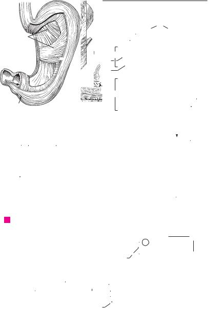
Digestive system 123
|
|
|
|
|
|
|
|
|
|
|
|
|
|
|
|
|
1 |
|
|
|
|
|
|
|
|
|
|
|
14 |
|
|
|
|
2 |
|
|
5 |
|
|
|
|
|
15 |
|
|
|
|
|
|
|
|
3 |
|
|
|
|
|
|
|
|
|
|
|
|
|
|
|
||||
|
|
|
|
|
9 |
16 |
|
|
|
|
|
|
|
|
|||
|
|
|
|
|
|
|
|
|
|
|
|
|
|
|
|||
|
|
|
|
|
|
|
11 |
|
|
|
|
|
|
4 |
|||
|
|
|
|
|
|
|
|
|
|
|
|
|
|
||||
|
|
|
|
|
|
|
|
|
|
|
|
|
|
|
|||
|
|
|
|
|
|
|
7 |
|
|
|
|
|
|
|
|
||
|
|
|
|
|
|
|
17 |
|
|
|
|
|
|
|
|
|
|
|
|
|
|
|
|
|
|
|
|
||||||||
|
8 |
|
|
|
|
|
|
|
|
5 |
|||||||
|
|
|
|
|
|
|
|
|
|
|
|
|
|
|
|||
|
|
|
|
|
|
13 |
|
|
10 |
|
|
|
|
|
|||
|
|
|
|
|
|
|
|
||||||||||
|
|
6 |
|
|
|
|
|
|
|
|
|
|
|
6 |
|||
2 |
|
9 |
|
|
|
9 |
|
|
|
|
|||||||
|
|
|
|
|
|
|
|
|
|||||||||
|
|
|
|
|
|
|
|
||||||||||
|
|
|
7 |
|
|
|
|
|
5 |
7 |
|
|
|
|
7 |
||
|
|
|
|
|
|
|
|
|
|
|
|
|
|
||||
|
|
|
|
|
|
|
|
|
|
|
|
|
4 |
|
|
||
|
|
|
|
|
|
|
|
|
|
|
|
|
|
|
|
|
|
|
1 |
|
|
|
|
|
|
|
|
|
6 |
8 |
|||||
|
|
|
|
|
|
|
|
|
|
|
|
||||||
|
|
|
|
|
|
|
|
|
|
|
|
|
|
3 |
|
|
|
|
|
|
|
|
|
|
|
|
|
||||||||
A |
Stomach musculature |
B |
Stomach wall, overview |
|
|
|||||||||
16 |
15 |
19 |
|
16 |
19 |
|||||||||
|
|
|
|
|
|
|||||||||
|
|
|
|
|
|
|||||||||
|
|
|
|
|
|
|
|
|
|
|
|
|
15 |
|
|
|
|
|
|
|
|
|
|
|
|
|
|
|
|
|
|
|
|
|
|
|
|
|
|
|
|
|
|
|
|
|
|
|
|
|
|
|
|
|
Mucosa of pylorus |
|
|
||
17 |
|
|
|
|
|
|
|
D |
|
|
||||
|
|
|
|
|
|
|
|
18 |
|
|
|
|
||
|
|
|
|
|
|
|
|
|
|
|
|
|
|
|
|
|
|
|
|
|
|
|
|
|
|
|
|
|
|
CTunica mucosa, fundus ventriculi
30
|
|
|
|
|
|
|
|
|
|
|
|
29 |
||
|
|
|
|
|
|
|
|
|
||||||
|
|
|
|
|
|
|
|
31 |
|
|||||
|
|
|
|
|
|
|
|
|
|
|
|
|
|
|
|
|
|
|
|
|
|
|
32 |
|
|||||
|
|
|
|
29 |
28 |
|
26 |
|||||||
|
|
|
|
|
||||||||||
|
|
|
|
|
|
|||||||||
|
|
|
|
|
|
|
25 |
|
|
|
|
|
|
|
|
|
|
|
|
|
|
|
|
|
|
|
|
|
|
|
|
|
|
|
|
24 |
|
|
|
|
|
|
|
|
|
|
|
|
|
|
|
22 |
|
|
|
|
|
|
|
29 |
|
|
21 |
|
|
|
|
|
|
|
||||
|
Intestinal canal |
|
|
|
|
|
|
|
|
|
Intestinal wall, histological section |
|||
E |
|
|
|
|
|
|
|
|
F |
|||||
9
10
11
12
13
14
15
16
17
18
19
20
21
22
23
24
25

|
124 |
Digestive system |
|
|
||||
|
|
|
|
DUODENUM. Initial segment of the small in- |
19 |
Ileocecal valve. Valva ileocaecalis (Valva ilealis). |
||
1 |
|
1 |
|
|||||
|
|
|
|
testine, about 25−30 mm long, between the py- |
|
Two-lipped valve at the entrance of the ileum |
||
2 |
|
|
|
|
lorus and the duodenojejunal flexure. A |
|
into the large intestine. D |
|
|
2 |
|
Superior part. Pars superior. Horizontal begin- |
19 a |
Papilla ilealis. B |
|||
|
|
|
|
|
ning part of the duodenum. A |
20 |
Opening of ileocecal valve. Ostium valvae |
|
3 |
|
3 |
|
Ampulla [[bulbus duodeni]]. Functional dilata- |
|
ilealis. Transverse, slitlike aperture of ileum at |
||
|
|
|
entrance into the large intestine in the cadaver. |
|||||
|
|
|
|
|
tion at the beginning of the duodenum. It is |
|
||
4 |
|
|
|
|
briefly visible in radiograms. A |
|
D |
|
|
4 |
|
Descending part. Pars descendens. Lateral, ver- |
21 |
Frenulum valvae ilealis. Fold formed by the |
|||
|
|
|
|
|
tical segment. A |
|
union of the lips of the ileocecal valve. B D |
|
|
|
|
|
|
|
|||
5 |
|
|
|
|
|
|||
5 |
|
Horizontal (inferior) part. Pars horizontalis |
21 a Ostium papillae ilealis. B |
|||||
|
|
|
|
|
(inferior). Horizontal segment below the head |
22 |
Vermiform appendix. Appendix vermiformis. |
|
6 |
|
|
|
|
of the pancreas. A |
|
Appendage of the cecum, usually 9 cm long, |
|
6 |
Ascending part. Pars ascendens. Segment of |
|
with abundant lymphatic tissue. B C D |
|||||
|
|
|||||||
|
23 |
Ostium appendicis vermiformis. Opening of |
||||||
7 |
|
|
|
|
duodenum lying to the left of the head of the |
|||
|
|
|
|
pancreas and ascending up to the duodenoje- |
|
vermiform appendix into the cecum. D |
||
|
|
|
|
|
junal flexure. A |
24 |
Aggregated lymphatic nodules of ver- |
|
8 |
7 |
|
Superior duodenal flexure. Flexura duodeni |
|
miform appendix. Folliculi lymphatici aggre- |
|||
|
|
|
|
|
superior. Flexure between the superior and |
|
gati appendicis vermiformis. Lymphatic tissue |
|
|
|
|
|
|
|
|||
9 |
|
|
|
|
horizontal parts of the duodenum medial to the |
|
within the wall of the appendix. |
|
|
|
|
|
gallbladder. A |
25 |
COLON. Portion of large intestine extending |
||
|
|
|
|
|
||||
|
|
|
8 Inferior duodenal flexure. Flexura duodeni in- |
|
from the ileocecal valve to the rectum. |
|||
10 |
|
|
|
|||||
|
|
26 |
Ascending colon. Colon ascendens. Segment of |
|||||
|
|
|
|
ferior. Flexure between the descending and |
||||
|
|
|
|
|
horizontal parts of the duodenum. A |
|
large intestine ascending retroperitoneally on |
|
11 |
9 |
|
Duodenojejunal flexure. Flexura duodenoje- |
|
the right side. C |
|||
|
27 |
Right colic flexure. Flexura coli dextra. Flexure |
||||||
|
|
|
|
|
junalis. Flexure between the duodenum and je- |
|||
|
|
|
|
|
||||
|
|
|
|
|
junum. A |
|
between the ascending and transverse colon. C |
|
12 |
|
|
|
|
|
|||
10 |
|
Suspensory muscle of duodenum. M. suspen- |
28 Transverse colon. Colon transversum. Trans- |
|||||
|
|
|||||||
|
|
|
verse segment of large intestine situated in- |
|||||
|
|
|
|
|
sorius duodeni. Bundle of smooth muscle which |
|
||
13 |
|
|
|
|
|
|||
|
|
|
|
|
traperitoneally between the right and left colic |
|||
|
|
|
|
attaches the duodenojejunal flexure to the dia- |
|
|||
|
|
|
|
|
phragm or to the celiac trunk. A |
|
flexures. C |
|
|
|
|
|
|
|
|||
14 |
11 |
|
Longitudinal folds of duodenum. Plicae longi- |
29 Left colic flexure. Flexura coli sinistra. Flexure |
||||
|
|
between the transverse and descending colon |
||||||
|
|
|
|
|
tudinales duodeni. Longitudinal mucosal folds |
|
||
|
|
|
|
|
|
below the left subphrenic space. In its vicinity |
||
15 |
|
|
|
|
on the left side of the posterior wall of the de- |
|
||
|
|
|
|
|
lies Cannon-Böhm’s ring, the boundary be- |
|||
|
|
|
|
scending part of the duodenum produced by the |
|
|||
|
|
|
|
|
pancreatic and bile ducts. A |
|
tween the cranial (vagus nerve) and sacral para- |
|
|
|
|
|
|
|
|||
16 |
|
|
|
|
|
sympathetics. C |
||
12 |
|
Greater duodenal papilla. Papilla duodeni |
|
|||||
|
30 Descending colon. Colon descendens. Segment |
|||||||
|
|
|
|
|
major. Elevation at the end of the longitudinal |
|||
|
|
|
|
|
|
of large intestine extending retroperitoneally on |
||
17 |
|
|
|
|
fold at the openings of the common bile duct |
|
||
|
|
|
|
|
the left side between the left colic flexure and |
|||
|
|
|
|
and pancreatic duct. A |
|
|||
|
|
|
|
|
|
sigmoid colon. C |
||
|
13 |
|
Lesser duodenal papilla. Papilla duodeni |
|
||||
18 |
|
31 |
Sigmoid colon. Colon sigmoideum. Portion of |
|||||
|
|
|
|
minor. Elevation located superior to the greater |
||||
|
|
|
|
|
duodenal papilla, usually at the opening of the |
|
colon lying intraperitoneally between the de- |
|
|
|
|
|
|
|
scending colon and the rectum. C |
||
19 |
|
|
|
|
accessory pancreatic duct. A |
|
||
|
|
|
|
32 |
Semilunar folds of colon. Plicae semilunares |
|||
14 |
|
Duodenal glands [[Brunner’s glands]]. Glan- |
||||||
|
|
|
coli. Crescentic contraction folds between two |
|||||
20 |
|
|
|
|
dulae duodenales. Mucous glands located pre- |
|
haustra comprising all layers of the intestinal |
|
|
|
|
|
dominantly in the submucosa of the duodenum. |
|
|||
|
|
|
|
|
|
wall. C D |
||
|
15 |
JEJUNUM. Middle segment of the small in- |
|
|||||
|
33 |
Haustra (sacculationes) coli. Outpocketings |
||||||
21 |
||||||||
|
|
|
|
testine. It extends about 2.5 meters from the |
||||
|
|
|
|
|
between two semilunar folds. C D |
|||
|
|
|
|
|
duodenojejunal flexure. A C |
|
||
|
|
|
|
|
34 |
Appendices epiploicae (omentales). Serosa- |
||
22 |
16 |
|
ILEUM. Terminal segment of small intestine, |
|||||
|
|
covered appendages containing adipose tissue, |
||||||
|
|
|
|
|
about 3.5 mm long. C |
|
located on the free and omental tenia. C |
|
|
|
|
|
|
|
|||
2317 LARGE INTESTINE. Intestinum crassum. About 1.5−1.8 m long; it extends from the cecum to the anus and consists of tenia, haustra and ap-
24pendices epiploicae.
18 CECUM. Caecum. Initial portion of the large in-
25testine, ca. 7 cm long, located below the opening of the ileum. C D

|
|
|
|
|
|
|
|
|
|
|
|
Digestive system 125 |
|
|||||||
|
246.21 |
216.28 |
|
|
|
|
|
|
|
|
|
|
|
|
||||||
|
|
|
|
|
|
|
|
|
|
|
|
1 |
||||||||
|
|
|
|
|
|
|
|
|
|
|
|
|
|
|
|
|
|
|
|
|
|
|
|
|
|
|
|
|
|
|
|
|
21a 19a |
|
2 |
||||||
|
|
|
|
|
|
|
|
|
|
|
|
|
||||||||
|
248.1 |
|
|
|
|
|
|
|
|
|
3 |
|||||||||
|
|
|
|
|
|
|
|
|
|
|
|
|
|
|
|
|
||||
|
|
|
|
|
|
|
|
|
|
|
|
|
|
|
|
|
||||
|
|
|
|
|
|
|
|
|
|
|
|
|
|
|
|
|
||||
|
7 |
|
|
|
|
|
|
|
|
|
|
|
|
|
|
|
|
|
4 |
|
|
3 |
|
|
|
|
|
|
|
|
|
|
|
|
|
|
|
||||
|
2 |
|
|
|
|
10 |
|
|
|
|
|
21 |
|
|
||||||
|
|
|
|
|
|
|
|
|||||||||||||
|
|
|
|
|
|
|
||||||||||||||
|
|
|
|
|
|
|
|
|
|
|
|
|
|
|
|
|
|
|
5 |
|
|
|
|
|
|
|
|
|
|
|
9 |
|
|
|
|
|
|
|
|
|
|
|
|
|
|
|
|
|
|
|
|
128.1 |
|
|
|
|
|
|
|
|
|
|
|
|
|
|
|
|
|
|
|
|
|
|
|
|
|
|
|
|
|
|
|
|
13 |
|
|
|
|
|
|
|
|
|
|
|
|
|
|
|
|
|
6 |
|
|
|
|
|
|
|
|
|
|
|
|
|
|
|
|
|
|
|
|||
|
|
|
|
|
|
|
|
|
|
|
|
|
|
|
|
|
|
|
||
4 |
11 |
|
|
|
|
|
6 |
|
|
|
|
|
|
|
|
|
|
|
|
|
|
|
|
|
|
|
|
|
|
|
|
|
|||||||||
|
|
|
|
|
|
|
|
|
|
|
7 |
|||||||||
|
|
|
|
|
|
|
|
|
|
|
|
|
|
|
|
|||||
|
12 |
|
|
|
|
|
|
|
|
|
|
|
|
|
|
|
|
|
|
|
|
|
|
|
|
|
|
|
|
|
|
|
|
|
|
|
|
|
|
|
|
|
|
|
|
|
|
|
|
|
|
|
15 |
|
|
|
|
|
|
|
|
|
|
|
|
|
|
|
|
|
|
|
|
|
|
|
|
|
|
|
|
8 |
|
|
5 |
|
|
|
|
|
|
|
|
|
|
|
|
|
||||||
|
|
|
|
|
|
|
|
|
|
|
|
|
|
|
||||||
|
|
|
|
|
|
|
|
|
|
|
|
|
|
|
||||||
|
8 |
|
|
|
|
|
|
|
|
|
|
|
|
|
22 |
|
|
|
9 |
|
|
|
|
|
|
|
|
|
|
|
|
|
|
|
|
|
|
|
|
|
|
|
|
|
|
|
|
|
|
|
|
|
|
|
|
|
|
|
|
|
|
|
APortal vein, inferior vena cava, aorta and duodenum
27
28
15
26
16
18
22 
31
CSmall and large intestines, anterior view
B Cecum in the living body
29
28
32
30 |
32 |
|
||
|
|
|
33 |
19 |
|
|
|
|
|
|
|
|
|
|
|
|
32 |
21 |
|
|
|
34 |
|
|
|
|
|
|
18 |
22
D Cecum in the cadaver
10
11
12
13
14
15
16
19
17
20
18
2319
20
21
22
23
24
25

|
126 |
Digestive system |
|
|
|
|
|||
|
|
|
Tunica muscularis. Bilayered muscle wall of |
21 |
M. rectococcygeus. Thin plate of smooth |
||||
1 |
|
1 |
|||||||
|
|
|
colon. B |
|
|
|
muscle extending from the 2nd to 3rd coccygeal |
||
2 |
|
2 |
Longitudinal layer. |
Stratum |
longitudinale. |
|
vertebrae to the rectum. C |
||
|
22 |
M. rectourethralis. Smooth muscle fibers ex- |
|||||||
|
|
|
Distinct outer layer of longitudinally oriented |
||||||
|
|
|
|
muscle of variable thickness. B |
|
|
tending from the membranous part of the |
||
3 |
|
3 |
Taeniae coli. About |
1 cm wide, thickened |
|
urethra to the rectum. C |
|||
|
23 |
Transverse rectal folds. Plicae transversae |
|||||||
|
|
|
|
bands of longitudinal muscle fibers. B |
|||||
4 |
|
4 |
Taenia mesocolica. Tenia located at the attach- |
|
recti. Usually three lateral transverse folds. The |
||||
|
|
middle is the largest (Kohlrausch) and projects |
|||||||
|
|
|
ment of the mesocolon in the posteromedial |
|
|||||
|
|
|
|
|
about 6 mm above the anus from the right side, |
||||
|
|
|
|
part of the ascending and descending colon. A |
|
||||
|
|
|
|
|
|||||
5 |
|
|
|
|
the others from the left. C |
||||
5 |
Taenia omentalis. Tenia of transverse colon lo- |
|
|||||||
24 |
ANAL CANAL. Canalis analis. Terminal segment |
||||||||
|
|
|
|
cated at the attachment of the greater omentum |
|||||
|
|
|
|
|
of the digestive tube beginning with the anal |
||||
6 |
|
|
|
in the posteromedial part of the ascending and |
|
||||
|
|
|
|
columns. D |
|||||
|
|
|
descending colon. A |
|
|
|
|||
|
6 |
Taenia libera. Free tenia located between the |
25 |
Anal columns. Columnae anales. Six to ten |
|||||
7 |
|||||||||
|
longitudinal folds provided with abundant |
||||||||
|
|
|
tenia mesocolica and tenia omentalis. A |
|
|||||
|
|
|
|
|
venous plexuses. D |
||||
|
7 |
Circular layer. Stratum circulare. Inner layer of |
|
||||||
8 |
26 |
Anal sinuses. Sinus anales. Recesses between |
|||||||
|
|
|
circularly coursing muscle fibers of the colon. B |
||||||
|
|
|
8 Tela submucosa. Displaceable layer between |
|
the anal columns. D |
||||
9 |
|
|
27 |
Anal valves. Valvulae anales. Small transverse |
|||||
|
|
|
the muscularis mucosa and the muscularis, con- |
||||||
|
|
|
|
folds bordering the anal sinuses distally. D |
|||||
|
|
|
|
sisting mainly of collagenous connective tissue |
|
||||
|
|
|
|
28 |
Anorectal line. Linea anorectalis. Upper border |
||||
10 |
|
|
|
containing nerves and blood vessels. B |
|||||
9 |
Tunica mucosa. Villus-free mucous coat of the |
|
of anal canal formed by the puborectal muscle at |
||||||
|
|
|
|
colon comprised of |
simple, goblet-cell rich, |
|
the level of the levator sling just above the anal |
||
11 |
|
|
|
|
columns. D |
||||
|
|
|
columnar epithelium, connective tissue and |
|
|||||
|
|
|
29 |
Anal pecten. [[Pecten analis]]. A lighter stripe |
|||||
|
|
|
|
lamina muscularis mucosae. B |
|
||||
|
|
|
|
|
|||||
12 |
10 |
Lamina muscularis mucosae. Layer of smooth |
|
between the anal valves and the anocutaneous |
|||||
|
line. It is firmly connected with the underlying |
||||||||
|
|
|
|
muscle cells between the lamina propria and |
|
tissues by fibrous longitudinal muscles. D |
|||
|
|
|
|
|
|||||
13 |
|
|
|
submucosa. Its contraction produces a wrin- |
|
||||
|
|
|
30 |
Anocutaneous line. [[Linea anocutanea]]. Lower |
|||||
|
|
|
kling of the mucosa. B |
|
|
||||
|
11 |
Intestinal glands. |
Glandulae |
intestinales. |
|
border of anal pecten at the level of the lower |
|||
14 |
|
margin of the internal anal sphincter. D |
|||||||
|
|
|
Tubular glands of the colonic mucosa. B |
|
|||||
|
|
|
31 External anal sphincter. M. sphincter ani exter- |
||||||
|
12 |
Solitary lymphatic nodules. Nodi lymphatici |
|||||||
|
|||||||||
|
|
nus. Circular layer of skeletal muscle lying on the |
|||||||
15 |
|
|
|
solitarii. Individual lymphatic nodules dis- |
|
||||
|
|
|
|
internal anal sphincter. C D See p. 174.14−17 |
|||||
|
|
|
|
persed throughout the lamina propria. B |
32 Anus. Lower opening of anal canal surrounded |
||||
|
|
|
|
||||||
16 |
13 |
RECTUM. Tenia-free segment, about 15 cm |
|||||||
|
by the subcutaneous and superficial parts of the |
||||||||
|
|
|
|
long, located between the sigmoid colon and the |
|
external anal sphincter. C D |
|||
|
|
|
|
anus. C |
|
|
|
||
|
|
|
|
|
|
|
|
||
1714 Sacral flexure. Flexura sacralis. Anteriorly concave bend in the rectum that conforms with the
18sacrum. C
|
15 |
Perineal flexure. Flexura perinealis. Anteriorly |
|
19 |
|
convex bend in the rectum located just above |
|
|
the anus. C |
||
|
|
||
|
16 |
Ampulla recti. Dilated portion of the rectum |
|
20 |
|||
|
above the anal canal. C |
||
|
|
|
17 Tunica muscularis. Muscular wall of rectum. C
2118 Longitudinal layer. Stratum longitudinale. Layer of longitudinally oriented muscle fibers
22distributed uniformly throughout the entire circumference of the rectum. C
2319 Circular layer. Stratum circulare. Inner layer of circularly oriented muscle fibers of the tunica muscularis; no semilunar folds form in this part
24of the rectum. C
20 Internal anal sphincter. M. sphincter ani inter-
25nus. Thickened muscular ring of the circular layer at the anus, ca. 1−2 cm in height. C

Digestive system 127
1
 178.5
178.5
5
4
11
6
6178.14
|
|
|
|
|
|
|
|
|
|
10 |
|
|
|
|
|
|
|
|
|
|
|
|
|
|
|||
A |
|
Right colic flexure |
|
|
|
|
|
|
|
|
|
|
|
|
|
|
|
|
|||||||||
|
|
|
|
|
|
|
|
|
|
|
|
|
|
|
|
|
|
|
|
|
|||||||
|
|
|
|
|
|
|
|
|
|
|
|
|
|
|
|
|
|
|
|
|
|
|
|
|
|
|
|
|
|
|
|
|
|
|
|
|
|
|
|
|
|
|
|
|
|
|
|
|
|
|
|
|
|||
|
|
|
|
|
|
|
|
|
|
1 |
|
|
|
|
|
|
|
|
|
|
|
|
|||||
|
|
|
|
|
|
|
|
|
|
|
|
|
|
|
|
|
|
|
|
|
|
||||||
|
|
|
|
|
|
|
|
|
|
|
|
|
|
|
|
|
|
|
|
|
|
|
|||||
|
|
|
|
|
|
|
|
|
|
|
|
|
|
|
|
|
|
|
|
|
|
|
|
|
|
|
|
|
|
|
|
|
|
|
|
|
|
|
|
|
|
|
|
|
|
3 |
|
|
|
||||||
|
|
|
|
|
|
|
|
|
|
|
|
|
|
|
|
Wall of colon, histological section |
|||||||||||
|
|
|
|
|
|
|
|
|
|
|
|
|
|
|
B |
||||||||||||
|
|
18 |
|
|
|
|
|
|
|
|
|
|
|
|
|
|
|
|
|
|
|
|
|
||||
|
|
|
|
|
|
|
|
|
|
19 |
|
|
|
|
|
|
|
|
|
|
|
|
|
|
|
|
|
|
|
14 |
|
|
|
|
174.9 |
28 |
|
|
|||||||||||||||||
|
|
|
|
|
|
|
|
|
|
|
|
|
|
|
|
||||||||||||
|
|
23 |
|
|
|
|
|
|
|
|
|
|
|
|
|
|
|||||||||||
|
|
|
|
|
|
25 26 |
|
|
|||||||||||||||||||
|
|
|
|
|
|
|
|
|
|
|
|
|
|
|
|
||||||||||||
|
|
|
|
|
|
|
|
|
|
|
|
|
|
|
|
|
|
|
|
||||||||
|
|
19 |
|
|
|
|
16 |
|
|
21 |
|
|
|
|
|
|
|
|
|
|
|
|
|
|
|||
|
|
|
|
|
|
|
|
|
|
|
|
|
|
|
|
|
|
|
|
|
|
||||||
|
|
|
|
|
|
|
|
|
|
|
|
|
|
|
|
|
|
|
|
|
|
|
|
|
|||
|
|
|
|
|
|
|
|
|
|
|
|
|
|
|
|
|
|
|
|||||||||
|
|
|
|
|
|
|
|
|
|
31 |
|
|
|
|
|
||||||||||||
|
|
|
|
|
|
|
|
|
|
|
|
|
|
|
|
||||||||||||
17 |
|
|
|
|
|
|
|
|
27 29 |
|
24 |
|
|||||||||||||||
15 |
|
|
|
|
|
|
|
|
|
|
|
31 |
|
|
|
|
|
|
|
|
|
|
|
|
|
|
|
|
|
|
|
|
|
|
|
|
|
|
|
|
|
|
|
|
|
|
|
|
|
|
|
|
|||
22 |
|
|
|
|
|
|
|
|
|
|
|
|
|
30 |
|
|
|||||||||||
|
|
|
|
|
|
|
|
|
|
||||||||||||||||||
|
|
|
|
|
|
|
|
|
|
|
|
|
|
|
|
|
|||||||||||
|
|
|
|
|
|
|
|
|
|
|
|
|
|
|
|
|
|
|
|
||||||||
|
|
|
|
|
|
|
|
32 |
20 |
|
|
|
|
|
|
|
|
|
32 |
|
|
||||||
|
|
|
|
|
|
|
|
|
|
|
|
|
|
|
|
|
|
|
|||||||||
|
|
|
|
|
|
|
|
|
|
|
|
|
|
|
|
|
|
||||||||||
|
|
|
|
|
|
|
|
|
|
|
|
|
|
|
|
|
|
|
|
|
|
|
|
||||
|
|
Rectum |
|
|
|
|
|
|
|
|
|
Anus, frontal section |
|
|
|||||||||||||
C |
|
|
|
|
|
|
|
D |
|
|
|||||||||||||||||
2
3
4
5
6
7
9
8
9
8
10
7
11
212
13
14
15
16
17
18
19
20
21
22
23
24
25

|
128 |
Digestive system |
|
|
||||
|
|
|
PANCREAS. Organ 13−15 cm in length that lies |
19 |
Accessory pancreatic duct. Ductus pancreati- |
|||
1 |
|
1 |
||||||
|
|
|
partly in the duodenal loop, partly behind the |
|
cus accessorius. Additional excretory duct usu- |
|||
|
|
|
|
omental bursa at the level of L1−2. A B |
|
ally found draining into the minor duodenal |
||
2 |
|
2 |
Head of pancreas. Caput pancreatis. It is nestled |
|
papilla (p. 124.13) above the major duodenal |
|||
|
|
|
|
within the loop of the duodenum. A |
|
papilla. B |
||
3 |
|
3 |
Uncinate process. Processus uncinatus. Hook- |
20 Accessory pancreas. (Pancreas accessorium). |
||||
|
|
Pancreatic tissue dispersed within the wall of |
||||||
|
|
|
|
shaped process passing behind the superior |
|
|||
|
|
|
|
|
the stomach or duodenum. |
|||
4 |
|
|
|
mesenteric vessels. A B |
|
|||
|
|
|
21 Endocrine pancreas. Pars endocrina pancreatis. |
|||||
|
4 |
Pancreatic notch. Incisura pancreatis. Groove |
||||||
|
|
|||||||
|
|
|
Part of the pancreas consisting of about 1 million |
|||||
|
|
|
|
between the uncinate process and the remain- |
|
|||
5 |
|
|
|
|
Langerhans‘ islets, which produce glucagon and |
|||
|
|
|
ing part of the head of the pancreas. A B |
|
||||
|
|
|
|
|
insulin. |
|||
|
|
|
5 Body of pancreas. Corpus pancreatis. Part of the |
|
||||
|
|
|
22 LIVER. Hepar. It is divided into segments on the |
|||||
6 |
|
|
||||||
|
|
|
pancreas lying mainly in front of the vertebral |
|||||
|
|
|
|
basis of the branchings of its blood vessels and |
||||
|
|
|
|
column. It arises from the dorsal anlage of the |
|
biliary ducts. The individual segments are not |
||
7 |
|
|
|
pancreas. A B |
|
|||
|
|
|
|
represented uniformly in the literature. The In- |
||||
|
6 |
Anterior surface. Facies anterior. Anterosupe- |
|
ternational Nomenclature Committee has |
||||
|
|
|||||||
8 |
|
|
|
riorly directed anterior (front) surface of the |
|
adopted Hjortsjö’s classification. C |
||
|
|
|
pancreas. A |
23 Diaphragmatic surface. Facies diaphragmatica. |
||||
|
|
|
|
|||||
|
|
|
|
|
|
|||
9 |
7 |
Posterior surface. Facies posterior. Posteriorly |
|
Surface of liver facing the diaphragm. C |
||||
|
|
|
directed dorsal (back) surface of the pancreas. B |
24 |
Superior part. Pars superior. The cranially |
|||
|
|
|
|
|||||
|
|
|
|
|
|
|||
10 |
8 |
Inferior |
surface. Facies inferior. Anteroinferi- |
|
directed superior portion of the diaphragmatic |
|||
|
|
|
orly directed inferior surface of the pancreas. It |
|
liver surface. C |
|||
|
|
|
|
|
||||
|
|
|
|
is bounded above by the root of the transverse |
25 |
Cardiac impression. Impressio cardiaca. Flat im- |
||
11 |
|
|
|
|||||
|
|
|
mesocolon. A |
|||||
|
|
|
|
pression made by the heart on the left side of the |
||||
|
9 |
Superior |
margin. Margo superior. Superior |
|
liver in front of the inferior vena cava. C |
|||
12 |
|
|
|
margin of the pancreas located between the |
26 |
Anterior part. Pars anterior. The anteriorly |
||
|
|
|
|
anterior and posterior surfaces. A B |
|
directed portion of the diaphragmatic liver sur- |
||
|
|
|
|
|
||||
13 |
10 |
Anterior |
margin. Margo anterior. Anterior |
|
face. C |
|||
|
|
|||||||
|
|
|
|
margin of the pancreas, which corresponds to |
27 |
Right part. Pars dextra. Portion of the diaphrag- |
||
|
|
|
|
|||||
14 |
|
|
|
the line of attachment of the transverse meso- |
|
matic surface directed toward the right side of |
||
|
|
|
colon (p. 178.5). It also forms the lower boun- |
|
the body. C |
|||
|
|
|
|
dary of |
the omental bursa at the posterior |
28 |
Posterior part. Pars posterior. The posteriorly |
|
15 |
|
|
|
abdominal wall. A |
||||
|
|
|
|
directed portion of the diaphragmatic surface. C |
||||
11 |
Inferior margin. Margo inferioris. Inferior mar- |
|
||||||
|
29 |
Bare area. Area nuda. [[Pars affixa]]. Bare por- |
||||||
16 |
|
|
|
gin of the pancreas situated between the lower |
||||
|
|
|
|
tion of the diaphragmatic surface not covered by |
||||
|
|
|
anterior and posterior surfaces. A |
|
||||
|
12 |
Tuber omentale. Prominence on the body of |
|
peritoneum. C |
||||
17 |
30 |
Groove for vena cava. Sulcus venae cavae. Deep |
||||||
|
|
|
pancreas |
near the head. It projects into the |
||||
|
|
|
|
|
groove for reception of the inferior vena cava. C |
|||
|
|
|
|
omental bursa and is caused by the vertebral |
|
|||
18 |
|
|
|
column. A B |
31 |
Fissure for lig. venosum. Fissura ligamenti venosi. |
||
|
13 |
Tail of pancreas. Cauda pancreatis. Upper left |
|
Groove for the venous ligament extending from |
||||
|
|
|||||||
19 |
|
|
|
tail of the pancreas that lies in contact with the |
|
the liver hilum to the inferior vena cava between |
||
|
|
|
|
the caudate lobe and left lobe. C |
||||
|
|
|
|
spleen. A B |
|
|||
|
|
|
|
|
|
|||
2014 Capsule of pancreas. Capsula pancreatis.
15 Exocrine pancreas. Pars exocrina pancreatis.
21The bulk of the pancreas where pancreatic juice is produced.
22 |
16 |
Pancreatic lobule. Lobulus pancreaticus. The |
|
|
macroscopically visible lobule of the pancreas. A |
|
17 |
Pancreatic duct. Ductus pancreaticus. Main ex- |
23 |
||
|
|
cretory duct of the pancreas opening on the |
|
|
greater duodenal papilla together with the bile |
24 |
|
duct. B |
18 Sphincter muscle of pancreatic duct. M.
25sphincter ductus pancreatici. Circular muscle before the duct opening. See p. 135 A

|
|
|
|
|
|
|
|
|
Digestive system |
129 |
|
|
|
|
|
|
|
|
|
|
|
|
|
|
|
|
|
|
|
|
5 |
|
|
|
|
|
|
|
|
1 |
|
|
|
|
12 |
|
9 |
16 |
|
|
|
2 |
|||
|
|
|
|
|
|
|
|
|
|
|
|
|
|
|
|
|
6 |
|
|
|
|
13 |
|
|
|
3 |
|
|
|
|
|
|
|
|
|
|
|
|
|||
4 |
|
|
|
|
|
|
|
|
|
|
|
4 |
|
2 |
|
|
|
|
|
|
|
|
|
|
|
|
|
|
|
|
|
|
|
|
|
|
|
|
|||
3 |
|
|
|
|
|
|
|
|
|
5 |
|||
11 |
8 |
10 |
|
|
|
||||||||
|
|
|
|
||||||||||
6
248.20220.1
|
|
|
|
|
|
|
|
|
|
|
|
|
7 |
|
|
|
|
|
|
|
|
|
|
|
|
|
|
|
|
|
|
|
|
|
|
|
|
|
|
|
8 |
A |
Duodenum and pancreas, anterior view |
|
|
|
|
|
|
|
|||||
|
|
|
|
|
|
|
|
||||||
|
7 |
9 |
12 |
17 |
19 |
|
|
|
|
|
|||
|
|
|
|
9 |
|||||||||
|
|
|
|
|
|
|
|
|
|
|
|
||
|
|
|
|
|
|
|
|
|
|
|
|
||
|
|
|
|
|
|
|
|
|
|
|
|
|
|
|
|
|
|
|
|
|
|
|
|
|
|
|
10 |
13 |
|
|
|
|
|
|
|
|
|
|
|
|
|
|
|
|
|
|
|
|
|
|
|
||||
|
|
|
4 |
|
|
|
|
124.13 |
11 |
||||
|
|
|
|
|
|
|
|
|
|
|
|||
|
|
|
|
|
|
|
|
|
|||||
|
|
|
|
|
|
|
|
|
|
|
|
|
|
|
|
|
|
5 |
17 |
|
|
|
|
124.12 |
12 |
||
|
Pancreas dissected, |
3 |
|
|
|
||||||||
|
|
|
|||||||||||
B |
|
|
|
|
|
|
|
|
|
||||
|
|
|
|
|
|
|
|
|
|||||
|
duodenum fenestrated, |
|
|
|
|
|
|
|
|
|
13 |
||
|
from behind |
|
|
|
|
|
|
|
|
|
|
||
|
|
|
|
|
|
|
|
|
|
||||
|
|
|
|
|
|
|
|
|
|
|
|
|
14 |
|
|
|
|
|
|
|
|
|
|
|
|
|
|
|
|
|
|
|
|
|
|
|
|
|
|
|
|
23 |
|
|
|
|
|
|
|
|
|
|
15 |
||
|
|
|
|
|
|
|
|
|
|
|
|||
178.22 |
|
|
|
|
26 |
|
|
|
|
|
|||
|
|
|
|
|
|
|
|
16 |
|||||
|
|
|
|
|
|
|
|
|
|
|
|
|
|
|
|
|
|
|
|
|
|
|
|
|
|
|
|
25 |
|
|
|
|
|
|
|
|
|
17 |
|||
|
|
|
|
|
|
|
|
|
|
||||
|
|
|
|
|
|
|
|
|
|
|
|
|
|
29 |
|
|
|
|
|
24 |
|
|
|
18 |
|||
|
|
|
|
|
|
|
|
|
|
|
|
|
|
|
|
|
|
|
|
|
|
|
|
|
|
|
19 |
|
|
|
|
|
|
|
|
|
|
|
|
|
|
|
|
|
|
|
|
|
|
|
|
|
|
|
|
|
|
|
|
|
|
|
|
|
|
|
|
|
20 |
|
|
28 |
|
|
|
|
29 |
27 |
|
||||
|
|
|
|
|
|
|
|||||||
|
|
|
|
21 |
|||||||||
|
|
|
|
|
|
|
|
|
|
|
|
|
|
|
|
|
|
|
|
30 |
|
|
|
|
|
|
|
|
|
|
|
|
|
|
|
|
|
|
|
22 |
|
|
|
|
|
|
|
|
|
|
|
|
|
|
|
178.2431
130.26 |
246.24 |
|
23 |
|
|
|
|||
|
Liver, superior view |
|
|
|
C |
|
178.23 |
24 |
|
|
|
|
|
|
|
|
|
|
|
|
|
|
|
|
|
|
|
|
25 |
|
|
|
|
|

|
130 |
Digestive system |
|
|
|
|||
|
|
|
Visceral surface. Facies visceralis. Posteroinfe- |
19 |
Anterior segment of liver. Segmentum an- |
|||
1 |
|
1 |
||||||
|
|
|
rior, partially concave surface of liver facing the |
|
terius. C D |
|
||
2 |
|
|
|
viscera. |
20 |
Posterior segment of liver. Segmentum post- |
||
|
|
|
Fossa of gallbladder. Fossa vesicae biliaris. |
|||||
|
2 |
|
erius. C D |
|
||||
|
|
|
|
Fossa on the visceral surface of the liver that |
21 Left lobe of liver. Lobus hepatis sinister. Its right |
|||
|
|
|
|
lodges the gallbladder. A |
||||
3 |
|
|
|
|
border corresponds to a line connecting the in- |
|||
|
3 |
Fissure for round ligament. Fissura ligamenti |
|
|||||
|
|
|
ferior vena cava and fundus of gallbladder. A B C |
|||||
4 |
|
|
|
teretis. Fissure on the visceral surface of the liver |
22 |
Medial segment. Segmentum mediale. C D |
||
|
|
|
that lodges the ligamentum teres hepatis. A |
|||||
|
|
|
23 |
Quadrate part. Pars quadrata. Part belonging |
||||
|
|
4 |
Round ligament of liver. Lig. teres hepatis. The |
|||||
|
|
|
to the quadrate lobe. D |
|||||
5 |
|
|||||||
|
|
|
connective tissue remains of the umbilical vein. |
|
||||
|
|
|
24 |
Lateral segment. Segmentum laterale. C D |
||||
|
|
|
|
B |
||||
|
|
|
|
25 Quadrate lobe. Lobus quadratus. Liver lobe sit- |
||||
6 |
5 |
Ligamentum venosum. [[Arantii]]. Venous liga- |
||||||
|
uated between the gallbladder, round ligament |
|||||||
|
|
|
ment of liver, the fibrous remains of the ductus |
|
||||
|
|
|
|
venosus. B |
|
and hilum. A B |
||
7 |
|
|
|
|
||||
|
|
|
26 |
Caudate lobe. Lobus caudatus. Liver lobe sit- |
||||
6 |
Hilum of liver. Porta hepatis. Fissure between |
|||||||
|
|
uated between the inferior vena cava, hilum and |
||||||
|
|
|
|
the caudate and quadrate lobes in which run the |
|
|||
8 |
|
|
|
hepatic artery proper, portal vein and hepatic |
|
ligamentum venosum. A B |
||
|
|
|
27 |
Papillary process. Processus papillaris. Portion |
||||
|
|
|
|
duct. A B |
||||
|
7 |
Tuber omentale. Bulge on the visceral surface |
|
of caudate lobe projecting caudally. A B |
||||
9 |
|
|||||||
28 |
Caudate |
process. Processus caudatus. |
||||||
|
|
|
of the left lobe to the left of the ligamentum ve- |
|||||
|
|
|
|
|||||
|
|
|
|
nosum. A B |
|
Parenchymal connection between the caudate |
||
10 |
|
|
|
|
||||
8 |
Esophageal impression. Impressio oe- |
|
and right lobes cranial to the hilum. A B |
|||||
|
|
|
|
sophagea. Groove on the left lobe where the |
29 |
Tunica serosa. Peritoneal covering containing |
||
11 |
|
|
|
liver is indented by the esophagus. A |
|
simple squamous epithelium. |
||
|
9 |
Gastric impression. Impressio gastrica. Im- |
30 |
Tela subserosa. Connective tissue layer beneath |
||||
|
||||||||
12 |
|
|
|
pression caused by contact of the stomach with |
|
the serosa. |
|
|
|
|
|
the visceral surface of the left lobe of the liver. A |
31 |
Tunica fibrosa. Immobile connective tissue |
|||
|
|
|
|
|||||
|
10 |
Duodenal impression. Impressio duodenalis. |
|
capsule of liver; thick, especially in the bare area |
||||
13 |
|
|||||||
|
|
|
Impression caused by contact of the duodenum |
|
not covered by peritoneum. |
|||
|
|
|
|
with the right side of the visceral surface of the |
32 Ligament of vena cava. [[Lig. venae cavae]]. |
|||
|
|
|
|
liver next to the neck of the gallbladder. A B |
||||
14 |
|
|
|
|
Connective tissue bridging over the inferior |
|||
|
|
|
|
|
||||
|
11 |
Colic impression. Impressio colica. Impression |
|
vena cava. |
|
|||
|
|
|
||||||
|
|
|
|
caused by contact of the colon with the visceral |
|
|
|
|
15surface of the right lobe of liver to the right of the gallbladder. A
1612 Renal impression. Impressio renalis. Impression caused by contact of the right kidney with the visceral surface of the right lobe. It overlaps
17the bare area. A
18 |
13 |
Suprarenal |
impression. Impressio |
su- |
|
prarenalis. Impression caused by contact of the |
|||
|
|
right suprarenal gland with the bare area on the |
||
19 |
|
right side near the inferior vena cava. A |
|
|
|
14 |
[Appendix fibrosa hepatis]. Connective |
tissue |
|
|
||||
20 |
band occasionally present at the upper end of |
|
the left lobe of the liver. A |
||
|
||
|
|
15 Inferior margin. Margo inferior. Border be-
21tween the diaphragmatic and visceral surfaces of the liver. A
2216 Incisura lig. teretis. Notch for round ligament at the lower border of the liver. A B
2317 Lobes of liver. Lobi hepatis. The four macroscopically visible lobes of the liver, which are described below.
2418 Right lobe of liver. Lobus hepatis dexter. Its border to the left lobe corresponds to the line
25connecting the inferior vena cava and fundus of the gallbladder. A B C

Digestive system 131
14
8 |
32 |
28 |
13 |
926
7 |
|
|
|
|
|
|
|
|
|
12 |
|||||
|
|
|
|
|
|
|
|
|
|
||||||
21 |
|
|
|
|
|
|
|
|
|
|
|||||
|
|
27 |
|
18 |
|||||||||||
|
|
|
|
|
|
|
|
|
|
|
|
|
|
|
|
|
|
|
|
|
|
|
|
|
|
|
|
|
|
||
|
|
|
|
|
|
|
|
|
6 |
|
|
|
|||
3 |
|
|
|
|
|
|
|
|
10 |
|
|||||
|
|
|
25 |
|
|
|
|||||||||
|
|
|
|
|
|
||||||||||
|
|
|
|
|
|
|
|
|
|
|
|
||||
15 |
|
|
|
|
|
|
|
|
|
2 |
|
||||
|
|
|
|
|
|
|
|
|
|
|
|
|
|
|
|
|
|
|
|
|
|
|
|
|
|
|
|
|
|
|
11 |
A |
Liver from behind and below |
|
|
|
|
|
|
|
|
||||||
|
|
|
|
|
|
|
|
|
|||||||
|
16 |
|
|
|
|
|
|
|
|
15 |
|||||
|
|
|
|
|
|
|
|
|
15 |
|
|
|
|
||
246.21 |
|
|
|
|
|
|
|
|
|
|
|
||||
|
|
|
|
|
|
|
|
|
|
|
|
|
|
18 |
21 |
|
|
|
|
|
|
|
|
|
|
|
|
|
|
||
|
|
|
|
|
|
|
|
|
|
|
|
|
|
||
5 |
|
|
|
|
|
|
|
|
|
|
|
|
|
24 |
|
|
|
|
|
|
|
|
20 |
19 |
22 |
||||||
|
|
|
|
|
|
|
|||||||||
26 |
|
|
|
|
|
|
|||||||||
7 |
|
|
|
|
|
|
|
|
|
|
|
|
|
|
|
28 |
|
|
|
|
|
|
|
|
|
|
|
|
|
|
|
27 |
|
|
|
|
|
|
|
|
|
|
|
|
|
|
|
|
|
|
|
|
|
|
|
|
|
|
|
|
|
||
6 |
|
|
|
|
|
|
|
|
|
|
|
|
|
|
|
|
|
|
|
|
|
|
|
|
|
|
|
|
|
||
21 |
|
|
|
|
|
|
|
|
|
|
Liver segments, |
|
|||
|
|
|
|
18 |
|
|
|
C |
|
||||||
|
|
|
|
|
|
|
|
|
|
|
anterior view |
|
|||
|
|
|
|
|
|
|
|
|
|
|
|
|
|
||
4 |
|
|
|
10 |
|||
|
|||
|
25 |
||
1
2
3
4
5
6
7
8
9
10
11
12
13
14
15
16
17
18
19
20
|
|
|
|
24 |
|
|
|
|
||
|
|
|
|
|
|
|
21 |
|||
|
|
16 |
|
22 |
20 |
|||||
|
|
|
|
|
|
|
|
|
||
178.22 |
134.1 |
|
|
23 |
|
|
22 |
|||
|
|
|
|
|
|
|
|
|
||
|
Hepatic porta |
|
|
|
|
|
|
|
||
|
|
|
|
|
|
|
||||
|
|
|
|
|
|
|
|
|
||
B |
|
|
|
|
19 |
|
23 |
|||
|
|
|
|
|
|
|
|
|||
|
|
|
|
|
|
|
|
|
|
|
|
|
|
|
|
|
|
|
|
|
|
|
|
|
|
|
|
|
|
|
|
24 |
|
|
|
|
|
|
Liver segments, |
|
|
|
|
|
|
|
|
|
D |
|
|
|
|
|
|
|
|
|
|
|
25 |
||||
|
|
|
|
|
|
posterior view |
|
|
|
|
|
|
|
|
|
|
|
|
|
|
|
132 |
Digestive system |
||
|
|
1 Hepatic lobules. Lobuli hepatis. They measure |
|
1 |
|
||
|
|
1−2 mm in size. A |
|
|
|
|
|
2Perivascular fibrous capsule. Capsula fibrosa
2perivascularis. Connective tissue sheath accompanying the liver vessels and biliary ducts until
3
their terminal branches. A
3 Interlobular arteries. Arteriae interlobulares. Branches of the hepatic artery proper between
4the liver lobules. A
4 Interlobular veins. Venae interlobulares.
5Branches of the portal veins between the liver lobules. A
6 |
5 Central veins. Venae centrales. Efferent veins in |
|
the center of the liver lobule. A |
||
|
6 Interlobular ductules. Ductuli interlobulares.
7Biliary drainage channels between the liver lobules. A
87 Ductuli biliferi. Biliary drainage channels which connect the interlobular ductules with the right and left hepatic ducts.
98 Common hepatic duct. Ductus hepaticus communis. Duct between the cystic duct and the
10 junction of right and left hepatic ducts. B
|
9 Right hepatic duct. Ductus hepaticus dexter. |
11 |
Duct arising from the right lobe of liver. B |
|
10 Anterior branch. Ramus anterior. Anterior branch of the right hepatic duct. B
1211 Posterior branch. Ramus posterior. Posterior branch of the right hepatic duct. B
1312 Left hepatic duct. Ductus hepaticus sinister. Duct arising from the left lobe of the liver. B
1413 Lateral branch. Ramus lateralis. Lateral branch of the left hepatic duct. B
1514 Medial branch. Ramus medialis. Medial branch of the left hepatic duct. B
15 Right duct of caudate lobe. Ductus lobi caudati
16dexter. Branch coming from the right half of the caudate lobe and usually leading into right he-
17patic duct. B
16Left duct of caudate lobe. Ductus lobi caudati sinister. Branch arising from the left half of the
18caudate lobe and emptying usually into the left hepatic duct. B
19
20
21
22
23
24
25

Digestive system 133
2
 5 4 3 6
A Two liver lobules
15 16
11
13
14
10 |
9 |
12 |
|
|
8 |
BBranches of the hepatic duct, anterior view
1
2
3
4
5
6
7
8
9
10
11
12
13
14
15
16
17
18
19
20
21
22
23
24
25