


ventricular diastolic pressure secondary to intravenous infusion of volume results in increased Em, Am, Em:Am ratio, and Sm velocities and decreases in TDI measured myocardial performance index (MPI) and TDI-derived isovolumic relaxation time (IVRT) (140). Right ventricular end diastolic pressure correlates significantly with Em, Am, Sm, TDI-derived IVRT, TDI calculated MPI, E:Em (trans tricuspid E wave to Em), with the pulsed-wave tissue Doppler derived IVRT, Sm, and Em being predictive of the pressure (140).
RV PW TDI
Increased Preload
Increased Sm, Em, and Am velocities
Decreased MPI
Decreased IVRT
Right ventricular longitudinal color TDI variables in systole and both phases of diastole are higher than those in the left ventricle. This is presumably because the right ventricle is dominated by longitudinal fibers whereas the left ventricle has a predominance of radial fibers.
Evaluation of Color-Flow Doppler
Normal Regurgitant Flows
Color-flow Doppler provides qualitative and semi-quantitative flow information. Aliasing may not always be abnormal because a low Nyquist limit may be exceeded, and spectral Doppler is often required in order to determine if flow velocity is truly high, turbulent, or reversed. It is common to see insufficiencies at several of the valves. The leaks are trivial to mild and not hemodynamically significant. These insignificant regurgitant jets in man never extend more than 1 cm from the closed valves (141,142). These same insufficiencies are found in dogs; however, the timing and descriptions of the jets are only reported in the horse (143,144).
Pulmonary insufficiency is reported in 25 to 75% of normal dogs, aortic insufficiency is seen less often in approximately 10 to 11% of dogs, tricuspid insufficiency is reported in 50% of dogs, and mitral insufficiency is reported in 15% of dogs (Figures 4.51, 4.52, 4.53, 4.54) (97,100,101,145). Combined aortic and pulmonary regurgitations are reported in only 4% of normal dogs, but most studies have not looked for, or reported combinations of insufficiencies (100). The incidence of these insignificant leaks is extremely variable in people, and this is probably also the case in animals (146,147). One study in beagles reported no dogs with tricuspid insufficiency (145). Kirberger (94) found only insufficient mitral valves in his study of 50 beagles and German Shepherd Dogs, while Gaber (97) found no incidence of mitral insufficiency in her study of 28 healthy normal dogs.
Trivial to mild insufficiencies may be seen at all valves in normal animals.
The jets do not extend more than 1 cm past the annulus.
Figure 4.51 Trivial pulmonary insufficiency is seen in 25 to 75% of normal dogs and has a high incidence in horses as well as seen in this transverse plane of the heart base in a horse. RV = right ventricle, PV = pulmonic valve, AO = aorta, LA = left atrium.
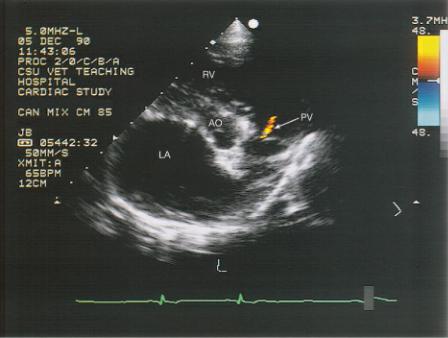
Figure 4.52 (A) Trivial aortic insufficiency is seen in 10 to 11% of dogs and is commonly seen in the (B) horse. RV = right ventricle, LV = left ventricle, AI = aortic insufficiency, LA = left atrium.
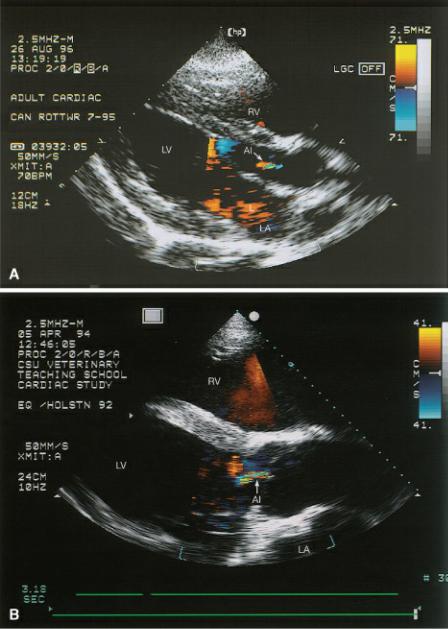
Figure 4.53 (A) Fifteen percent of dogs have trivial mitral regurgitation and (B) a large number of horses have these clinically insignificant leaks. LV = left ventricle, LA = left atrium, MR = mitral regurgitation.
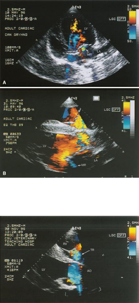
Figure 4.54 Tricuspid insufficiency is reported in as much as 50% of dogs and has a high incidence in horses as seen in this image. RV = right ventricle, LV = left ventricle, AO = aorta, TR = tricuspid regurgitation.
When spectral Doppler was used to interrogate the small regurgitant jets in beagles, the velocities

were less than those seen with regurgitation secondary to pathology and less than would be expected from normal pressure gradients within the heart (145). Signal strength is also weak when interrogating these small jets (100,145). There is usually no corresponding murmur to identify the presence of these clinically insignificant leaks (100,145).
There were small transient leaks across all valves in 40 clinically normal thoroughbred and thoroughbred cross horses with an incidence of mitral regurgitation in 67.5%, tricuspid regurgitation in 77.5%, aortic regurgitation in 47.5%, and pulmonary regurgitation in 40%. Another group comprised of 15 normal horses of various breeds had a 26% incidence of mitral regurgitation, 46% incidence of tricuspid regurgitation, 26% incidence of pulmonary insufficiency, and 80% occurrence of aortic insufficiency (144). Most of these leaks were associated with valve closure early in systole (148). Color-flow regurgitations is considered to be valve-closure related if it does not last for more than one frame. These findings were seen in athletic horses that may show a higher incidence of small regurgitations than in a general population of horses. Valvular regurgitation is seen frequently in healthy athletic humans (149). Regurgitation at the tricuspid valve is often indistinct while regurgitant flow at the mitral valve usually has discrete jet formation. When stricter criteria are used, with regurgitation only counted as present if it lasted more than 50% of systole, then only 2 horses of 40 had what could be classified as tricuspid regurgitation, and no horses had mitral regurgitation. In horses some mitral regurgitant jets extended as far as 9 cm into the left atrial chamber (148).
Pathological regurgitation can be semi-quantitatively assessed by measuring the size of the colorflow jet within the atria in the case of AV valvular insufficiency or within the outflow tract or ventricle in the case of aortic or pulmonic insufficiency (150,151). Information about quantifying pathological insufficiencies is presented in Chapter 5.
Color M-mode can be obtained during a color-flow exam. This helps separate the events of diastole and systole in order to better time the color-flow information. After turning on color-flow Doppler, place an M-mode cursor over the area of interest and activate M-mode. Figure 4.55 shows how color M-mode identifies the timing of aliased flow.
Use color M-mode to help define the timing of color flow patterns.
Figure 4.55 Timing of systolic and diastolic color flow Doppler can easily be identified by using color-flow M-mode images. (A) Normal color-flow M-mode shows a nonaliased red signal during systole within the aortic valve on this aortic M-mode image. (B) Aortic insufficiency is ruled out based upon a lack of turbulence during diastole, but systolic turbulence is present in this heart with aortic stenosis. AO = aorta, LA = left atrium.