

Histological slides (exam)
.pdfHistology Atlas
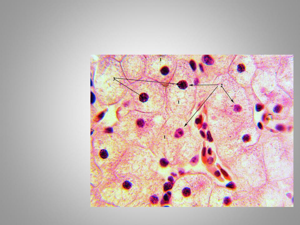
Common cell morphology. Liver cells (hepatocytes)
Hematoxylin and eosin staining.
1.– Cytoplasm
2.– Nuclei
3.– Nucleoli
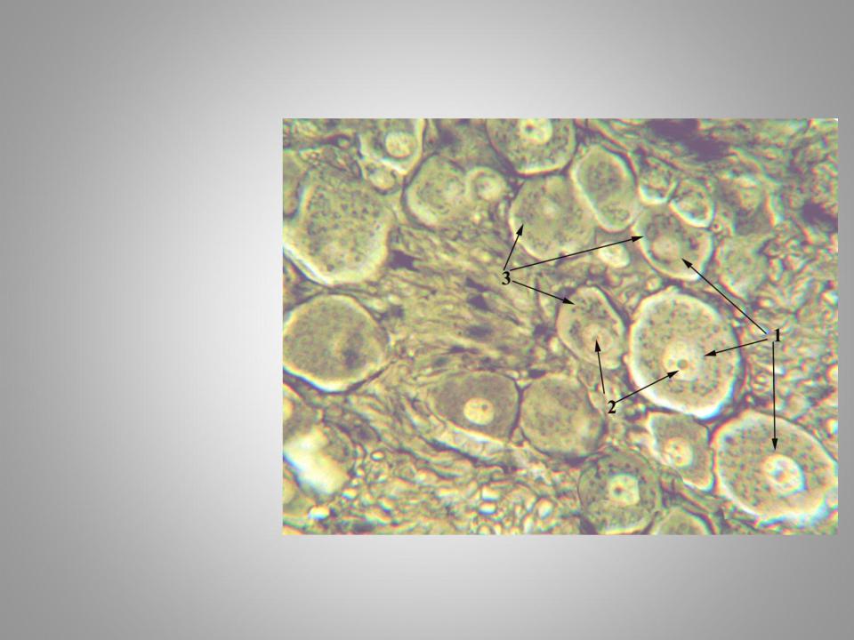
Golgi complex in spinal ganglion neurons
Osmium tetroxide staining.
1.Nucleus
2.Nucleous
3.Golgi complex
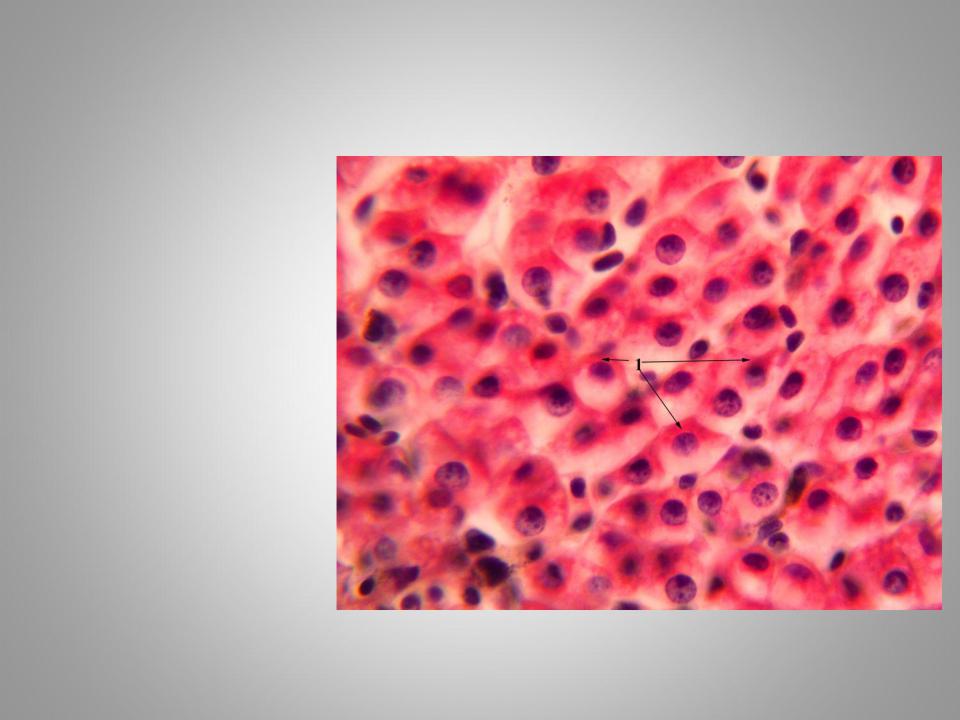
Glycogen inclusions in liver cells
Staining by Best’s
Carmine
1. Clumps of glycogen
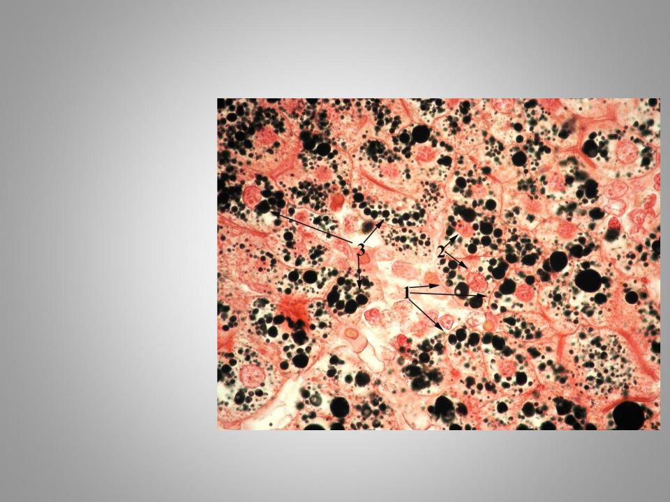
Lipid inclusions in liver cells
To identify the fat inclusions slide fixed with
osmium tetroxide and stained by safranin.
1.Cells boundaries
2.Nuclei
3.Lipid inclusions
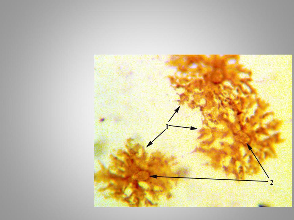
Pigment inclusions in pigment cells (melanocytes)
Slide not stained
1.Processes of melanocytes
2.Nuclei
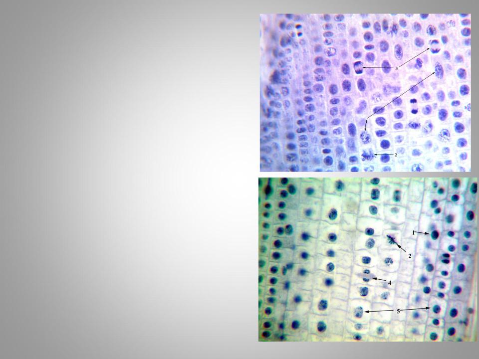
Mitosis
Slide is stained with iron hematoxylin.
1.The beginning of prophase
2.Metaphase
3.Anaphase
4.Telophase
5.Interphase
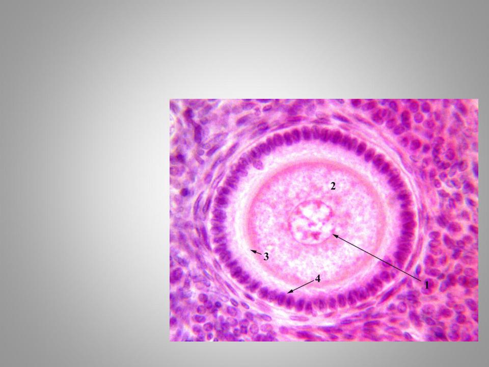
Ovum
1.Nucleus
2.Yolk inclusions
3.Zona pellucida
4.Corona radiata
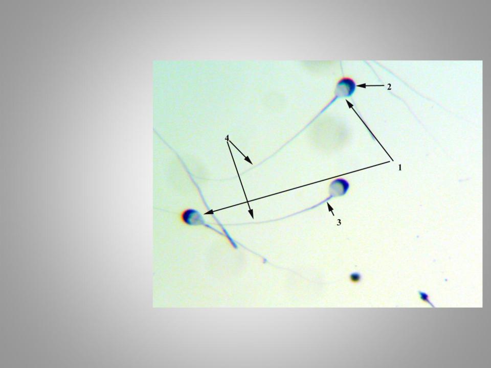
Spermatozoa
Iron hematoxylin staining.
1.Nucleus
2.Acrosome
3.Neck
4.Tail
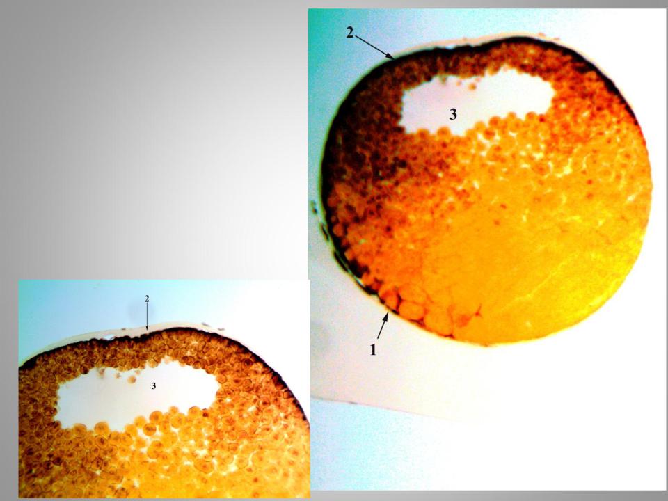
Blastula
Hematoxylin and pikrofucsin staining.
1.Vegetative pole
2.Аnimal pole
3.Blastocoele