

Histological slides (exam)
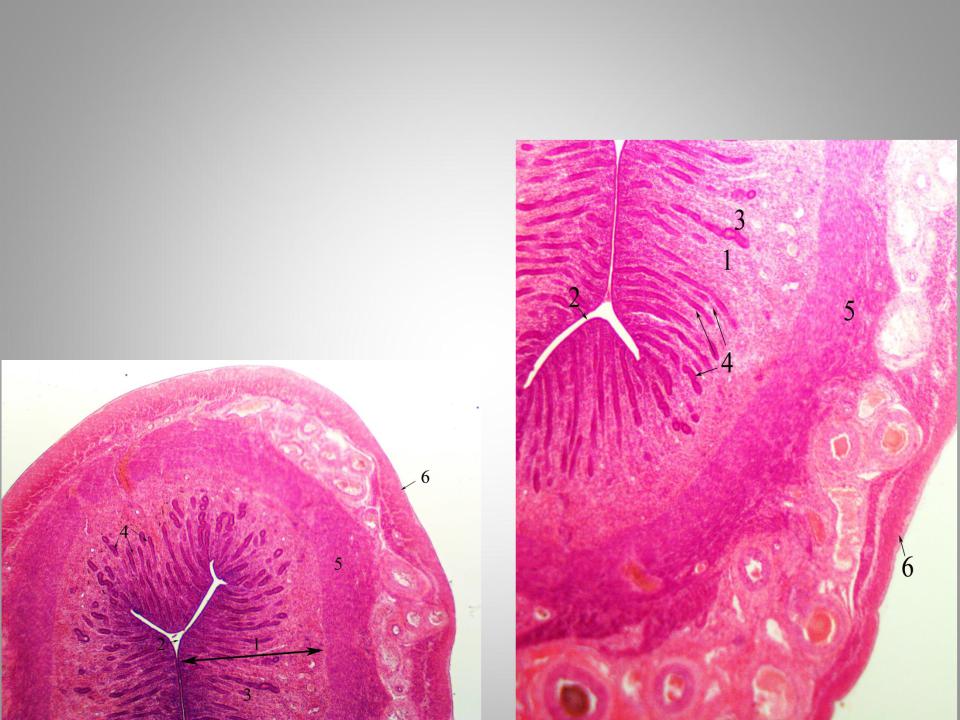
.pdfProstate gland
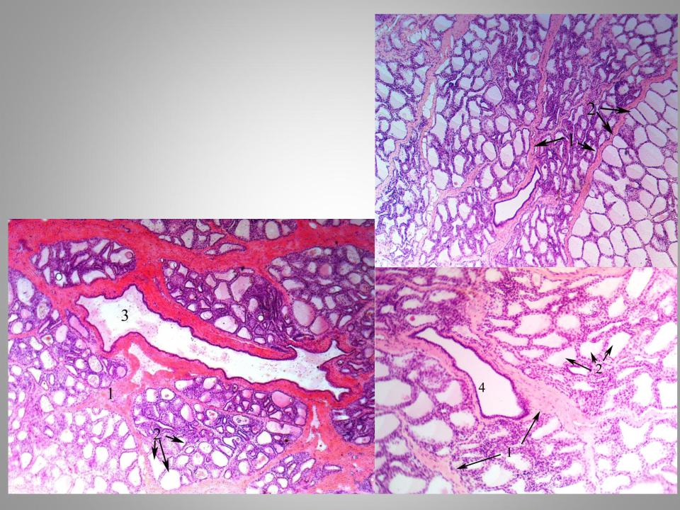
Hematoxylin and eosin staining.
1.Fibromuscular septa
2.Smooth muscle cells
4. Urethra
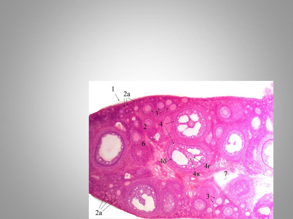
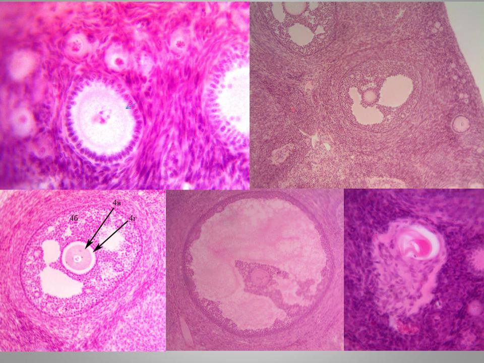
Ovary
Hematoxylin and eosin staining.
1.Tunica albuginea 2a. Primordial follicles
2.Primary follicle
3.Secondary follicle 4б – granulosa sells 4в – oocyte
4гzona pellucida
5.Tertiary (Graafian ) follicle
6.Atretic follicle
7.Medulla
 2
2
3
4
4 |
|
5 |
6 |
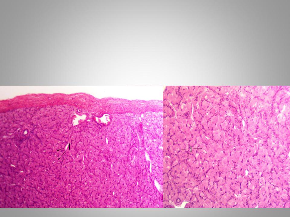
Corpus luteum
Hematoxylin and eosin staining.
1. Luteal cells
Uterus
Hematoxylin and eosin staining.
1 . Endometrium
2.Simple columnar epithelium
3.Lamina propria of endometrium
4.Simple tubular glands
5 Myometrium Submucous layer Vascular layer Outer layer 6-Perimetrium
Mammary gland
Hematoxylin and eosin staining.
1.Interlobular connective tissue
2.Alveoli
3.Lactiferous duct
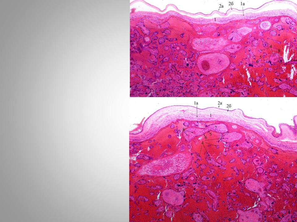
Fetal part of placenta
Hematoxylin and eosin staining.
1.Chorionic plate
2.Amniotic membrane 2а - amniotic epithelium
2б – extraembryonic mesoderm of amniotic membrane
4.Chorionic villi
5.Cytotrophoblast
6.Syncytyotrophoblast
7.Extraembryonic mesoderm of chorionic villi
8.Fetal blood vessels
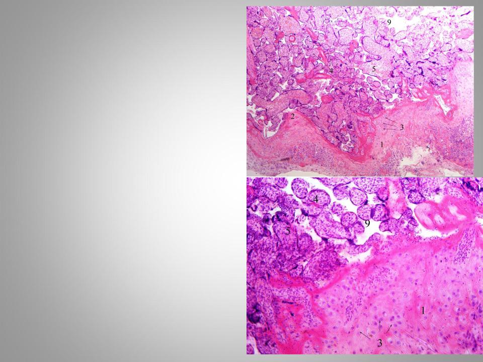
Maternal part of placenta
Hematoxylin and eosin staining.
1.Basal lamina of endometrium
2.Septae
3.Decidual cells
4.Chorionic villi
5.Connective tissue
6.Blood vessels
9. Lacunae filled with maternal blood