

Histological slides (exam)
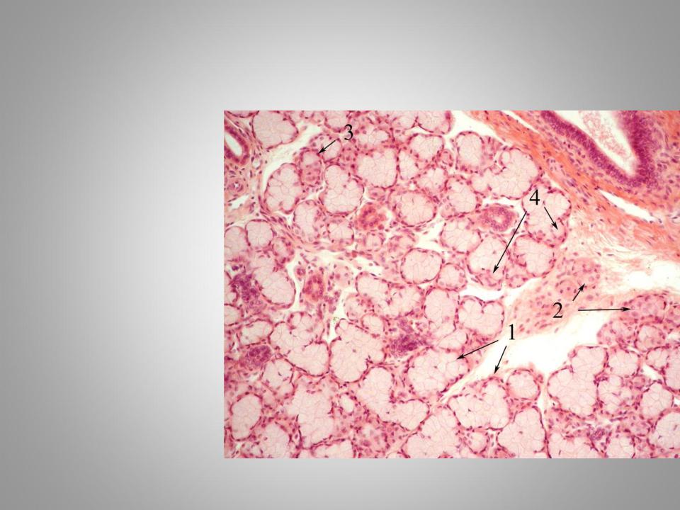
.pdfParotid gland
Hematoxylin and eosin staining.
1.Connective tissue capsule
2.Connective tissue septa
3.Interlobular duct
*- serous acini
4.Serous cells
5.Mioepitelial cells
6.Intercalated duct
7.Striated duct
Mixed salivary gland
Hematoxylin and eosin staining.
1.Mucouse secretory portions
2.Serous secretory portions
3.Mixed secretory portions
4.serous demilune
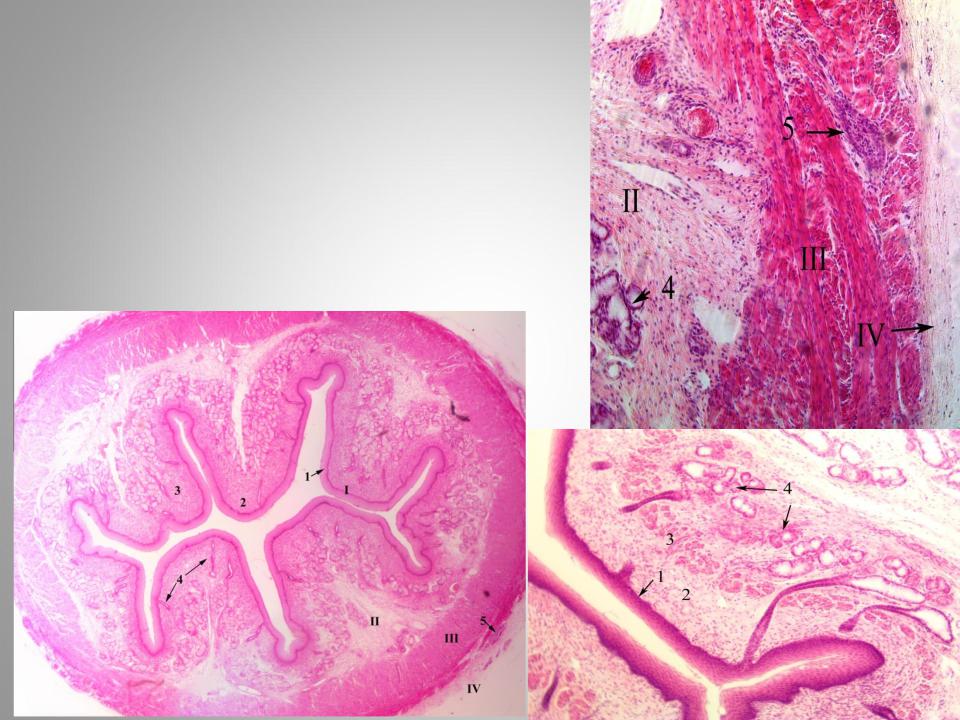
Esophagus (transversal section)
Hematoxylin and eosin staining.
I- Mucosa
1.non-keratinized stratified epithelium
2.lamina propria
3.muscularis mucosae
IISubmucosa
4.esophageal glands proper IIIMuscularis
5.myenteric plexus (or Auerbach's plexus) IV - Adventitia
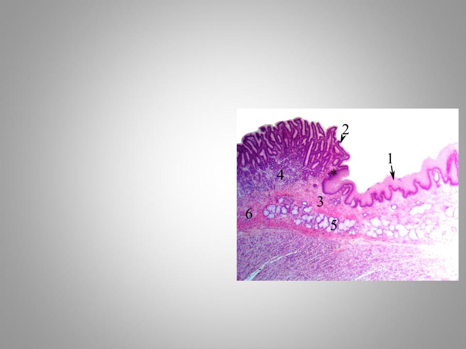
Esophago-gastric yunction
Hematoxylin and eosin staining.
*- The junction of the esophagus with the stomach
1.Stratified squamous epithelium
2.Simple columnar epithelium of stomach
3.Lamina propria of the esophagus
4.Cardiac glands of the stomach
5.Esophageal glands proper
6.Submucosa of the stomach
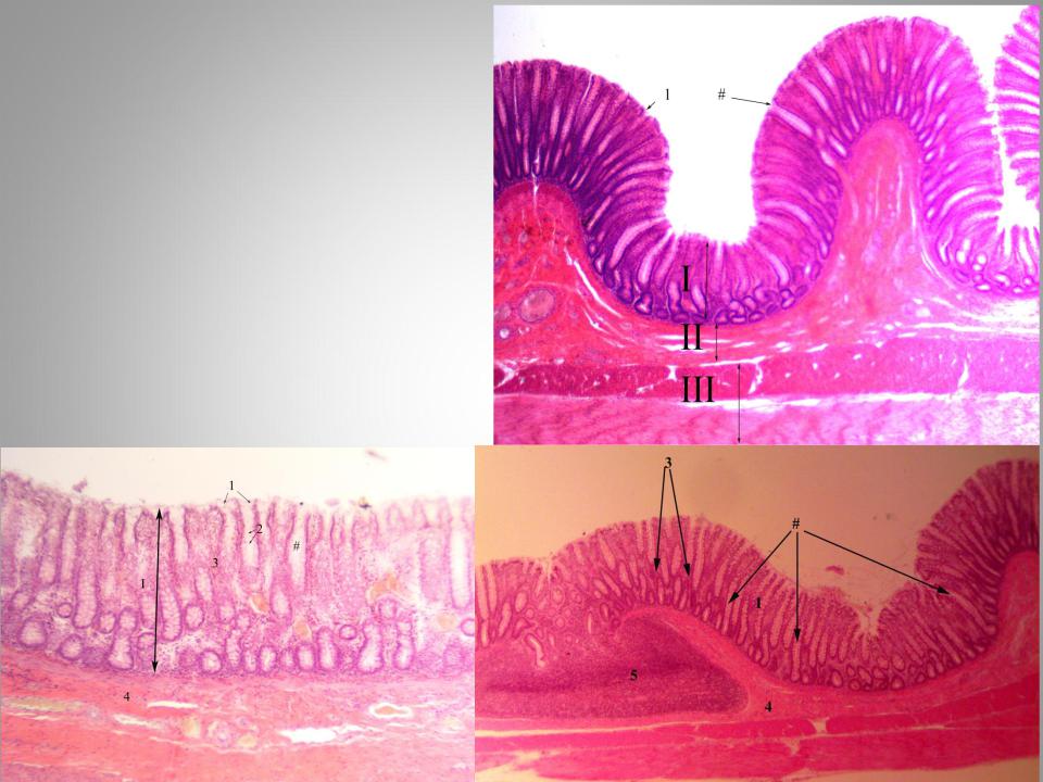
Fundus of the stomach
Congo red staining.
*- Folds composed of the mucosa and submucosa
I- Mucosa
IISubmucosa
IIIMuscularis IVSerosa
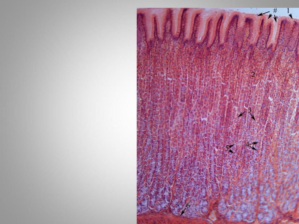
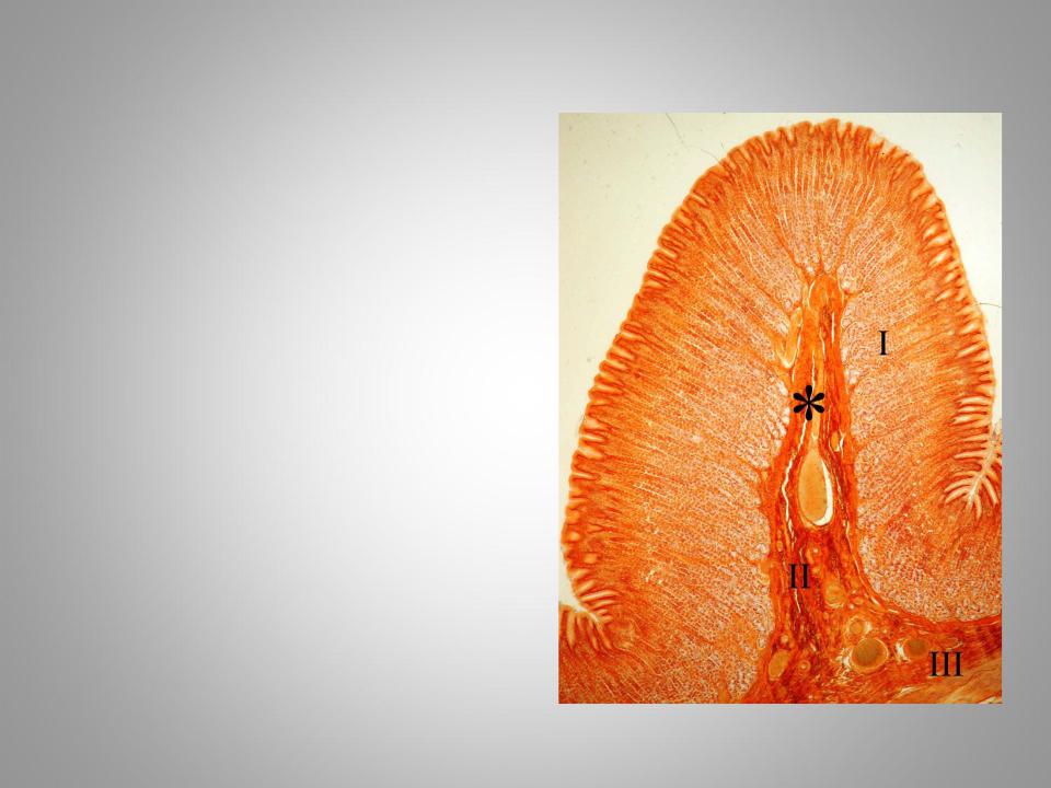
Fundus of the
stomach
Congo red staining.
#- gastric pits
1.Simple columnar epithelium
2.Lamina propria
3.Fundic glands
4.Parietal cells
5.Chief cells
6.Muscularis mucosae
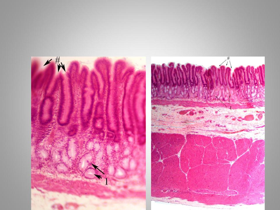
Pyloric part of stomach
Hematoxylin and eosin staining.
#- gastric pits
1.Pyloric glands
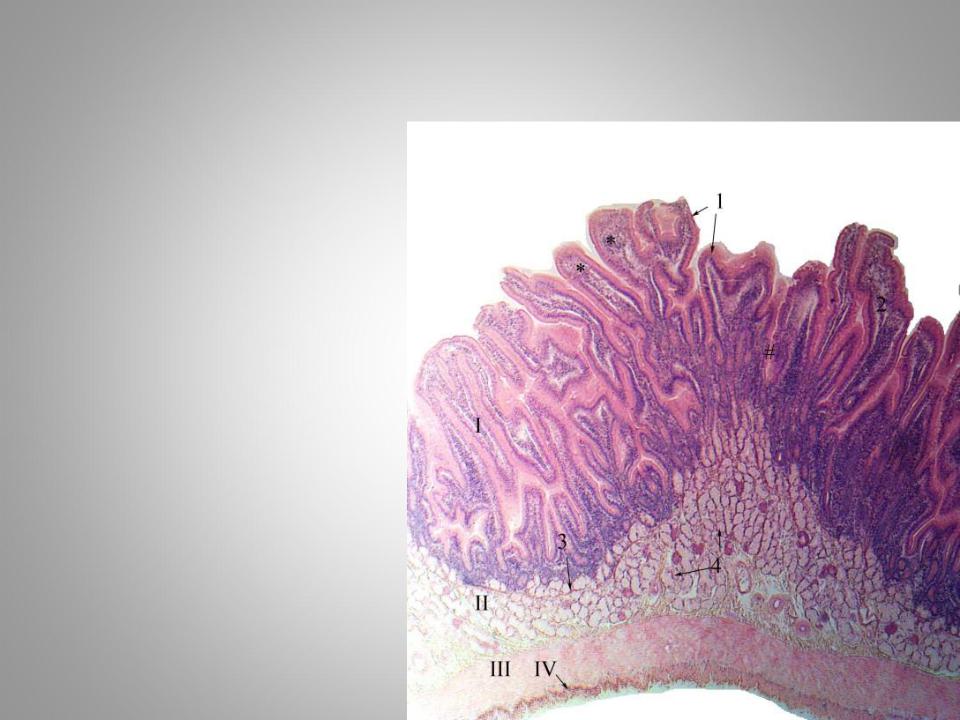
Duodenum
Hematoxylin and eosin staining.
I – Mucosa *- Ворсинки
1.Simple columnar epithelium
2.Lamina propria
3.Muscularis mucosae II – Submucosa
4.Brunner’s glands
IIIMuscularis
IVSerosa
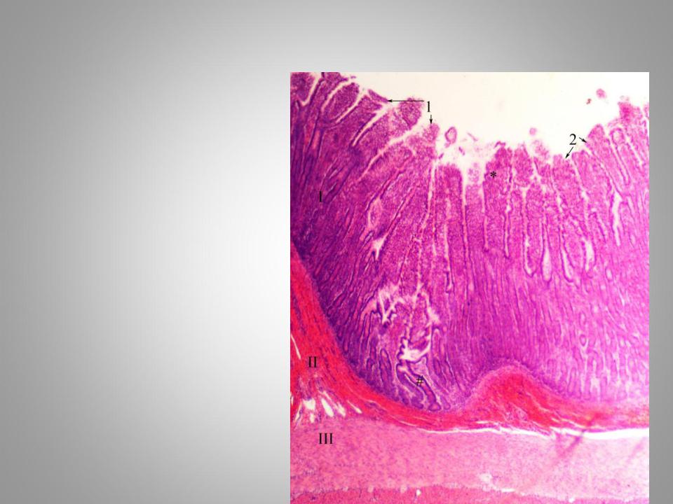
Small intestine (jejunum or ileum)
Hematoxylin and eosin staining.
I – Mucosa *- villi
#- crypts
1.Simple columnar epithelium
2.Goblet cells
IISubmucosa
IIIMuscularis
IVSerosa
Large intestine
Hematoxylin and eosin staining.
I – Mucosa #-crypts
1.Simple columnar epithelium
2.Goblet cells
3.Lamina propria
IISubmucosa
4.Connective tissue with vessels
5.Lymphoid follicles
IIIMuscularis
IVSerosa