

Histological slides (exam)
.pdfUnmyelinated nerve fibers
Hematoxylin and eosin staining.
*- Unmyelinated nerve fibers 1. Nuclei of Shwann cells
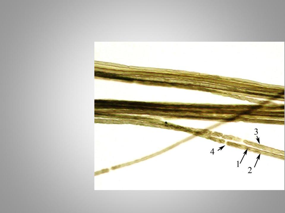
Myelinated nerve fibers
Staining with osmic acid
1.Axial cylinder
2.Myelin sheath
3.Schwann sheaths
4.Nodes of Ranvier
5.Clefts of SchmidtLantermann
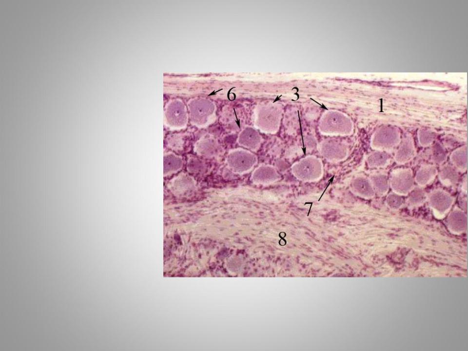
Spinal ganglion
Hematoxylin and eosin staining.
1.Сonnective tissue capsule
3. Pseudounipolar neurons of spinal ganglion
6.Glial satellite cells
7.Сonnective tissue capsule of neurons
8.Nerve fibers
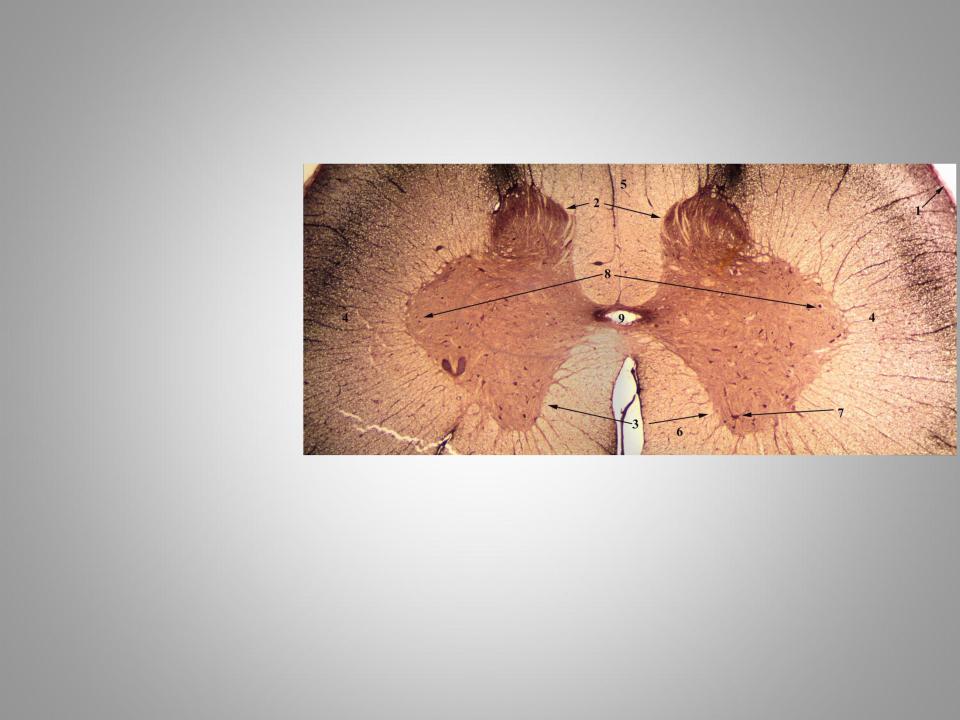
Spinal cord
Silver nitrate staining by Ramón y Cajal method
1.Pia mater
2.Dorsal horns
3.Ventral horns
4.Lateral columns
5.Ventral columns
6.Dorsal columns
7.Motor neurons
8.Intercalated neuron
9.Central canal
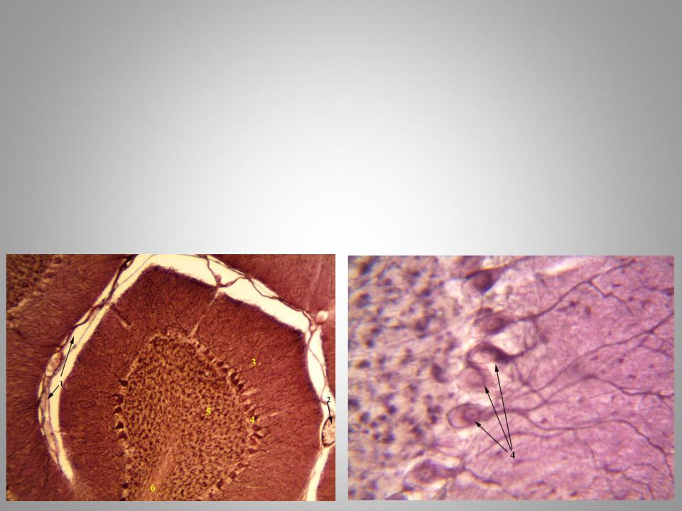
Cerebellum
Silver nitrate staining by Ramón y Cajal method
1.Pia mater
2.Blood vessels
3.Molecular layer of cortex
4.Ganglionic layer of cortex
5.Granular layer of cortex
6.White matter
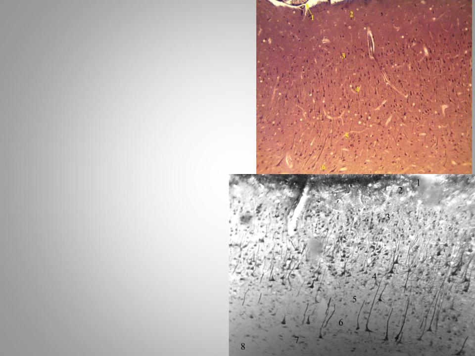
Cortex of the human brain
Silver nitrate staining by
Ramón y Cajal method
1.Pia mater with blood vessels
2.Molecular layer
3.Outer granular layer
4.Outer pyramidal layer
5.Inner granular layer
6.Betz cells( layer of large pyramidalcells)
7.Layer of polymorphic cells
8.White matter
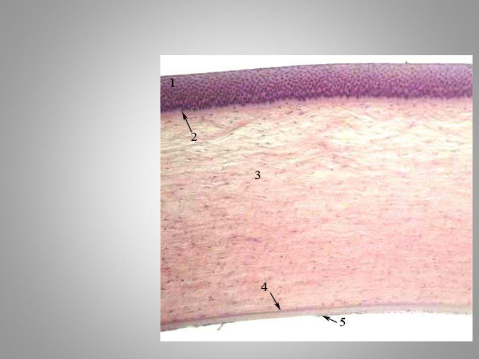
Cornea
Hematoxylin and eosin staining.
1.Anterior epithelium (stratified nonkeratinized squamous)
2.Bowman’s membrane
3.Substantia propria
4.Descemet’s membrane
5.Posterior epithelium (simple squamous)
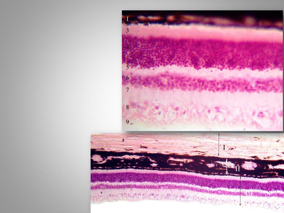
The posterior wall of the eyeball
Hematoxylin and eosin staining.
І Sclera
ІІ Choroid
ІІІ Retina:
1.Pigment epithelium
2.Outer limiting membrane
3.Layer of rods and cones
4.Outer nuclear layer
5.Outer plexiform layer
6.Inner nuclear layer
7.Inner plexiform layer
8.Layer of ganglion cells
9.Layer of optic nerve fibers 10.Layer of inner limiting membrane
Organ of Corti
Hematoxylin and eosin staining.
*-Cochlea duct
#- Scala vestibuli ##- Scala tympani
1.Vestibular membrane
2.Сосудистая полоска
3.Basilar membrane
4.Spiral ligament
5.Spiral limbus
6.Vestibular lip of spiral limbus
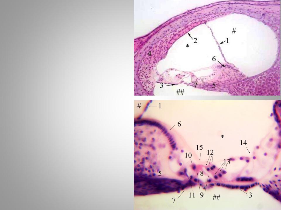
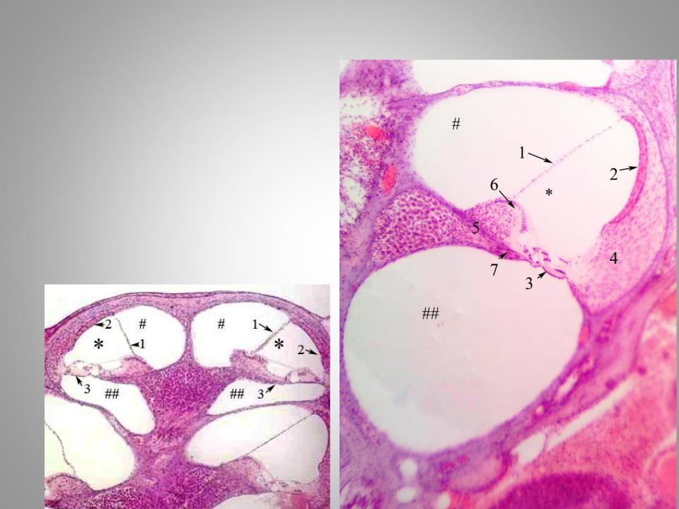
Organ of Corti
Hematoxylin and eosin staining.
*-Cochlea duct
#- Scala vestibuli ##- Scala tympani
1.Vestibular membrane
2.Stria vascularis
3.Basilar membrane
4.Spiral ligament
5.Spiral limbus
6.Vestibular lip of spiral limbus
7.Tympanic lip of limbus
8.Tunnel
9.Columnar cells
10.Inner hair (sensory) cells 11.Inner phalangeal cells 12.Outer hair (sensory) cells 13.Outer phalangeal cells 14.Hensen's cells 15.Tectorial membrane