

Histological slides (exam)
.pdfFibrous cartilage
Hematoxylin and eosin staining.
1.Bundles of collagen fibers
2.Ground substance
3.Chondrocytes
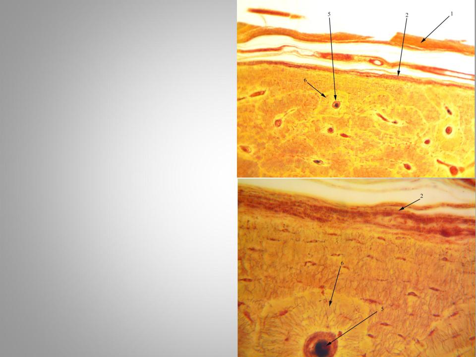
Сompact bone (transversus section)
Slide is stained by Schmorl’s method with Thionine and picric acid )
1.Periosteum
2.Outer circumferential lamellae
3.Endost
4.Inner circumferential lamellae
5.Oteonal canals
6.Osteons lamellae
7.Interstitial lamellae
8.Osteocytes
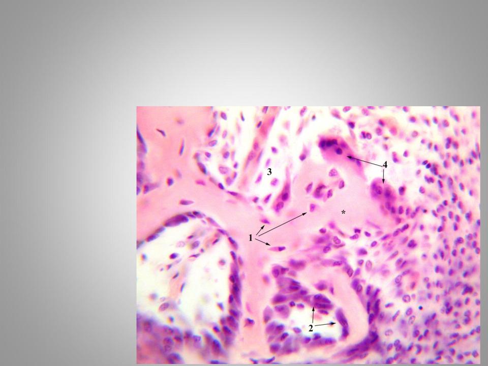
Intramembranous ossification(direct ossification)
Hematoxylin and eosin staining.
*- bone trabeculae
1.Оsteocytes
2.Оsteoblasts
3.Меsenchyme
4.Оsteoclasts
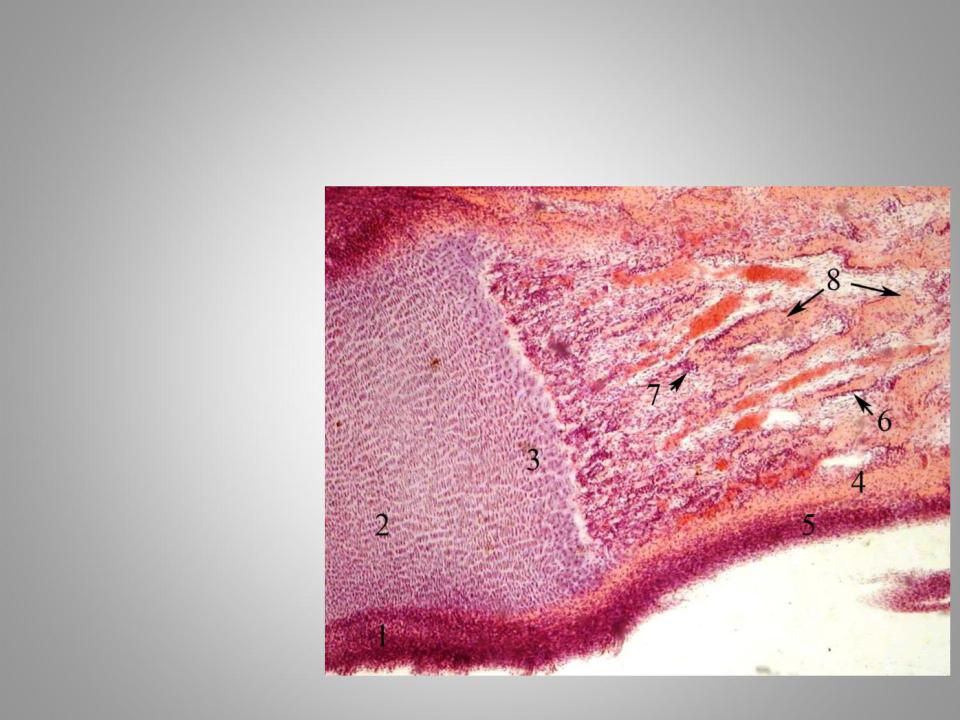
Endochondral ossification (indirect ossification)
Hematoxylin and eosin staining.
1.Perichondrium
2.Columnar cartilage zone(zone of proliferation)
3.Resorbing cartilage zone
4.Bony collar (periosteal bone)
5.Periosteum
6.Osteoblasts
7.Оsteocytes
8.Endochondral bone
Smooth muscle tissue of
urinary bladder
Hematoxylin and eosin staining.
1.Smooth myocytes
2.Connective tissue
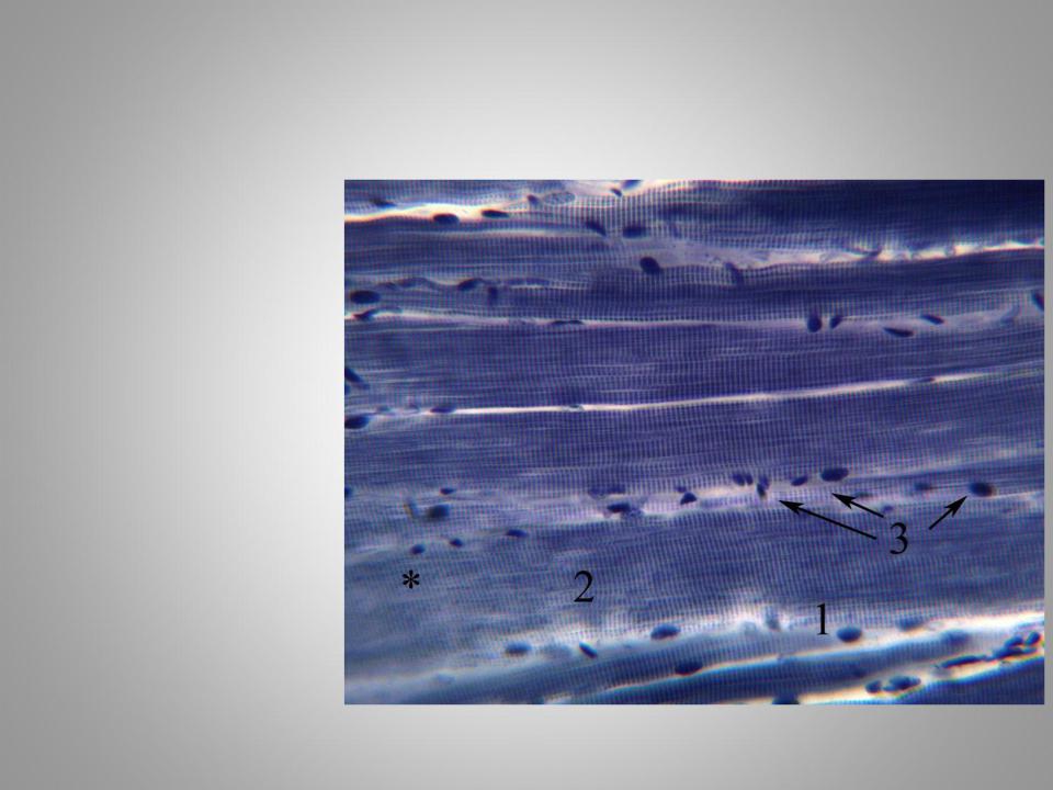
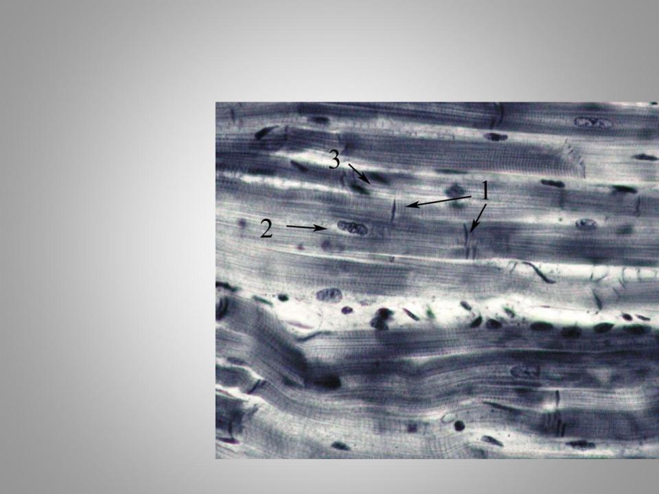
Striated skeletal muscle of the tongue
Iron hematoxylin staining.
1.Connective tissue
2.Transverse striations of the fibers
3.Nuclei of muscle fibers
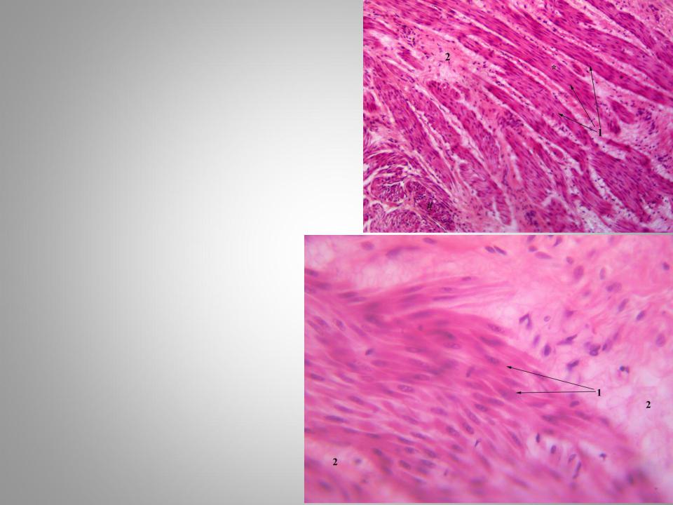
Cardiac muscle tissue
Iron hematoxylin staining.
1.Intercalated disks
2.Nuclei of cardiomyocytes
3.Anastomoses between cardiomyocytes
4.Connective tissue
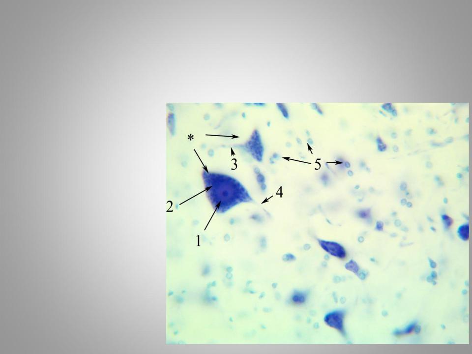
Basophilic substance (Nissl body, chromophilic substance) in the neurons of spinal cord
Methylene blue staining by the Nissl method.
*- neurons
1.Nucleus
2.Basophilic substance (Nissl body)
3.Dendrites
4.Аxon
5.Neuroglial cells

Neurofibrills in the nerve cells of the spinal cord
Silver nitrate impregnation
1.Neurofibrills
2.Neurons nuclei
3.Neurons processes