

Histological slides (exam)
.pdfPrimitive streak
Hematoxylin staining.
1.Ectoderm
2.Endoderm
3.Primitive groove
4.Primitive streak
5.Mesoderm
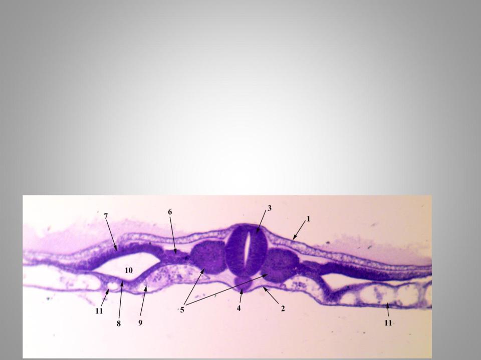
Embryo axial organs
Iron hematoxylin staining.
1.Ectoderm
2.Endoderm
3.Neural tube
4.Notochord
5.Somites
6.Nephrotome
7.Somatopleurea(parietal layer of splanchnotome)
8.Splanchnopleura (visceral layer of splanchnotome)
9.Mesenchyme
10.Coelom
11.Blood vessels
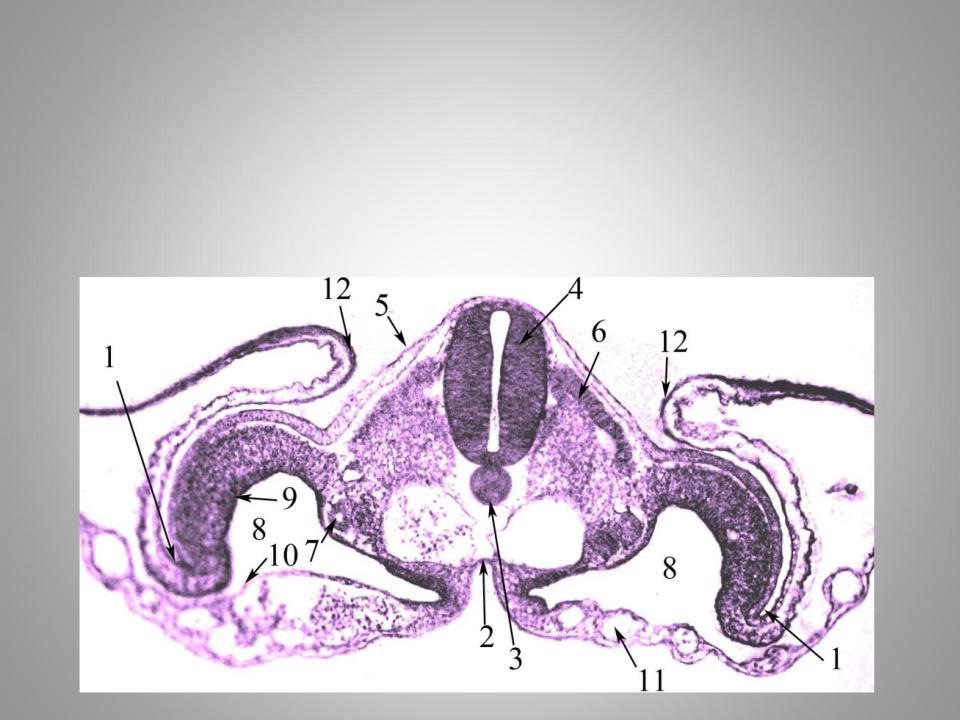
Iron hematoxylin staining.
1.Body flexion
2.Intestinal endoderm
3.Notochord
4.Neural tube
5.Skin ectoderm
6.Dermatomes
Body flexion
7.The formation of the renal tubules from the nephrotome
8.Coelom
9.Parietal layer of splanchnotome
10.Visceral layer of splanchnotome
11.Blood vessels
12.Amnionic fold
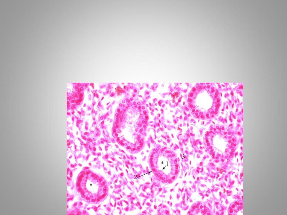
Simple low prismatic (cuboidal) epithelium
Hematoxylin and eosin staining.
* - epithelial cells
1.Apical pole
2.Basal pole
3.Connective tissue
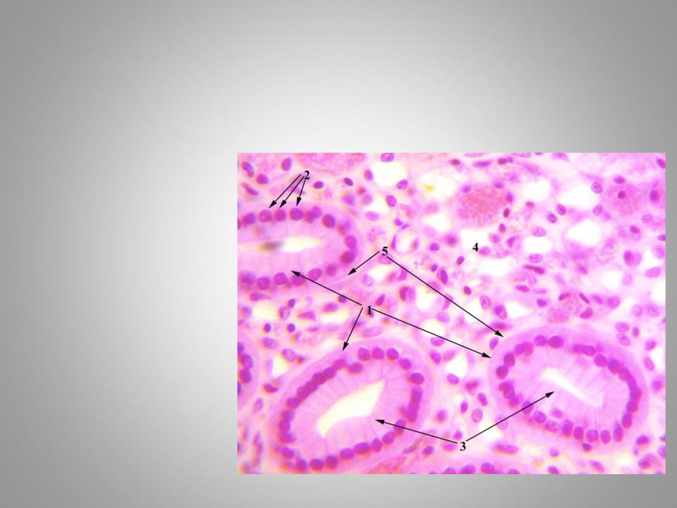
Simple high prismatic (columnar) epithelium
Hematoxylin and eosin staining.
1.Epithelial cells
2.Basal pole
3.Apical pole
4.Blood vessels
5.Basal lamina
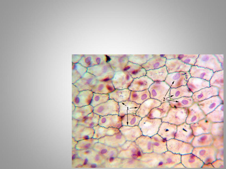
Simple squamous coelomic epithelium - mesothelium
Film slide of rabbit gastrohepatic omentum.
Silver nitrate impregnation and hematoxylin staining.
1.Cells boundaries
2.Nuclei
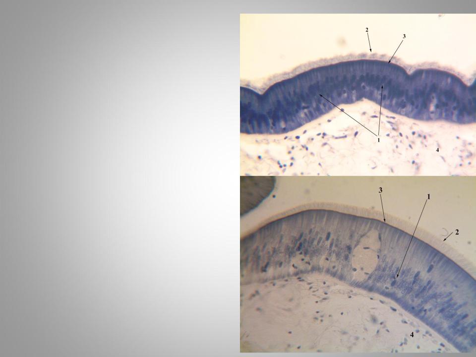
Simple pseudostratified columnar ciliated epithelium
Slide is stained with iron hematoxylin.
1.Nuclei
2.Microvilli
3.Basal bodies of microvilli
4.Connective tissue
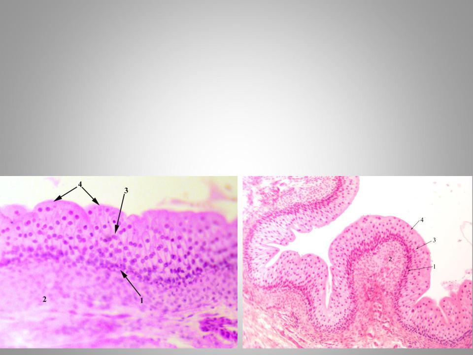
Transitional epithelium of the urinary bladder
Hematoxylin and eosin staining.
1.Basal layer
2.Connective tissue
3.Intermediate cell layer
4.Surface cell layer
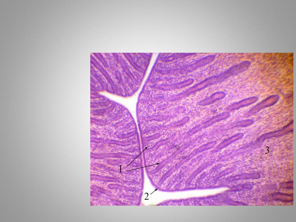
Simple tubular uterus glands
Hematoxylin and eosin staining.
1.Glands
2.Cuboidal epithelium
3.Сonnective tissue

Unicellular endoepithelial glands ( Leydig cells) and simple alveolar extraepithelial glands
Slide is made from Axolotl skin.
Hematoxylin and eosin staining.
1.Leydig cells
2.Сonnective tissue
3.Simple alveolar unbranched extraepithelial glands
4.Excretory duct