

Histological slides (exam)
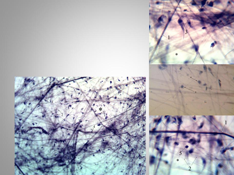
.pdfChick embryo mesenchyme
Slide is stained with iron hematoxylin.
* – Mesenchyme
1.Cells nuclei
2.Cells processes
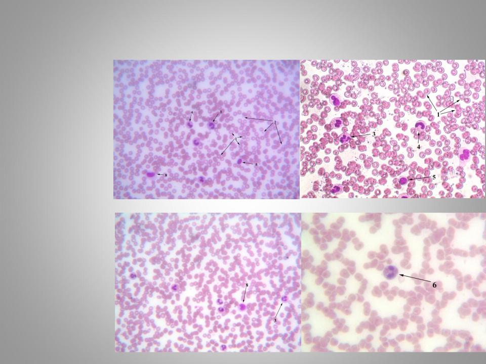
Human blood (smear)
Blood smear is stained with Hematoxylin and eosin or by Romanowsky-
Giemsa’s method.
1.Erythrocytes
2.Platelets
3.Segmented neutrophils
4.Banded neutrophils
5.Lymphocytes
6.Eosinophils
7.Basophils
8.Monocytes
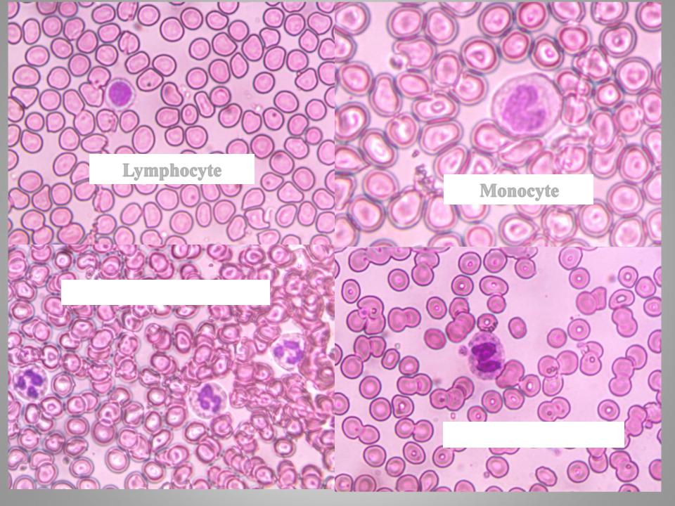
Lymphocyte
Monocyte
Segmented neutrophils
Eosinophil
Loose connective tissue
Iron hematoxylin staining.
*- ground substance
1.Collagen fibers
2.Elastic fibers
3.Fibroblasts
4.Macrophages
5.Tissue’s lymphocytes
6.Plasma cells
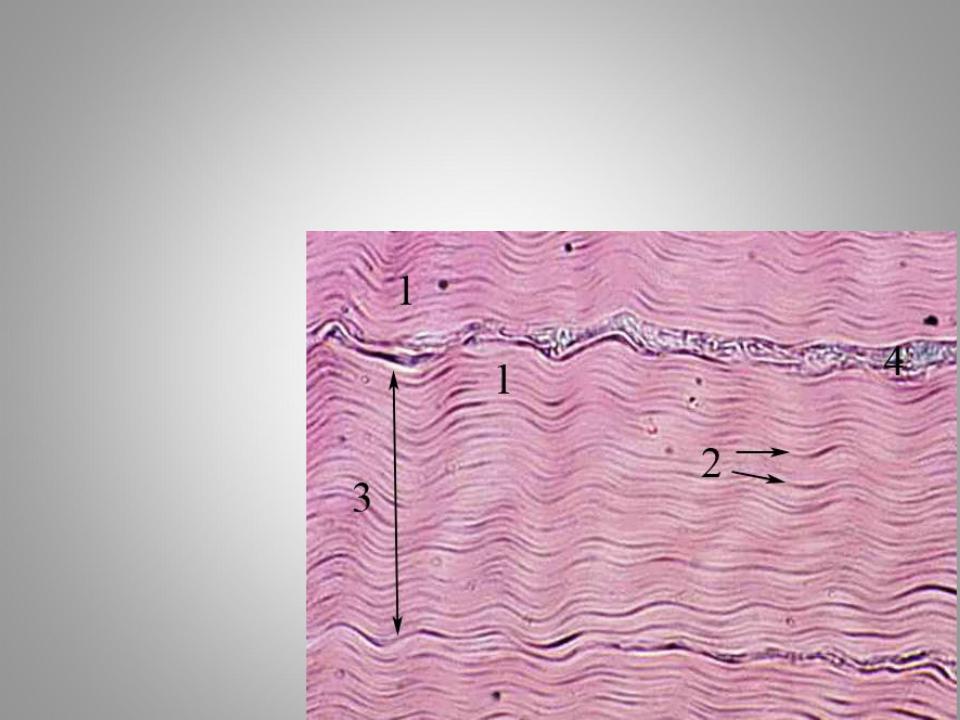
Tendon (longitudinal section)
Hematoxylin and eosin staining.
1.Primary bundles of collagen fibers
2.Tendinocyte nuclei
3.Secondary bundles of collagen fibers (tendon fascicles)
4.Endotendineum
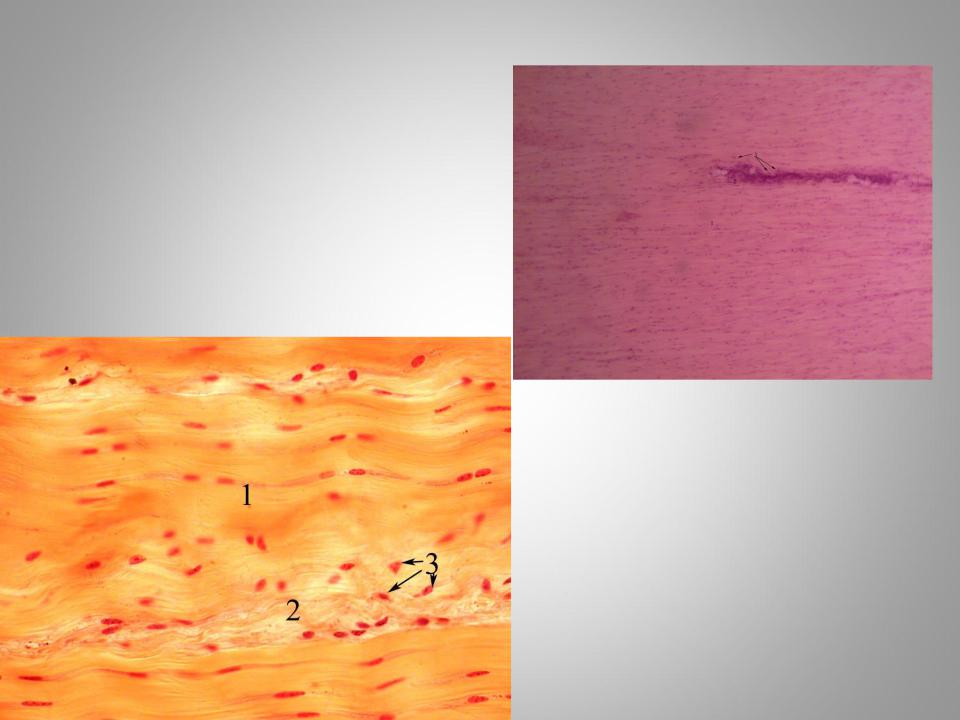
Ligament(longitudinal section)
Slide is stained with picric acid, acid fuchsin and hematoxylin.
1.Elastic fibers
2.Collagen fibers
3.Fibroblasts
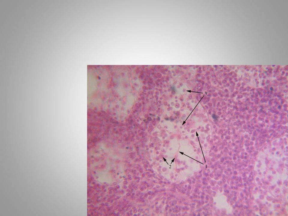
Reticular tissue of lymph node
Hematoxylin and eosin staining.
*- Reticular cells
1.Cytoplasm and processes
2.Cells nuclei
3.Limphocytes
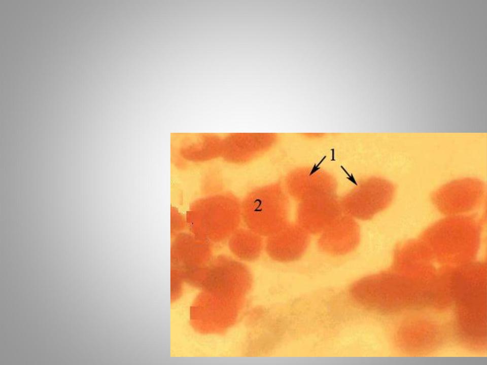
Adipose tissue
To identify the fat inclusions slide is stained with sudan and
Hematoxylin
1.Adipocytes
2.Lipid drop
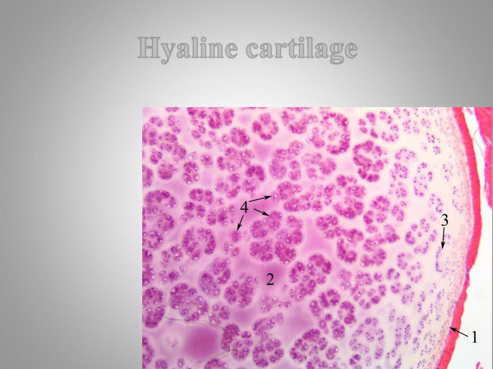
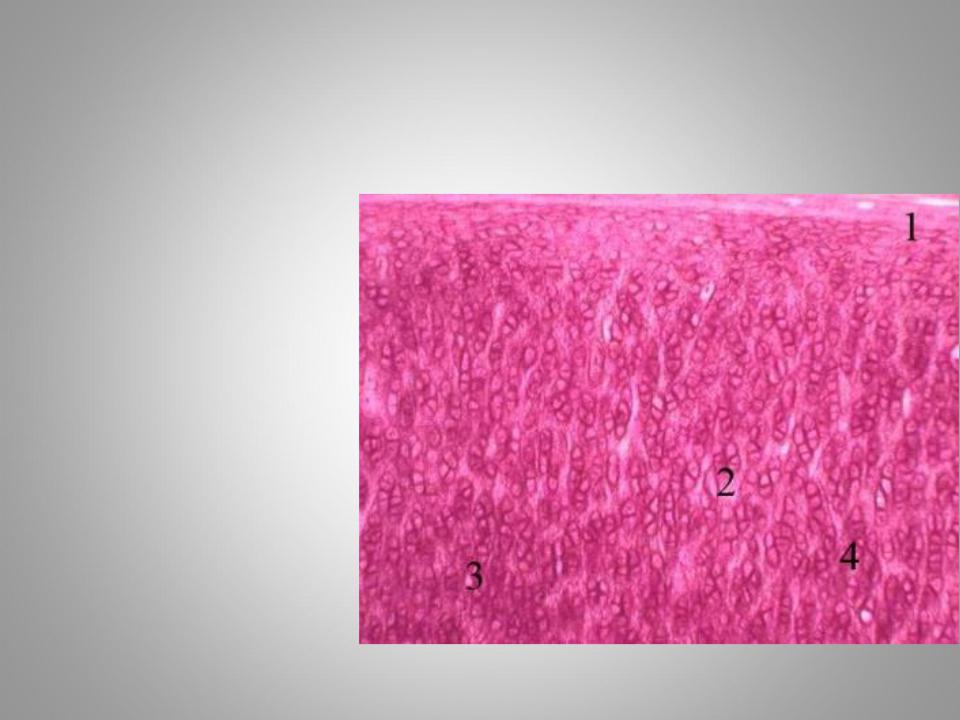
Hyaline cartilage
Hematoxylin and eosin staining.
1.Perichondrium
2.Extracellular matrix
3.Zone of young chondrocytes
4.Mature chondrocytes (isogenous groops)
Elastic cartilage
Slide is stained with orcein and hematoxylin.
1.Perichondrium
2.Ground substance
3.Elastic fibers
4.Isogenic cells groops