


Chapter 4
Rhodophyta
RHODOPHYCEAE
The Rhodophyceae, or red algae, comprise the only class in the division Rhodophyta. The Rhodophyceae are probably one of the oldest groups of eukaryotic algae. The red algae are most likely directly descended from a cyanome in the Glaucophyta (see Chapter 3). It is likely that the first red alga evolved into an ecological niche that was unoccupied by cyanobacteria, the only extant photosynthetic alga that evolved oxygen. This ecological niche would have been in waters with a pH less than 5, which, for some unknown reason, cyanobacteria are not able to inhabit (Brock, 1973). Indeed, modern phylogenetic studies utilizing nucleic-acid sequencing have shown that Cyanidium, an alga that lives in acidic waters, is probably the oldest extant red alga (Oliveira and Bhattacharya, 2000).
The Rhodophyceae lack flagellated cells, have chlorophyll a, phycobiliproteins, floridean starch as a storage product, and thylakoids occurring singly in the chloroplast.
A majority of the seaweeds are red algae, and there are more Rhodophyceae (about 4000 species) than all of the other major seaweed groups combined. Although marine red algae occur at all latitudes, there is a marked shift in their abundance from the equator to colder seas. There are few species in polar and subpolar regions where brown and green algae predominate, but in temperate and tropical regions they far outnumber these groups. The average size of the plants also
differs according to geographical region. The larger species of fleshy red algae occur in cool-temperate areas, whereas in tropical seas the Rhodophyceae (except for massive calcareous forms) are mostly small, filamentous plants. The Rhodophyceae also have the ability to live at greater depths in the ocean than do members of the other algal classes. They live at depths as great as 200 m, an ability related to the function of their accessory pigments in photosynthesis. About 200 species of Rhodophyceae are found in freshwater, where they do not reach as great a size as the red seaweeds (Skuja, 1938). The majority of freshwater red algae occur in running waters of small to mid-sized streams (Sheath and Hambrook, 1988). Few red algae occur at currents of less than 30 cm s 1. This fast flow probably favors red algae because loosely attached competitors are washed out and because of a constant replenishment of nutrients and gases.
Cell structure
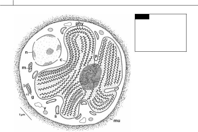
The major features of a red algal cell (Fig. 4.1) are a chloroplast with one thylakoid per band and no chloroplast E.R., floridean starch grains in the cytoplasm outside the chloroplast, no flagella, pit connections between cells in filamentous genera, and a eukaryotic type of nucleus (Scott et al., 1980).
Cell walls
Cellulose forms the microfibrillar framework in most rhodophycean cell walls, although in the
90 EVOLUTION OF THE CHLOROPLAST
Fig. 4.1 Semidiagrammatic drawing of a cell of Porphyridium cruentum. (c) Chloroplast; (g) Golgi;
(m)mitochondrion; (mu) mucilage;
(n)nucleus; (p) pyrenoid; (phy) phycobilisomes; (s) starch; (v) vesicle. (Adapted from Gantt and Conti, 1965.)
haploid phase of the Bangiales (Bangia and Porphyra) a -1,3 linked xylan (polysaccharide composed of xylose residues) performs this function (Frei and Preston, 1964). Unicellular red algae have an amorphous matrix of sulfated polysaccharides without cellulose surrounding the cells (Arad et al., 1993). The amorphous polysaccharides or mucilages occur between the cellulose microfibrils in the rest of the red algae. The two largest groups of amorphous mucilages are the agars (Fig. 1.11) and the carrageenans (Fig. 4.15). These mucilages may constitute up to 70% of the dry weight of the cell wall. Cuticles, composed mostly of protein, can occur outside the cell wall (Craigie et al., 1992).
Chloroplasts and storage products
Chloroplasts are usually stellate with a central pyrenoid in the morphologically simple Rhodophyceae (Fig. 4.1), whereas in the remainder of the Rhodophyceae they are commonly discoid (Fig. 4.3). In the Rhodophyceae with apical growth, the chloroplasts usually originate from small colorless proplastids with few thylakoids in the apical
cell (Lichtlé and Giraud, 1969). Chloroplasts are surrounded by the two membranes of chloroplast envelope with no chloroplast endoplasmic reticulum present (Figs. 4.1, 4.2, 4.3). Thylakoids occur singly inside the chloroplasts. The phycobilin pigments are localized into phycobilisomes on the surface of the thylakoids, a situation similar to that in the cyanobacteria.
Chlorophyll a is in the chloroplasts. There have been erroneous reports of chlorophyll d occurring in the chloroplast. It has been shown that the chlorophyll d in these studies came from the cyanobacterium Acaryochloris marina, an epiphyte on red algae (Murakami et al., 2004).
The phycobiliproteins include R-phycocyanin, allophycocyanin, and three forms of phycoerythrin, the phycoerythrins being present in the greatest amount, giving the algae their pinkish color. B-phycoerythrin is present in the more primitive red algae and has been found in Pophyridium (Fig. 4.1), Rhodosorus (Fig. 4.24 (a)), Rhodochorton (Fig. 4.31) and Smithora. R-phycoerythrin occurs in most higher red algae, and C-phycoerythrin occurs in
RHODOPHYTA 91
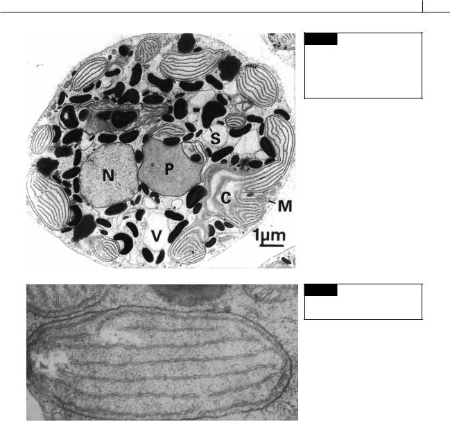
Fig. 4.2 Transmission electron micrograph of a section of a cell of
Rhodella violacea. (C) Chloroplast;
(M) mitochondrion; (N) nucleus; (P) pyrenoid; (S) starch; (V) vacuole. (From Marquardt et al., 1999.)
Fig. 4.3 Chloroplast in a carpospore of Polysiphonia. (From Tripodi, 1974.)
Porphyridium (Fig. 4.1), Porphyra (Fig. 4.27), and Polysiphonia (Figs. 4.44, 4.45). The phycobiliproteins are in phycobilisomes on the surface of thylakoids (Fig. 4.1). The phycobilisomes are spherical if both phycoerythrin and phycocyanin are present. The phycobilisomes are discoid if only phycocyanin is present (Gantt, 1969).
Complementary chromatic adaptation occurs in the red algae. Orange and red light stimulate the production of long-wavelength absorbing phycocyanin, while green light stimulates the formation of short-wavelength absorbing phycoerythrin (Sagert and Schubert, 1995). The color will vary.
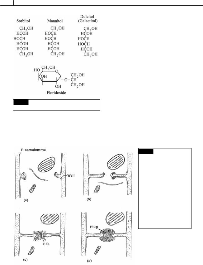
Floridoside (O- -D-galactopyranosyl-(1,2)-glyc- erol) is the major product of photosynthesis in the red algae, although mannitol, sorbitol, digeneaside, and dulcitol also occur (Fig. 4.4) (Barrow et al., 1995; Karsten et al., 2003). The concentration of floridoside increases in red algal cells as the salinity of the medium increases (Reed, 1985). This change in floridoside concentration is thought to compensate, at least in part, for the changes in external osmolarity, thereby preventing water from leaving the algal cells as the salinity increases. The levels of floridoside can be as high as 10% of the tissue dry-weight in some
92 EVOLUTION OF THE CHLOROPLAST
Fig. 4.4 Chemical structure of low molecular
polysaccharides that occur in the red algae.
marine red-algal thalli. The first observable product of photosynthesis is phosphoglyceric acid, as is the case in higher plants. Floridoside appears after 30 seconds of illumination and after 2 hours floridoside is the major product of
photosynthesis. Floridoside apparently has the same function as sucrose, the common product of photosynthesis in green algae and higher plants.
Floridean starch (Fig. 1.28) is the long-term storage product, occurring as grains in the cytoplasm outside of the chloroplast (Fig. 4.1). Floridean starch is similar to the amylopectin of higher plants, staining red-violet with iodine. In the more primitive Rhodophyceae the starch grains are clustered as a sheath around the pyrenoid of the chloroplast, whereas in the more advanced Rhodophyceae the starch grains are scattered in the cytoplasm (Hara, 1971; Lee, 1974).
Pit connections
Pit connections occur between the cells in all of the orders except the Porphyridiales, and the haploid phase of the Bangiales. It has been pointed out that the term “pit connection” is inappropriate because the structure is neither a “pit” nor a “connection”; however, because the term has been used for so long, it is probably best to retain it. A pit connection consists of a proteinaceous plug core in between two thallus cells (Figs. 4.5, 4.6).
Fig. 4.5 Semidiagrammatic drawing of the formation of a pit connection in a red alga. (a) The cross wall begins to furrow inward with the wall precursors found in vesicles derived from the cytoplasm; (b) the cross wall septum is complete, leaving an opening (aperture) in the center; (c) endoplasmic reticulum lies across the opening in the wall, and electron-dense material condenses in this area; (d) the pit connection is formed, consisting of a plug with the plasmalemma continuous from cell to cell. (Adapted from Ramus, 1969, 1971; Lee, 1971.)