

Neutron Scattering in Biology - Fitter Gutberlet and Katsaras
.pdf50 N. Niimura
neutron density was observed to extend from the position of the O atom of the carboxyl group labeled E35Oε1, suggesting that this carboxyl oxygen atom is a protonated atom. On the other hand, in Fig. 3.6b, it is seen that around this oxygen (E35Oε1) there is a water molecule at pH 7.0, but no indication of hydrogen (deuterium) atoms. The fact that this catalytic site is deprotonated explains why lysozyme has significantly reduced activity at pH 7.0. The results indicate that a water molecule around the carboxyl oxygen atom at pH 7.0 is kicked out as a result of the protonation of this oxygen atom at pH 4.9, and suggests that it is in fact this enzymatically-active proton which is subsequently transferred to the oxygen atom of the substrate (sugar) during the hydrolysis process. Mason et al. have carried out a neutron di raction study of lysozyme at pH 4.2 using a triclinic crystal. They report a protonated carboxylate group of Glu35 [26].
3.3 Hydrogen Bonding
3.3.1 Weak and Strong Hydrogen Bonding
Hydrogen bonds play important roles in countless biological processes. The interaction energy of the hydrogen bond is intermediate between those involving covalent and van der Waals forces. Hydrogen bonds are directional and form several kinds of networks in biological macromolecules. However, since it is not easy to determine the positions of all the hydrogen atoms in protein molecules using X-rays or NMR alone, detailed discussions of hydrogen bonds, X–H–Y (in which X and Y are the hydrogen donor and acceptor, respectively), have often been limited, because of the absence of detailed positional information of H atoms.
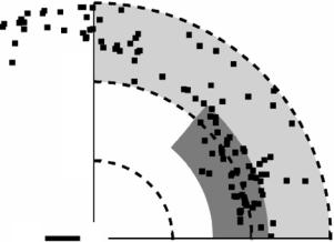
Along with other investigators, Baker and Hubbard have extensively discussed H-bonds in globular proteins, using hydrogen atom positions predicted from atomic coordinates derived from high-resolution protein X-ray data [27]. Hydrogen atoms were added to the various protein models at their calculated positions, but only those that could be unambiguously defined by the protein geometry. As a matter of fact, no hydrogens were placed on amino or hydroxyl groups, such as those in Ser, Thr, Tyr, or Lys side chains. Thus, in their conclusions, the authors stressed the necessity of high-resolution neutron di raction studies. Recently performed high-resolution neutron results meet their suggestion. Figure 3.7 shows one example, taken from the study of a mutant form of rubredoxin. In this diagram, the H-bonds, X–H(D)–Y have been plotted with the H(D) atom at the origin, the X–H(D) bond defining the horizontal axis, and the Y atom distributed in the (x, y) plane [20]. The resulting figure is consistent with the concept of the weak and strong H-bonds as proposed by Desiraju and Steiner [28]:
Strong H-bonds: 1.5 ˚A < d[H–Y] < 2.2 ˚A, 130◦ < angle[X–H–Y] < 180◦
3 Neutron Protein Crystallography |
51 |
[Å]
3
2
1
X |
D |
|
[Å] |
|
1 |
2 |
3 |
Fig. 3.7. The distribution of Y atoms in X–H(D)–Y hydrogen bonds, with the position of the H(D) atom fixed at the origin. The component of H(D)–Y along the X–H(D) direction is plotted along the horizontal axis, and the component of H(D)–Y perpendicular to X–H(D) is plotted in the vertical direction. The region of strong hydrogen bonds is indicated by the gray area, while weak hydrogen bonds are shown in the light gray area
Weak H-bonds: 2.2 ˚A < d[H–Y] < 3.0 ˚A, 90◦ < angle[X–H–Y] < 180◦
In Fig. 3.7, strong and weak H-bonds are indicated by gray and light gray areas, respectively. It can be seen that high-resolution neutron protein crystallography has allowed to examine H-bonds in more detail, and that numerous “weak H-bonds” in a protein structure can be identified with this method.
3.3.2 Bifurcated Hydrogen Bonds
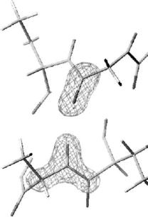
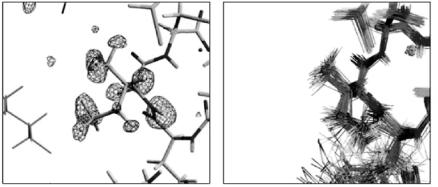
In our studies of several small proteins, all the hydrogen bonds between the C=O and N–H groups in the helices of the proteins have been surveyed including their hydrogen atom positions. Figure 3.8 shows one example of how a conventional hydrogen bond in a α-helix of myoglobin is seen in a neutron di raction experiment. The hydrogen positions as well as carbon, nitrogen and oxygen positions have been refined. This figure clearly indicates that the location of the experimental H positions with neutron data is usually unambiguous. However, we have found several exceptions to the conventional picture of H-bonds in α-helices.
When the hydrogen bonds in the α-helices were refined individually, several bifurcated hydrogen bonds were found. The occurrence of bifurcated hydrogen bonds in the α-helices of proteins has been proposed earlier, based on an
52 N. Niimura
Thr 67
C
O
D
N
C C
Thr 70
Ala 71
Fig. 3.8. Hydrogen bond in a α-helix. |Fo| − marked atoms were omitted for the calculation
|Fc| omit nuclear density map. The of Fc for the omit-map
analysis of calculated hydrogen atom positions from atomic coordinates derived from high-resolution X-ray data [27, 29]. However, it is somewhat risky to discuss the detailed structure of bifurcated hydrogen bonds based solely on those predictions. In high-resolution neutron protein crystallography, the H atoms of the polypeptide backbone can be identified and refined unambiguously. In the case of myoglobin a positional refinement with loosened restraints for the planarity of the peptide plane was performed, i.e., the O–C–N–H group was allowed to deviate from a planar trans-configuration. The result is that the O–C–N–H torsion angle showed deviations up to 15◦ from planarity with an average value equal to 179.2◦ and a standard deviation of 6.3◦. These values are in good agreement with those from ultra-high-resolution X-ray structure determinations [30, 31].
3.4 H/D Exchange
In a recently-completed analysis of wild type rubredoxin [19], the protein solution used was subjected to a H2O/D2O exchange prior to the growth of the crystals. Out of a total of 74 hydrogen/deuterium atoms at potentially exchangeable sites, 24 atoms did not have significant positive (deuterium) peaks at the expected positions. Of those, 11 atoms were bound to nitrogen
3 Neutron Protein Crystallography |
53 |
(a) |
(b) |
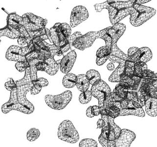
Fig. 3.9. |Fo| − |Fc| omit maps in the β-sheet region (a) and near the iron–sulfur cluster. (b) Negative densities are marked with arrows
atoms of the main chain, implying that those positions are not fully accessible to the H/D exchange process. Moreover, five of them have prominent negative (hydrogen) peaks. Figure 3.9a, b show |Fo| − |Fc| omit maps around those H atoms, calculated without contributions from any H and D atoms bonded to the main chain N atoms. Of the five residues whose backbone N–H groups did not exchange with D, Val4, Cys5, and Tyr12 are located at the central-sheet of the protein, while Cys38 and Ala43 are in the region of the unique FeS4 redox site. Those atoms all form hydrogen bonds with neighboring oxygen atoms of the main chain. In Fig. 3.9a, b, negative densities are at those H atoms positions even though most other H/D positions have positive densities. Those H atoms (negative densities) did not engage in H2O/D2O exchange in D2O presumably because of the poor solvent accessibility to those positions.
In order to obtain quantitative information about the distribution of hydrogen/deuterium populations in the rubredoxin molecule, the occupancies of the H and D atoms bonded to the main chain N atoms were refined. In this least-squares refinement, a D atom and an H atom were constrained to be in the same position, and their B-factors were set equal to the values of the N atoms to which they were bonded. During this refinement, the sum of the two occupancies of the H and D atoms was not constrained to be 1, but after the refinement these occupancies were recalculated and reset to give a sum of 1.
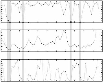
The result of this population refinement is shown in Fig. 3.10. The five atoms mentioned above have quite small (or nearly zero) values of the H/D exchange ratio. Comparing this result with the distribution of B-factors and accessible surface area (ASA) of the main chain atoms, it is seen that the H/D atoms having small H/D exchange ratios also have small B-factor values and small ASAs (Fig. 3.10, middle and bottom). These results not only show
54 N. Niimura
Temperature H/D Exchange
ASA
(Hratio D)to
) 2
(Afactor
) 2 (A
H: in backbone amide NH
1.0
0.8
0.6
0.4
0.2
0.0
0 |
10 |
20 |
30 |
40 |
50 |
20
15
10
5
0
0 |
10 |
20 |
30 |
40 |
50 |
50
40
30
20
10
0
0 |
10 |
20 |
30 |
40 |
50 |
|
|
|
Residue No. |
|
|
Fig. 3.10. H/D exchange ratio (top), B-factor (middle) and accessible surface area (ASA) (bottom) of main chain
that those atoms are located in the interior of the protein molecule, but also suggest that the regions around those atoms have such a rigid structure so that solvent molecules are unable to contact them. The same H/D exchange analyzes have been carried out on mutant form of rubredoxin and myoglobin and very similar results have been observed [18, 20].
It is interesting to compare the H/D exchange of wild type rubredoxin obtained by neutron protein crystallography with the one obtained by NMR [32]. Generally speaking, the trend is that an amide hydrogen bond, which has a fast H/D NMR exchange rate will show a high H/D exchange ratio in the neutron di raction experiment, and conversely a slow rate of NMR exchange also corresponds to a low ratio in the neutron di raction. However, a few exceptions can be found. Although a certain N–H bond has a slow H/D exchange rate according to NMR data, its H/D exchange ratio from neutron di raction is very high and it was found that a conformational change around the amide hydrogen has occurred in the crystallization process. Figures 3.11a, b show such an example near residue Ile7 of wild type rubredoxin. In the solution structure of Ile7 determined by NMR (Fig. 3.11b), the amide hydrogen atom is completely surrounded by the sidechain of Ile7 and shielded from the water [31], H/D exchange under such conditions should be slow. On the contrary, in the present neutron di raction crystal structure (Fig. 3.11a), Cδ of Ile7 is
3 Neutron Protein Crystallography |
55 |
(a) |
|
Neutron |
|
|
(b) |
|
NMR* |
|
|
|
|
|
|
|
|
|
|
|
|
|
|
|
Wild-type rubredoxin |
|
|
Zinc-substituted rubredoxin |
|
|||||
|
|
|
|
|
|
|
*P. R. Blake et. al., Protein Sci 1(1992) 1508 |
|||
|
|
|
|
|
CYS8 |
|
|
|
CYS8 |
|
|
|
|
|
|
|
|
|
|
|
|
|
|
|
|
|
Fe3+ |
|
|
|
|
H |
|
|
|
Cg 2 |
|
|
|
Cg 2 |
Zn2+ |
||
|
|
|
D |
|
|
|
||||
|
ILE7 |
Cb Ca |
|
|
|
|
|
|||
|
|
|
|
Cb Ca |
|
|||||
|
|
|
Cg 1 |
|
|
ILE7 |
H |
|||
|
|
|
D |
|
|
|
|
|||
|
|
Cd |
|
|
Cg 1 |
|
||||
|
|
|
|
|
|
|
|
|||
Cd
Nz
LYS2
Neighbor molecule
Contours: Omit map on H and D atoms of ILE7
Blue: σ=+3.5, Red; σ=3.5
Fig. 3.11. (a) The omit map on H and D atoms of Ile7 of wild-type rubredoxin determined by neutron di raction. (b) The portion of Ile7 of wild type rubredoxin (zinc-substituted) determined by NMR
bent to the outside of the proteins, the amide hydrogen atom is exposed to water and the H/D exchange ratio should be high.
3.5 Hydration in Proteins
3.5.1 Experimental Observation of Hydration Molecules
The hydration structure of myoglobin has been studied by Schoenborn et al. and the hydration layer structure such as radial distribution function of water around protein atoms were obtained [34–36]. In our recent study of myoglobin, the hydration structure of individual water molecules was presented [37]. Figure 3.12 displays one region of the hydration structure around myoglobin. It shows that all of the hydration water molecules are completely isolated. It is also remarkable that a sulfate group can be clearly distinguished in this map. In some cases, hydrogen (deuterium) atoms in water molecules can be clearly identified in triangular (boomerang) shaped peaks and the formation of the hydrogen bonds between two water molecules can be recognized as well. At the same time it is interesting to note that, near the two triangular-shaped contours, a spherically shaped water molecule can be found (Fig. 3.12). Moreover, water molecules with other shapes, such as ellipsoidal (stick-shaped) ones, have been found in other places. The interpretation of these shapes will be discussed in Sect. 3.5.2 [37].
56 N. Niimura
D2O
D2O
D2O
SO4
D2O
D2O
Fig. 3.12. Protein–protein contact region in the case of myoglobin. 2|Fo| − |Fc| nuclear density map contoured at positive and negative values. The 2|Fo|−|Fc| X-ray electron density map for the water molecules (D2O) is superimposed. The triangularshaped neutron contours correspond to D2O molecules
3.5.2 Classification of Hydration
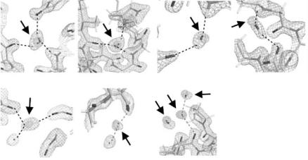
We have categorized observed water molecules into the following classes based on their appearance in Fourier maps: (i) triangular shape, (ii) ellipsoidal stick shape, and (iii) spherical shape. Moreover the second category, ellipsoidal stick shapes can be further sub-classified as (iia) short and (iib) long. We found that this classification conveniently reflects the degree of disorder and/or dynamic behavior of a water molecule. A typical example of the (i) triangular shape is shown in Fig. 3.13a-1,2, in which the contours indicate 2|Fo| − |Fc| maps calculated from neutron and X-ray data, respectively. The oxygen positions observed by X-ray and neutron scattering coincide within experimental error. In this case, the two deuterium atoms and the oxygen atom of the water molecule are H-bonded to nearby O/N and deuterium atoms, respectively. Thus, it can be seen that the orientation of this water molecule is well-defined. In fact, triangular shaped contours correspond to the most highly-ordered water molecules in our maps.
A typical example of a short ellipsoidal stick shape (iia) is shown in Fig. 3.13b-1,2. The oxygen position observed by X-rays is located at one end of the neutron Fourier peak, and only one deuterium atom could be observed.
|
3 |
Neutron Protein Crystallography |
57 |
|
(a)-1 |
(a)-2 |
(b)-1 |
(b)-2 |
|
(c) |
(d)-1 |
(d)-2 |
Fig. 3.13. 2|Fo| − |Fc| nuclear density maps of water molecules of hydration for myoglobin and the rubredoxin mutant observed by neutron protein crystallography. Examples shown are those of those of: (a) triangular shape, (b) short ellipsoidal shape, (c) long ellipsoidal shape and (d) spherical shape. In these maps, the contours correspond to neutron peaks, while the superimposed contours correspond to oxygen peaks from X-ray data (marked by arrows). Observed (located) atoms from the neutron data are shown as stick diagrams. Note that in Fig. 3.13a all atoms of the central D2O molecule are visible, whereas in the other diagrams only some of the solvent atoms have been located: O, D (Fig. 3.13b), two D (Fig. 3.13c)and O only (Fig. 3.13d)
The observed D and O atoms are H-bonded to neighboring O/N and D atoms, respectively, but the other deuterium atom was not identified because of the molecular rotation (or packing disorder) around the fixed O–D bond. Thus, short ellipsoidal stick shaped peaks are interpreted to represent water molecules rotationally disordered around an O–D bond. A typical example of the long ellipsoidal stick-shaped peak (iib) is shown in Fig. 3.13c. The O position observed by X-rays (but not by neutrons) is located in the middle of the neutron Fourier peak, and the two D atoms are clearly observed in the neutron map. The entire appearance is that of an elongated stick. In this case, the two D atoms are H-bonded to neighboring O and/or N atoms, but the O atoms of the D2O molecule cannot be identified because of the molecular rotation or packing disorder around the D–D axis. Finally, a typical example of the spherical-shaped peak (iii) is shown in Fig. 3.13d-1,2. Only the center of gravity of this type of water molecule can be defined because its orientation is totally disordered. A spherical peak in a neutron Fourier map always means that the whole water molecule is freely rotating, even if X-ray results (which only show the O atom) reveal no hint of this disorder.
58 N. Niimura
Although the above classification has been carried out based on the appearance of peaks in Fourier maps, it was found that the shapes are strongly correlated with the existence of hydrogen bonds, which fix the positions of atoms of water molecules. Most of the triangular-shaped water molecules are fixed at three atoms (D, O, D), while ellipsoidal ones are fixed at two atoms (D, D or D, O). In contrast, some spherical shaped water molecules are not fixed by any observed H-bonds. The average number of “anchor points” of triangular, ellipsoidal and spherical-shaped water molecules are 2.3, 1.3, and 0.3, respectively. In the three proteins of myoglobin, wild type and mutant form of rubredoxin, the average populations of triangular, ellipsoidal, and spherical shapes are 29%, 16%, and 55%, respectively [37].
3.5.3 Dynamic Behavior of Hydration
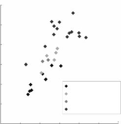
The dynamic behavior of water molecules becomes clearer when the B-factors obtained by neutron and X-ray experiments are plotted against each other as shown in Fig. 3.14, in which the B-factors obtained from the neutron analysis are the averaged values from three atoms (D, O, and D), while those from the X-ray analysis are those of the O atoms only. The B-factors of oxygen atoms obtained by X-rays are in the range from 13 to 45 ˚A2. It is observed that the
] 2
factor-B [A(neutron)
60
50
40
30
20
10
0
Triangular peak
Short allipsoidal peak
Long allipsoidal peak
Spherical peak
10 |
20 |
30 |
40 |
50 |
60 |
|
B-factor (X-rays) [A2] |
|
|
||
Fig. 3.14. Correlation between the B-factors of hydration water molecules obtained from neutron and X-ray scattering data for the rubredoxin mutant. Neutron B- factors were obtained using the average scattering lengths of D, O, and D atoms from every water molecule, regardless of shape; while in the case of X-ray B-factors, only those from O atoms were included
3 Neutron Protein Crystallography |
59 |
small, intermediate, and large B-factors from the X-ray analysis correspond to water molecules having the triangular, ellipsoidal, and spherical shapes, respectively. The spherical peak in the neutron Fourier map always means that the whole water molecule is freely rotating, even though the X-ray results (which show only the O atom) reveal no hint of this disorder.
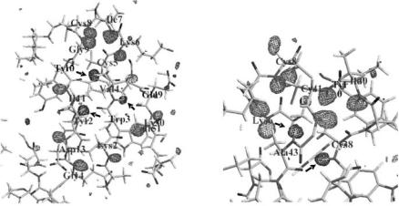
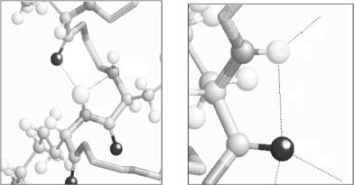
The construction of a data base of hydrogen and hydration in proteins is now under way. The positional coordinates of all hydrogen atoms and hydration water molecules determined by neutron protein crystallography are stored according to the usual PDB format. The main function of the hydrogen hydration data base (HHDB) is (i) to extract the structural information relevant to hydrogen atoms, such as the stereochemical atomic configuration in the vicinity of a selected hydrogen atom, (ii) the search of all the H-bonds between main chains, the main chain and the side chain, and side chains and (iii) the statistical classification of H-bonds. During the analysis of H-bonds by the use of HHDB, very unfamiliar types of H-bonds have been discovered as shown in Fig. 3.15. Figure 3.15a shows that the nitrogen atom of amide (Lys78) in the main chain is an acceptor of H atoms of the neighbor amide (Lys79) (the H bond length H–N is 2.18 ˚A), and Fig. 3.15b shows that the H- bond is formed between the amide (Lys2) N–H and O=C in Lys2 (the H-bond length H–O is 2.50 ˚A).
3.6 Crystallization
One fundamental problem in neutron crystallography is the di cultly in obtaining large single crystals. It is really true that neutron protein crystallography necessitates the use of large protein crystals, the volume of which should be larger than 1 mm3 currently. Usually such a large single crystal is di cult
(a) (b)
2.22
|
36 |
LYS:2:A |
LEU:76:A |
|
|
2.0B |
LYS:7B:A |
|
2.1B |
|
|
|
|
|
|
|
2.50 |
|
LYS:79:A |
|
|
|
LYS:2:A |
|
|
1.B7 |
Fig. 3.15. Very unfamiliar types of H-bonds found in (a) myoglobin and (b) mutant form of rubredoxin by operating the hydrogen and hydration data base (HHDB)