

Biomechanics Principles and Applications - Schneck and Bronzino
.pdf12 |
Biomechanics: Principles and Applications |
FIGURE 1.5 Comparison of predictions of Katz two-level composite model with the experimental data of Bonfield and Grynpas. Each curve represents a different lamellar configuration within a single osteon, with longitudinal fibers A, 64%; B, 57%; C, 50%; D, 37%; and the rest of the fibers assumed horizontal. (From Katz JL, Mechanical Properties of Bone, AMD, Vol. 45, New York, American Society of Mechanical Engineers, 1981. With permission.)
σij (t )= ∫t |
Cijkl (t − τ) |
d kl (τ) |
dτ |
(1.17) |
|
dτ |
|||||
− ∞ |
|
|
|
where σij(t) and εkl(τ) are the time-dependent second rank stress and strain tensors, respectively, and
Cijkl(t – τ) is the fourth-rank relaxation modulus tensor. This tensor has 36 independent elements for the lowest symmetry case and 12 nonzero independent elements for an orthotropic solid. Again, as for linear
elasticity, a reduced notation is used, i.e., 11 → 1, 22 → 2, 33 → 3, 23 → 4, 31 → 5, and 12 → 6. If we apply Eq. (1.17) to the case of an orthotropic material, e.g., plexiform bone, in uniaxial tension (compression) in the 1 direction [Lakes and Katz, 1974], in this case using the reduced notation, we obtain
σ1(t )= ∫t |
|
(t − τ) |
d 1(τ) |
|
(t − τ) |
d 2 (τ) |
|
(t − τ) |
|
d 3(τ) |
|
|
|
|
||||
C11 |
+ C12 |
+ C13 |
|
|
dτ |
(1.18) |
||||||||||||
dτ |
dτ |
|
dτ |
|||||||||||||||
|
− ∞ |
|
|
|
|
|
|
|
|
|
|
|
||||||
|
|
|
|
|
|
|
|
|
|
|
|
|
|
|
|
|
|
|
|
(t )= ∫t |
|
(t − τ) |
d 1(τ) |
|
|
(t − τ) |
d 2 (τ) |
|
(t − τ) |
d 3(τ) |
|
|
|
||||
σ2 |
C21 |
|
+ C22 |
+ C23 |
|
= 0 |
(1.19) |
|||||||||||
|
dτ |
|
||||||||||||||||
|
− ∞ |
|
|
dτ |
|
|
|
|
|
dτ |
|
|
|
|
||||
|
|
|
|
|
|
|
|
|
|
|
|
|
|
|
|
|
|
|
for all t, and

Mechanics of Hard Tissue |
|
|
|
|
|
|
|
|
|
|
13 |
|||
|
(t )= ∫t |
|
(t − τ) |
d 1(τ) |
|
(t − τ) |
d 2 (τ) |
|
(t − τ) |
d 3(τ) |
|
|
|
|
σ3 |
C31 |
+ C32 |
+ C33 |
|
dτ = 0 |
(1.20) |
||||||||
dτ |
dτ |
dτ |
||||||||||||
|
− ∞ |
|
|
|
|
|
|
|
|
|||||
|
|
|
|
|
|
|
|
|
|
|
|
|
|
|
for all t.
Having the integrands vanish provides an obvious solution to Eqs. (1.19) and (1.20). Solving them
|
[d (2τ) ] |
|
[d (3τ) ] |
|
|
|
|
|
|||
simultaneously for |
|
|
and |
|
|
and substituting these values in Eq. (1.17) yields |
|
||||
dτ |
dτ |
|
|
||||||||
|
|
|
|
|
|
|
|
|
|||
|
|
|
|
|
σ1(t )= ∫t |
E1 |
(t − τ) |
d 1(τ) |
dτ |
(1.21) |
|
|
|
|
|
|
dτ |
||||||
|
|
|
|
|
|
− ∞ |
|
|
|
|
|
where, if for convenience we adopt the notation Cij Cij (t – τ), then Young’s modulus is given by
|
C31 − |
(C21C33 |
C23 ) |
|
C21 − (C31C22 |
C32 ) |
|
||||
|
[ |
|
|
|
] |
[ |
|
|
] |
|
|
E1 (t − τ)= C11 + C12 |
[ |
|
] |
+ C13 |
[ |
|
] |
(1.22) |
|||
(C21C33 |
C23 )− C32 |
|
(C22C33 |
C32 )− C23 |
|
||||||
In this case of uniaxial tension (compression), only nine independent orthotropic tensor components are involved, the three shear components being equal to zero. Still, this time-dependent Young’s modulus is a rather complex function. As in the linear elastic case, the inverse form of the Boltzmann integral can be used; this would constitute the compliance formulation.
If we consider the bone being driven by a strain at a frequency ω, with a corresponding sinusoidal stress lagging by an angle δ, then the complex Young’s modulus E*(ω) may be expressed as
E * (ω)= E′(ω)+ iE′′(ω) |
(1.23) |
where E′(ω), which represents the stress–strain ratio in phase with the strain, is known as the storage modulus, and E″(ω), which represents the stress–strain ratio 90° out of phase with the strain, is known as the loss modulus. The ratio of the loss modulus to the storage modulus is then equal to tan δ. Usually, data are presented by a graph of the storage modulus along with a graph of tan δ, both against frequency. For a more complete development of the values of E′(ω) and E″(ω), as well as for the derivation of other viscoelastic technical moduli, see Lakes and Katz [1974]. For a similar development of the shear storage and loss moduli, see Cowin [1989].
Thus, for a more complete understanding of bone’s response to applied loads, it is important to know its rheologic properties. There have been a number of early studies of the viscoelastic properties of various long bones [Sedlin, 1965; Smith and Keiper, 1965; Laird and Kingsbury, 1973; Lugassy, 1968; Black and Korostoff, 1973]. However, none of these was performed over a wide enough range of frequency (or time) to completely define the viscoelastic properties measured, e.g., creep or stress relaxation. Thus it is not possible to mathematically transform one property into any other to compare results of three different experiments on different bones [Lakes and Katz, 1974].
In the first experiments over an extended frequency range, the biaxial viscoelastic as well as uniaxial viscoelastic properties of wet cortical human and bovine femoral bone were measured using both dynamic and stress relaxation techniques over eight decades of frequency (time) [Lakes et al., 1979]. The results of these experiments showed that bone was both nonlinear and thermorheologically complex, i.e., time–temperature superposition could not be used to extend the range of viscoelastic measurements. A nonlinear constitutive equation was developed based on these measurements [Lakes and Katz, 1979a].

14 |
Biomechanics: Principles and Applications |
FIGURE 1.6 Comparison of relaxation spectra for wet human bone, specimens 5 and 6 [Lakes et al., 1979] in simple torsion; T = 37°C. First approximation from relaxation and dynamic data. Human tibial bone, specimen 6.
Human tibial bone, specimen 5, Gstd = G(10 s). Gstd(5) = G(10 s). Gstd(5) = 0.590 × 10⁶ lb/in.2. Gstd(6) × 0.602 × 106 lb/in.2. (Courtesy Journal of Biomechanics, Pergamon Press.)
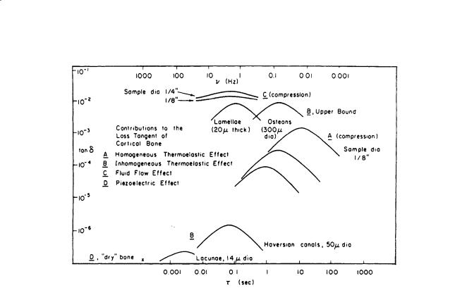
In addition, relaxation spectrums for both human and bovine cortical bone were obtained; Fig. 1.6 shows the former [Lakes and Katz, 1979b]. The contributions of several mechanisms to the loss tangent of cortical bone is shown in Fig. 1.7 [Lakes and Katz, 1979b]. It is interesting to note that almost all the major loss mechanisms occur at frequencies (times) at or close to those in which there are “bumps,” indicating possible strain energy dissipation, on the relaxation spectra shown on Fig. 1.6. An extensive review of the viscoelastic properties of bone can be found in the CRC publication Natural and Living Biomaterials [Lakes and Katz, 1984].
Following on Katz’s [1976, 1980] adaptation of the Hashin-Rosen hollow fiber composite model [1964], Gottesman and Hashin [1979] presented a viscoelastic calculation using the same major assumptions.
1.7 Related Research
As stated earlier, this chapter has concentrated on the elastic and viscoelastic properties of compact cortical bone and the elastic properties of trabecular bone. At present there is considerable research activity on the fracture properties of the bone. Professor William Bonfield and his associates at Queen Mary and Westfield College, University of London and Professor Dwight Davy and his colleagues at Case Western Reserve University are among those who publish regularly in this area. Review of the literature is necessary in order to become acquainted with the state of bone fracture mechanics.
An excellent introductory monograph which provides a fascinating insight into the structure-property relationships in bones including aspects of the two areas discussed immediately above is Professor John Currey’s The Mechanical Adaptations of Bones, published in 1984 by Princeton University Press.
Mechanics of Hard Tissue |
15 |
FIGURE 1.7 Contributions of several relaxation mechanisms to the loss tangent of cortical bone. A: Homogeneous thermoelastic effect. B: Inhomogeneous thermoelastic effect. C: Fluid flow effect. D: Piezoelectric effect [Lakes and Katz, 1984]. (Courtesy CRC Press.)
Defining Terms
Apatite: Calcium phosphate compound, stoichiometric chemical formula Ca5(PO4)3 ·X, where X is OH– (hydroxyapatite), F– (fluorapatite), Cl– (chlorapatite), etc. There are two molecules in the basic crystal unit cell.
Cancellous bone: Also known as porous, spongy, trabecular bone. Found in the regions of the articulating ends of tubular bones, in vertebrae, ribs, etc.
Cortical bone: The dense compact bone found throughout the shafts of long bones such as the femur, tibia, etc. also found in the outer portions of other bones in the body.
Haversian bone: Also called osteonic. The form of bone found in adult humans and mature mammals, consisting mainly of concentric lamellar structures, surrounding a central canal called the Haversian canal, plus lamellar remnants of older Haversian systems (osteons) called interstitial lamellae.
Interstitial lamellae: See Haversian bone above.
Orthotropic: The symmetrical arrangement of structure in which there are three distinct orthogonal axes of symmetry. In crystals this symmetry is called orthothombic.
Osteons: See Haversian bone above.
Plexiform: Also called laminar. The form of parallel lamellar bone found in younger, immature nonhuman mammals.
Transverse isotropy: The symmetry arrangement of structure in which there is a unique axis perpendicular to a plane in which the other two axes are equivalent. The long bone direction is chosen as the unique axis. In crystals this symmetry is called hexagonal.

16 Biomechanics: Principles and Applications
References
Ashman RB, Rho JY. 1988. Elastic modulus of trabecular bone material. J Biomech 21:177.
Black J, Korostoff E. 1973. Dynamic mechanical properties of viable human cortical bone. J Biomech 6:435. Bonfield W, Grynpas MD. 1977. Anisotropy of Young’s modulus of bone. Nature, London 270:453. Bumrerraj S. 1999. Scanning Acoustic Microscopy Studies of Human Cortical and Trabecular Bone, M.S.
(BME) project (Katz, JL, advisor), Case Western Reserve University, Cleveland, OH.
Choi K, Goldstein SA. 1992. A comparison of the fatigue behavior of human trabecular and cortical bone tissue. J Biomech 25:1371.
Chung DH, Buessem WR. 1968. In Vahldiek, FW and Mersol, SA (Eds.), Anisotropy in Single-Crystal Refractory Compounds, Vol. 2, p. 217. New York, Plenum Press.
Cowin SC. 1989. Bone Mechanics. Boca Raton, FL, CRC Press.
Cox HL. 1952. The elasticity and strength of paper and other fibrous materials. Br Appl Phys 3:72. Crolet JM, Aoubiza B, Meunier A. 1993. Compact bone: numerical simulation of mechanical character-
istics. J Biomech 26:(6)677.
Currey JD. 1964. Three analogies to explain the mechanical properties of bone. Biorheology (2):1. Currey JD. 1969. The relationship between the stiffness and the mineral content of bone. J Biomech
(2):477.
Currey J. 1984. The Mechanical Adaptations of Bones. Princeton, NJ, Princeton University Press. Gottesman T, Hashin Z. 1979. Analysis of viscoelastic behavior of bones on the basis of microstructure.
J Biomech 13:89.
Hashin Z, Rosen BW. 1964. The elastic moduli of fiber reinforced materials. J Appl Mech (31):223. Hashin Z, Shtrikman S. 1963. A variational approach to the theory of elastic behavior of multiphase
materials. J Mech Phys Solids (11):127.
Hastings GW, Ducheyne P (Eds.). 1984. Natural and Living Biomaterials, Boca Raton, FL, CRC Press. Herring GM. 1977. Methods for the study of the glycoproteins and proteoglycans of bone using bacterial
collagenase. Determination of bone sialoprotein and chondroitin sulphate. Calcif Tiss Res (24):29. Hogan HA. 1992. Micromechanics modeling of Haversian cortical bone properties. J Biomech 25(5):549. Katz JL. 1971a. Hard tissue as a composite material: I. Bounds on the elastic behavior. J Biomech 4:455. Katz JL. 1971b. Elastic properties of calcified tissues. Isr J Med Sci 7:439.
Katz JL. 1976. Hierarchical modeling of compact haversian bone as a fiber reinforced material. In Mates, RE and Smith, CR (Eds.), Advances in Bioengineering, pp. 17–18. New York, American Society of Mechanical Engineers.
Katz JL. 1980. Anisotropy of Young’s modulus of bone. Nature 283:106.
Katz JL. 1981. Composite material models for cortical bone. In Cowin SC (Ed.), Mechanical Properties of Bone, Vol. 45, pp. 171–184. New York, American Society of Mechanical Engineers.
Katz JL, Meunier A. 1987. The elastic anisotropy of bone. J Biomech 20:1063.
Katz JL, Meunier A. 1990. A generalized method for characterizing elastic anisotropy in solid living tissues. J Mater Sci Mater Med 1:1.
Katz JL, Ukraincik K. 1971. On the anisotropic elastic properties of hydroxyapatite. J Biomech 4:221. Katz JL, Ukraincik K. 1972. A fiber-reinforced model for compact haversian bone. Program and Abstracts
of the 16th Annual Meeting of the Biophysical Society, 28a FPM-C15, Toronto.
Keaveny TM, Hayes WC. 1993. A 20-year perspective on the mechanical properties of trabecular bone.
J Biomech Eng 115:535.
Knets IV. 1978. Mekhanika Polimerov 13:434.
Laird GW, Kingsbury HB. 1973. Complex viscoelastic moduli of bovine bone. J Biomech 6:59. Lakes RS. 1993. Materials with structural hierarchy. Nature 361:511.
Lakes RS, Katz JL. 1974. Interrelationships among the viscoelastic function for anisotropic solids: application to calcified tissues and related systems. J Biomech 7:259.
Lakes RS, Katz JL. 1979a. Viscoelastic properties and behavior of cortical bone. Part II. Relaxation mechanisms. J Biomech 12:679.

Mechanics of Hard Tissue |
17 |
Lakes RS, Katz JL. 1979b. Viscoelastic properties of wet cortical bone: III. A nonlinear constitutive equation. J Biomech 12:689.
Lakes RS, Katz JL. 1984. Viscoelastic properties of bone. In Hastings, GW and Ducheyne, P (Eds.), Natural and Living Tissues, pp 1–87. Boca Raton, FL, CRC Press.
Lakes RS, Katz JL, Sternstein SS. 1979. Viscoelastic properties of wet cortical bone: I. Torsional and biaxial studies. J Biomech 12:657.
Lang SB. 1969. Elastic coefficients of animal bone. Science 165:287.
Lipson SF, Katz JL. 1984. The relationship between elastic properties and microstructure of bovine cortical bone. J Biomech 4:231.
Lugassy AA. 1968. Mechanical and Viscoelastic Properties of Bone and Dentin in Compression, thesis, Metallurgy and Materials Science, University of Pennsylvania.
Maharidge R. 1984. Ultrasonic Properties and Microstructure of Bovine Bone and Haversian Bovine Bone Modeling, thesis, Rensselaer Polytechnic Institute, Troy, NY.
Park JB. 1979. Biomaterials: An Introduction. New York, Plenum Press.
Pellegrino ED, Biltz RM. 1965. The composition of human bone in uremia. Medicine 44:397. Piekarski K. 1973. Analysis of bone as a composite material. Int J Eng Sci 10:557.
Reuss A. 1929. Berechnung der Fliessgrenze von Mischkristallen auf Grund der Plastizitatsbedingung für Einkristalle, A. Zeitschrift für Angewandte Mathematik und Mechanik 9:49–58.
Rho JY, Ashman RB, Turner CH. 1993. Young’s modulus of trabecular and cortical bone material; ultrasonic and microtensile measurements. J Biomech 26:111.
Rho JY, Roy ME, Tsui TY, Pharr GM. 1999. Elastic properties of microstructural components of human bone tissue as measured by indentation. J Biomed Mater Res 45:48.
Ryan SD, Williams JL. 1989. Tensile testing of rodlike trabeculae excised from bovine femoral bone.
J Biomech 22:351.
Sedlin E. 1965. A rheological model for cortical bone. Acta Orthop Scand 36(suppl 83).
Smith R, Keiper D. 1965. Dynamic measurement of viscoelastic properties of bone. Am J Med Elec 4:156. Townsend PR, Rose RM, Radin EL. 1975. Buckling studies of single human trabeculae. J Biomech 8:199. Turner CH, Rho JY, Takano Y, Tsui TY, Pharr GM. 1999. The elastic properties of trabecular and cortical bone tissues are simular: results from two microscopic measurement techniques. J Biomech 32:437.
Van Buskirk WC, Ashman RB. 1981. The elastic moduli of bone. In Cowin, SC (Ed.), Mechanical Properties of Bone, AMD Vol. 45, pp. 131–143. New York, American Society of Mechanical Engineers.
Vejlens L. 1971. Glycosaminoglycans of human bone tissue: I. Pattern of compact bone in relation to age. Calcif Tiss Res 7:175.
Voigt W. 1966. Lehrbuch der Kristallphysik, Teubner, Leipzig 1910; reprinted (1928) with an additional appendix. Leipzig, Teubner, New York, Johnson Reprint.
Wagner HD, Weiner S. 1992. On the relationship between the microstructure of bone and its mechanical stiffness. J Biomech 25:1311.
Wainwright SA, Briggs WD, Currey JD, Gosline JM. 1982. Mechanical Design in Organisms. Princeton, NJ, Princeton University Press.
Weiner S, Traub W. 1989. Crystal size and organization in bone. Conn Tissue Res 21:259.
Yoon HS, Katz JL. 1976a. Ultrasonic wave propagation in human cortical bone: I. Theoretical considerations of hexagonal symmetry. J Biomech 9:407.
Yoon HS, Katz JL. 1976b. Ultrasonic wave propagation in human cortical bone: II. Measurements of elastic properties and microhardness. J Biomech 9:459.
Further Information
Several societies both in the United States and abroad hold annual meetings during which many presentations, both oral and poster, deal with hard tissue biomechanics. In the United States these societies include the Orthopaedic Research Society, the American Society of Mechanical Engineers, the Biomaterials Society, the American Society of Biomechanics, the Biomedical Engineering Society, and the Society

18 |
Biomechanics: Principles and Applications |
for Bone and Mineral Research. In Europe there are alternate year meetings of the European Society of Biomechanics and the European Society of Biomaterials. Every four years there is a World Congress of Biomechanics; every three years there is a World Congress of Biomaterials. All of these meetings result in documented proceedings; some with extended papers in book form.
The two principal journals in which bone mechanics papers appear frequently are the Journal of Biomechanics published by Elsevier and the Journal of Biomechanical Engineering published by the American Society of Mechanical Engineers. Other society journals which periodically publish papers in the field are the Journal of Orthopaedic Research published for the Orthopaedic Research Society, the Annals of Biomedical Engineering published for the Biomedical Engineering Society, and the Journal of Bone and Joint Surgery (both American and English issues) for the American Academy of Orthopaedic Surgeons and the British Organization, respectively. Additional papers in the field may be found in the journal
Bone and Calcified Tissue International.
The 1984 CRC volume, Natural and Living Biomaterials (Hastings, G.W. and Ducheyne, P., Eds.) provides a good historical introduction to the field. A more advanced book is Bone Mechanics (Cowin, S.C., 1989); the second edition was published by CRC Press in 2001.
Many of the biomaterials journals and society meetings will have occasional papers dealing with hard tissue mechanics, especially those dealing with implant–bone interactions.

Mechanics of Hard Tissue |
19 |
Appendix
The Voigt and Reuss moduli for both transverse isotropic and orthotropic symmetry are given below:
Voigt Transverse Isotropic
K V = 2(C11 + C12 )+ 4(C13 + C33 )
|
9 |
|
|
|
|
|
|
(1.A1) |
||
|
|
|
(C11 + C12 )− 4C13 + 2C33 +12(C44 + C66 ) |
|
|
|
||||
|
|
GV = |
|
|
|
|
||||
|
|
|
|
|
|
|
||||
|
30 |
|
|
|
|
|
|
|
||
Reuss Transverse Isotropic |
|
|
|
|
|
|
||||
K R = |
C33 (C11 + C12 )− 2C132 |
|
|
|
|
|
|
|||
(C11 + C12 − 4C13 + 2C33 ) |
|
|
|
|
|
|
|
|||
|
|
|
5[C33 (C11 + C12 )− 2C132 ]C44C66 |
|
|
|
(1.A2) |
|||
|
|
|
|
|
|
|
||||
GR = |
2{[C33(C11 + C12 )− 2C132 ](C44 + C66 )+[C44C66 (2C11 + C12 )+ 4C13 + C33 |
] 3} |
|
|||||||
Voigt Orthotropic |
|
|
|
|
|
|
||||
|
K V = |
C11 + C22 + C33 + 2(C12 + C13 + C23 ) |
|
|
|
|
|
|
|
|
|
|
|
|
|
|
|
|
|||
|
9 |
|
|
|
] |
|
|
|
||
|
GV = [ |
(C12 |
+ C13 |
|
|
(1.A3) |
||||
|
|
C11 + C22 + C33 + 3(C44 + C55 + C66 )− |
+ C23 ) |
|
|
|||||
|
15 |
|
|
|
|
|
|
|
||
Reuss Orthotropic
K R |
= |
|
∆ |
− 2(C11C23 + C22C13 + C33C12 ) |
|
|
C11C22 |
+ C22C33 + C33C11 |
|
|
|||
|
|
+ 2(C12C23 + C23C13 + C13C12 )− (C122 + C132 + C232 ) |
|
|
||
GR = 15 4{(C11C22 + C22C33 + C33C11 + C11C23 + C22C13 + C33C12 ) |
(1.A4) |
|||||
|
[ |
|
]} |
∆ |
|
|
|
|
− C12 |
(C12 + C23 )+ C23 (C23 + C13 )+ C13 (C13 + C12 ) |
|
||
+ 3(1 C44 +1 C55 +1 C66 ))
where ∆ is given in Eq. (1.15).


2
Mechanics of Blood Vessels
|
2.1 |
Assumptions.......................................................................... |
21 |
|
|
|
Homogeneity of the Vessel Wall • Incompressibility of the |
||
|
|
Vessel Wall • Inelasticity of the Vessel Wall |
• Residual Stress |
|
|
|
and Strain |
|
|
Thomas R. Canfield |
2.2 |
Vascular Anatomy................................................................. |
22 |
|
2.3 |
Axisymmetric Deformation |
23 |
||
Argonne National Laboratory |
||||
2.4 |
Experimental Measurements |
25 |
||
|
||||
Philip B. Dobrin |
2.5 |
Equilibrium........................................................................... |
25 |
|
Hines VA Hospital and Loyola |
2.6 |
Strain Energy Density Functions ........................................ |
27 |
|
University Medical Center |
|
Isotropic Blood Vessels • Anisotropic Blood Vessels |
||
2.1 Assumptions
This chapter is concerned with the mechanical behavior of blood vessels under static loading conditions and the methods required to analyze this behavior. The assumptions underlying this discussion are for ideal blood vessels that are at least regionally homogeneous, incompressible, elastic, and cylindrically orthotropic. Although physiologic systems are nonideal, much understanding of vascular mechanics has been gained through the use of methods based upon these ideal assumptions.
Homogeneity of the Vessel Wall
On visual inspection, blood vessels appear to be fairly homogeneous and distinct from surrounding connective tissue. The inhomogeneity of the vascular wall is realized when one examines the tissue under a low-power microscope, where one can easily identify two distinct structures: the media and adventitia. For this reason the assumption of vessel wall homogeneity is applied cautiously. Such an assumption may be valid only within distinct macroscopic structures. However, few investigators have incorporated macroscopic inhomogeneity into studies of vascular mechanics [17].
Incompressibility of the Vessel Wall
Experimental measurement of wall compressibility of 0.06% at 270 cm of H2O indicates that the vessel can be considered incompressible when subjected to physiologic pressure and load [2]. In terms of the mechanical behavior of blood vessels, this is small relative to the large magnitude of the distortional strains that occur when blood vessels are deformed under the same conditions. Therefore, vascular
Work sponsored by the U.S. Department of Energy Order Contract W-31-109-Eng-38.
0-8493-1492-5/03/$0.00+$.50 © 2003 by CRC Press LLC