


197
8
Single-Molecule Detection and Manipulation in Nanotechnology and Biology
Christopher L. Kuyper, Gavin D. M. Jeffries, Robert M. Lorenz, and Daniel T. Chiu
8.1
Introduction
The set of tools for visualizing the world at the molecular and nanoscopic level has been both broadened and refined in the past decades. Investigating individual molecules is becoming more routine, and a wide array of methods have revealed exciting new information that otherwise is unobtainable from ensemble studies. Examples of these tools include the family of scanning probe microscopies (SPM), such as scanning tunneling microscopy (STM) and atomic force microscopy (AFM), which have offered us exquisite details and visualizations of single atoms and molecules on surfaces [1–5]. The use of optical techniques to detect and study single molecules has also gained prominence in recent years owing to their ease of implementation and their ability to probe biological systems in solution under physiologically relevant conditions. Platforms such as confocal optics, epifluorescence detection, and total internal-reflection fluorescence (TIRF) microscopy have become increasingly common due to relatively straightforward design, impressive detection sensitivities, and easy integration into commercially available microscope systems [6–9]. Optically based single-molecule experiments are not only limited to detection and visualization; optical traps serve as tools for measuring forces and movements of individual biomolecules in solution [10–14]. Due to the broad range of optical techniques that have been developed for the detection and manipulation of single molecules, which range from near-field microscopy to the use of nonlinear optical methods [15–19], we will not be able to discuss all these examples in this chapter. Instead, we begin with an introduction of the common techniques used in optically based single-mole- cule detection (e.g., confocal, epifluorescence, TIRF) and manipulation (e.g., optical trapping) that are pertinent to biology and nanotechnology. We then continue with a discussion of a few select areas to which these techniques have been applied, both to illustrate the past accomplishments and the future potential of optically based sin- gle-molecule methodologies.
Nanofabrication Towards Biomedical Applications. C. S. S. R. Kumar, J. Hormes, C. Leuschner (Eds.) Copyright 2005 WILEY-VCH Verlag GmbH & Co. KGaA, Weinheim
ISBN 3-527-31115-7

1988 Single-Molecule Detection and Manipulation in Nanotechnology and Biology
8.2
Optical Detection of Single Molecules
8.2.1
Detecting Single Molecules with Confocal Fluorescence Microscopy
In confocal microscopy [Fig. 8.1(A)], coherent laser radiation is collimated and reflected off a dichroic mirror to fill the back aperture of a high numerical aperture (NA) objective (NA > 1.2). Exiting from the objective, laser light is focused to a dif- fraction-limited spot that has a beam waist of ~200–350 nm in diameter as defined by the Rayleigh criterion, which depends on the wavelength and NA of the objective. Along the same axis of the incoming light, fluorescence emitted from the focal point in the sample plane is recollected by the objective and passed through the dichroic mirror and a bandpass filter, which ensures that no extraneous Rayleigh scattering, or laser radiation, impinges on the detector. Prior to the bandpass filter, the fluorescent signal is focused by a tube lens into a pinhole located at the image plane of the microscope. To achieve maximum spatial resolution and signal collection efficiency, choice of the correct pinhole diameter is critical [20, 21]. In singlemolecule experiments, optimally designed configurations with 1006 high-NA objectives typically use pinholes 20–100 mm in diameter and produce an axial resolu-
tion of ~0.5–1 mm. With a waist of 200–350 nm and axial length of 1 mm, the ellipsoidal detection volume is on the order of ~10–15 L. As a result of extremely small detection volumes, background sources such as Raman scattering of water are reduced to improve the level of single-molecule signal. To record the signal, avalanche photo-
diodes are commonly used owing to remarkable quantum efficiency (QE, >70%) and low dark noise (< 25 counts s–1), which along with low background give impressive signal-to-noise ratios (SNRs) as compared to other optical methods (e.g., epifluorescence). While confocal designs typically do point detection, the use of a scanning mirror or a high-resolution piezoelectric stage can produce images with single-molecule sensitivity. Although these images display high SNRs, full image capture takes minutes, which is slow compared to wide-field detection methods using a CCD camera (see Sections 8.2.2 and 8.2.3).
The distinct advantages of confocal microscopy have led to a wide array of studies related to single-molecule spectroscopy (SMS). Single-molecule sensitivity is an important advantage in fluorescence correlation spectroscopy, which detects and correlates individual bursts from molecules diffusing into and out of the detection volume to give diffusional information about any fluorescent species [22–29]. Furthermore, Zare and coworkers reported real-time observations of single molecules diffusing through the probe volume with 2 ms resolution [30, 31]. Although a key advantage of confocal detection lies in its ability to study freely diffusing molecules, it also has permitted easy study of single molecules immobilized in solids, polymer matrices, and gels, and on surfaces. With remarkable time resolutions and detection sensitivities, confocal experiments can study biological processes that occur on the millisecond time scale; specifically, single-enzyme kinetics [32, 33], conformational changes in enzymes and DNA [34–37], ribozyme function and dynamics [38, 39],
8.2 Optical Detection of Single Molecules 199
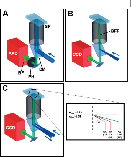
Figure 8.1. Three commonly used optical methods for detecting single molecules:
(A) confocal point detection, (B) epifluorescence imaging, and (C) total internal-reflection fluorescence (TIRF) microscopy. In all three geometries, directed laser light is reflected off a dichroic mirror (DM) into the back aperture of the high numerical aperture (NA) objective and is then imaged onto the sample plane (SP). Single-molecule fluorescence is collected through the same objective and further filtered by a bandpass filter (BF). In confocal detection,
the spectrally filtered light is further passed through a pinhole (PH) placed at the primary image plane. Both epifluorescence and TIRF designs require laser light to be focused at the back focal plane of the objective (BFP).
For detection, confocal setups typically use avalanche photodiodes (APD), while epifluorescence and TIRF microscopy use high sensitivity CCD cameras; (D) shows the critical angle needed to achieve objective-type TIRF with an NA of 1.4 or 1.45.

200 8 Single-Molecule Detection and Manipulation in Nanotechnology and Biology
protein complexes [40, 41], detection of DNA in nanopore technology [42], as well as orientational dynamics and optical properties of single dye molecules [43–47]. Not only can properties of single molecules be investigated, but the surrounding nanoenvironments can also be probed. For instance, heterogeneous rotational dynamics and lifetimes of single molecules in polymer films can be used to investigate molecular environments present within different regions of the film [48, 49]. Sensitive to changes in hydrophobicity, a new solvatochromic dye covalently bound to a polypeptide, for example, was used to monitor protein–protein interactions at the single-molecule level [50]. Inherent versatility, fast time resolution, and high SNR will continue to bolster the use of confocal optics in SMS; however, in contrast to epifluorescence and TIRF designs, which can study multiple point sources at once, confocal detection can only detect point source objects one at a time.
8.2.2
Visualizing Single Molecules with Epifluorescence Detection
Epifluorescence configurations are notably the most popular designs in optical microscopy. Lasers are commonly used for illumination in single-molecule studies, although recent reports describe single-molecule detection using mercury lamps and LEDs [51, 52]. Similar to confocal designs, epifluorescence illumination and fluorescence collection is carried out using the same high-NA objective and results in an area of illumination typically 50–100 mm in diameter for an objective with 1006 magnification. With proper collimation optics, lamps and LEDs produce even illumination over a wide area because of the noncoherent nature of such light sources. To achieve even, wide-area illumination with lasers, however, some additional optics are required. For instance, a spinning holographic diffuser or ground glass plate is placed in the beam line prior to the objective to disrupt the coherence of the laser light and to eliminate specular patterns at the image plane [13, 53]. Unfortunately, these designs can suffer unnecessary loss of incoming laser light to reflection and scattering. A more efficient design [Fig. 8.1(B)] focuses laser light at the back focal plane of the high-NA objective, which results in emergence of a collimated laser beam at the object plane and provides wide-field illumination of the sample [54]. For imaging, charge-coupled device (CCD) cameras are most common and can reach time resolutions of tens to hundreds of milliseconds for single-molecule experiments, limited by the available signal and sensor readout time. While, per pixel, time resolution with CCD imaging is two to three orders of magnitude slower than with confocal point detection, multiple molecules can be studied simultaneously. In epifluorescence, background from out-of-focus light and Raman scattering from a large detection volume, however, does limit the detection of single molecule signal. One strategy to further reduce background is to use dyes that fluoresce in the red [55]. Fortunately, detection sensitivity steadily improves as newly developed back-thinned CCD cameras produce images with low noise and can achieve QEs of ~90%. Advances towards higher-QE chips with lower readout noise and faster readout speeds will continue to improve epifluorescence detection.

8.2 Optical Detection of Single Molecules 201
Epifluorescence detection can provide information about immobilized single molecules as well as individual species that move over distances of microns. Some interesting biological applications include: (i) The study of the brownian diffusion and the dynamics of single DNA molecules [56–62], (ii) the direct imaging of individual molecular motors (e.g., myosin and kinesin) and bioparticles moving along the surface of glass coverslip [63–66], (iii) the observation of discrete rotations of an active F1 ATPase molecule [67], and (iv) the monitoring of the rates of enzymatic digestion of DNA molecules by k-exonuclease [68]. Moreover, with adequate signal, low noise, and stable optics, positions of fluorescent species can be accurately determined down to ~10 nm through Gaussian fitting of the fluorescent profile [68]. For single molecules, point-source fluorescence is below the diffraction limit (~200–350 nm) and can be highly magnified (100–5006) to result in an imaged spot that is spread over several micron-sized pixels (~7–25 mm diameter) with each pixel corresponding to sample distances of tens of nanometers. Remarkably, diffusional information from imaging single-molecule movements over time can be recorded in sol-gel films, porous materials, and in lipid bilayer membranes [53, 69–71]. Because of low SNR, single-molecule trajectories are typically accurate only to within 100–200 nm. While epifluorescence designs offer great advantage for studying molecules many microns above the coverslip, detection efficiencies for studies on surfaces are quite inferior to results produced using TIRF microscopy.
8.2.3
Total Internal-Reflection Fluorescence (TIRF) Microscopy
In comparison with point-detection techniques like confocal microscopy and widefield visualization of single molecules in free solution using epifluorescence, TIRF microscopy boasts impressive reduction of background and noise to produce widefield images of single molecules on surfaces. Total internal reflection occurs at an interface between high and low refractive index materials when incident light is directed at an angle greater than the critical angle defined by the two media. At the interface, light penetrates into the low refractive medium to produce an evanescent field that falls off exponentially, thus defining an illumination thickness of ~100–150 nm, depending on the wavelength used and the refractive indices of the two media [72]. Conveniently, excitation of any fluorescent species will only occur within the thin evanescent field. With a high power density within the evanescent field, which enhances single-molecule signal, and a dramatic reduction of background due to a decreased probe volume, improved detection efficiencies with TIRF are notable in comparison to epifluorescence.
Two commonly used TIRF configurations are (i) objective-type and (ii) prismbased. Objective-type TIRF not only requires the laser beam to be focused at the back focal plane of the objective like epifluorescence (see Section 8.2.2), but to ensure TIR the beam must also be directed towards the outer edge of the high-NA objective such that light is then incident at an angle greater than the critical angle [Fig. 8.1(C) with inset]. The critical angle in experiments using a glass coverslip (n = 1.51) and aqueous solution (n = 1.33) is ~62 , thus requiring use of objectives with

202 8 Single-Molecule Detection and Manipulation in Nanotechnology and Biology
an NA of 1.4 (H~68 ) or 1.45 (H~74 ) to achieve TIR. Practically, the extra 6 provided by an NA 1.45 objective makes implementing TIRF a relatively straightforward task. Prism-based configurations, as the name implies, use a prism placed on top of the sample to direct an incoming beam at the necessary angle to achieve TIR. Unfortunately, these designs are not as versatile as objective-type TIRF because the location of the prism restricts the types of systems that can be studied. Although in a side-by-side comparison, Ambrose and coworkers reported better signal-to-back- ground ratios (SBRs) for prism-based TIRF, objective-type TIRF produced significantly more photons from the single molecules [73]. In addition, objective-type TIRF designs improved both SNR and SBR by up to a factor of four when compared with epifluorescence [54, 74].
Even though single-molecule TIRF experiments are limited to studying molecules immobilized or close to surfaces, the enhancements of sensitivity prove quite useful for many applications. By degrading the focus of emission from single fluorophores and by changing the polarization of the excitation beam, orientation of emission dipoles can be determined from each molecule in all three dimensions; molecules with the dipole oriented in the z-axis showed a doughnut-like intensity profile [75, 76]. Furthermore, because of the thin sampling plane, TIRF has been used for isolated detection of individual binding events between molecules on the surface and free molecules in solution. For example, rates of absorption and conformational changes of individual k-DNA molecules binding to clean, fused-silica surfaces can be affected by pH and buffer composition [77]. In addition, TIRF microscopy can observe binding of biotinylated DNA to streptavidin-coated coverslips and covalent attachment of DNA to linkers attached to the surface, and has permitted real-time visualization of protein–protein interactions [78, 79]. Other examples include monitoring of the movements of kinesin on microtubules [80, 81], and myosin along the surface of a coverslip [82, 83]. Even a single, fluorescently labeled RNA polymerase can be visualized in real-time while moving along a single strand of DNA [84]. Analyzing the rates of movement for the myosin, kinesin, and RNA polymerase uncovered new kinetic information about each system. Typically in TIRF, experiments look at signals from the solid/liquid interface, but lateral diffusion of dyes along the interface of two immiscible liquids can also be investigated [85].
8.2.4
Single-Molecule Surface-Enhanced Resonance Raman Spectroscopy
First reported in the 1990s by two groups, Nie and coworkers and Kneipp et al. [86, 87], single-molecule surface-enhanced resonance Raman spectroscopy (SM-SERRS) has emerged as a promising new method for studying single molecules. These early works reported impressive enhancement factors on the order of 1014–1015 for single dye molecules (e.g., rhodamine 6G and crystal violet) adsorbed onto nanoclusters of silver particles. Surface-enhanced Raman analysis of single molecules offers several unique advantages: (i) in contrast to fluorescence spectroscopy, which excites molecular electronic states, Raman spectroscopy probes vibrational modes to elucidate structural information about a single molecule; (ii) SM-SERRS detection boasts

8.3 Single-Molecule Manipulations Using Optical Traps 203
spectra with impressive signals that can be two to three orders of magnitude brighter than fluorescence detection; and (iii) molecules studied using SM-SERRS can be observed for longer periods prior to photodestruction, due to use of an excitation wavelength that is to the red of the absorption wavelength of the dye and quenching of the excited state by metallic nanoclusters. Since the initial experiments, more papers have emerged, including studies on the significant enhancement factors in SM-SERRS [88], detection of single dye molecules in LangmuirBlodgett monolayers [89–91], SM-SERRS raster-scanned images of single molecules [89, 92], monitoring of surface dynamics [93], investigations of single-molecule magnets [94, 95], and real-time observation of single protein molecules [96–99]. For the interested reader, we recommend the comprehensive reviews by Zander, Kneipp et al. [100, 101].
Notably, Hofkens and coworkers reported the use of SM-SERRS to study enhanced green fluorescent protein (EGFP), which consists of a unique arrangement of three amino acid residues that produce a highly fluorescent complex [99]. EGFP and the family of GFPs are known to convert between a protonated and deprotonated state, and for on/off blinking, the protonated form is believed to cause dark periods in fluorescence observation of the protein [102]. Real-time SM-SERRS spectra of EGFP could be obtained with a time resolution of 5 s and agreed (–10 cm–1) with bulk EGFP data. Interestingly, the group observed specific frequency jumps over time that were believed to correspond with protonated and deprotonated forms of the molecule. SM-SERRS represents a union of spectroscopy and nanotechnology that provides detailed structural information on single molecules which would be unattainable using other methods. While SM-SERRS is limited in that spectra cannot be collected without the adsorption of molecules to suitable metallic nanoparticles, this method will undoubtedly continue to bring exciting new discoveries to the field of SMS.
8.3
Single-Molecule Manipulations Using Optical Traps
8.3.1
Force Studies Using Single-Beam Gradient Traps
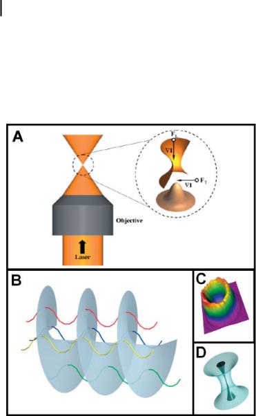
Data collection with techniques such as confocal, epifluorescence, or TIRF microscopy relies on detection of single-dye-molecule fluorescence induced by light illumination. Lasers, however, are not only limited to exciting fluorescence; focused coherent radiation can be used to probe single molecules through mechanical manipulation with a laser optical trap. First reported by Ashkin and coworkers, a single-beam gradient trap consists of a Gaussian (TEM00 mode) laser beam that is focused tightly onto the sample through a high-NA objective (e.g., NA 1.3) [103]. A particle in the vicinity of the focal spot will experience an attractive force that is directly proportional to the strength of the electric field and the polarizability of the molecule. As shown in Fig. 8.2(A), a particle will be held tightly within the three-dimensional
204 8 Single-Molecule Detection and Manipulation in Nanotechnology and Biology
(3D) intensity gradient by the transverse force (FT) that arises from the Gaussian intensity profile of the laser and the longitudinal force (FL) from the tight focusing by the high-NA objective. In opposition to this attractive gradient force, however, is the scattering force, which is mainly in the direction of beam propagation and experimentally makes trapping in the z-direction more difficult than in the x–y plane.
Through careful consideration of force balance along with clever integration of optical traps into novel detection schemes, force and displacement measurements
Figure 8.2. (A) Expanded drawing depicting the three-dimensional laser focus of a singlebeam gradient trap. A Gaussian laser beam is directed through a high-NA objective to produce a tightly focused intensity gradient (,I). A particle in the vicinity of the intensity gradient experiences a transverse force (FT) from the TEM00 mode of the laser and a longitudinal force (FL) from the tight focusing of the objective. (B) The Laguerre-Gaussian doughnut
mode (LG10) exhibits destructive interference along the beam axis due to the rotating phase of the beam. The colored ribbons represent individual light waves that comprise the rotating phase of the wavefront shown in gray.
(C) The resulting intensity profile produced from the mode results in a doughnut-like gradient that maintains a dark core (D) when focused by a high-NA objective.
8.3 Single-Molecule Manipulations Using Optical Traps 205
have been collected on a variety of individual biological molecules, particularly molecular motors. The pliant nature of the optical trap permits measurement of exquisitely small forces (~10-12 N) produced from biomolecules, which transduce chemical energy into a mechanical energy used to propel against the applied force of the trap. To measure forces generated by single molecules, a micron-sized polymer bead (usually polystyrene) is typically chemically or biochemically attached to the biomolecule and acts as a handle for subsequent optical manipulation. Nanometer-scale bead displacements, which arise from biomolecular forces, are sensed by a feedback mechanism that increases the laser power to a level needed to restore the position of the slightly displaced bead. Calibration of the displacements and power fluctuations provides the means to monitor detailed information about the energetics and motion of molecular motors at the single-molecule level. These exciting advances are discussed in a number of reviews [10–13, 104–106].
8.3.2
Optical Vortex Trapping
Since the initial observations of Ashkin and coworkers [103], advances in the area of optical trapping have progressed considerably, both in the area of single-molecule force measurements and in the development of more efficient and diverse types of traps to manipulate a broader spectrum of particles in the microscopic and nanoscopic scale, with emphasis towards biological applications. The single-beam gradient trap that uses a TEM00 laser mode is based on a balance of the gradient force that traps the particle and the opposing scattering force, which decreases the trapping efficiency of the gradient trap. To minimize this scattering force, Ashkin discussed the use of higher-order laser modes [107]. His investigation included the Laguerre–Gaussian doughnut mode (LG10 ), which has a dark core [108]. The unusual look of this mode is a result of destructive interference in the beam along the optical axis, generated by the rotating phase of the beam, as illustrated in Fig. 8.2B [109]. Figure 8.2(C) shows the resulting intensity gradient.
In recent years, the development of these tailored beams has become a broad and active area of research [110]. Investigations into the generation of these beams (initially of the LG10 doughnut mode) for optical trapping were undertaken by He et al. [109] using a computer-generated hologram (CGH) and later taken further by Gahagan et al. [111] and Arlt et al. [112]. The utility of CGHs was initially illustrated in 1992 by Bazhenov et al. [113], showing that holographic elements could induce a screw dislocation in the light wavefront. These CGH patterns acted as a diffractive optical element (DOE) in the beam path to produce the desired mode, which could then be selected and used. This same dislocation can be created by helically shifting the wavefront [114]. When focused down by a high-NA objective, the beam maintains its dark core (Fig. 8.2(D)).
In conventional laser tweezers, the types of particles that could be trapped must have a higher refractive index than the surrounding medium. Although the trapping of low-refractive-index particles was shown to be possible using a TEM00 beam, these schemes require the rapid scanning of the laser focus around the particle or

206 8 Single-Molecule Detection and Manipulation in Nanotechnology and Biology
utilize the interference pattern between two phase-shifted Gaussian beams [115, 116]. With the LG10 mode, both low-index and high-index particles can be held stably at the laser focus [117, 118]. Theoretical calculations of trapping efficiencies and potentials have been reported [119], which indicate that trapping efficiencies are on average greater using a LG10 beam than a TEM00 beam [120, 121]. The lateral trapping forces are reported not to change, and the increase in efficiency is largely due to the lack of scattering force that leads to more efficient trapping in the longitudinal direction.
Utilizing laser modes other than the usual TEM00 is advantageous in fields where the particle is nontransparent or very sensitive to photodamage. Reducing photodamage will allow a greater selection of biological materials to be optically manipulated, as well as decrease heating in the sample. Another observation of vortex trapping is the ability to generate movement of the trapped particle by the rotating phase of the beam [122, 123]. This transfer of angular momentum to absorptive particles permits the controlled rotation of the trapped particle, a useful tool in cellular studies [124, 125].
8.3.3
Optical Arrays
Some exciting work in the area of optical trapping has been in the development of optical tweezer arrays. Multiple traps can be constructed by adopting a time-sharing scheme, in which computer-controlled galvanoor piezoelectric mirrors scan rapidly the laser beam so as to park the laser focus in multiple locations at the object plane [115]. This scheme relies on the finite diffusion time it takes the particle to leave its originally trapped position and is not ideal for the trapping of small particles or a large number of particles. To overcome this limitation, holographic optical tweezer arrays (HOT arrays) were developed and can be generated in either two or three dimensions. Using the same holographic techniques utilized in creating the optical vortex trap, multiple optical traps can be generated from a single laser beam. These traps can be stationary or dynamic and can be conventional optical tweezers, vortex traps, or traps based on other higher-order laser modes. A number of methods exist to produce these HOT arrays, including the use of liquid crystal displays (LCDs) [126], spatial light modulators (SLMs) [127], and diffractive optics (physical and holographic) [128]. The use of LCDs or SLMs involves splitting a single beam into several separate beams, where each beam can have identical or different properties in their optical wavefronts [129]. The individual beams, when focused down by a high-NA objective, generate trapping forces in three dimensions [127]. One interesting application of HOT arrays is in the formation of nanoscopic materials, both physical (i.e., permanent) and virtual (i.e., transient). The ability to align particles into an ordered and predefined array, which can be controlled dynamically in time, should lead to interesting applications in areas of biomanipulation, organization, and construction, as well as in microfabrication and nanotechnology [130].
8.4 Applications in Single-Molecule Spectroscopy 207
8.4
Applications in Single-Molecule Spectroscopy
8.4.1
Conformational Dynamics of Single DNA Molecules in Solution
Owing to the intercalation of multiple fluorophores, single DNA molecules with lengths ranging from ~5 to ~60 mm can be easily visualized in solution using fluorescence microscopy. Although useful information can be obtained from simply observing DNA motions in solution, exciting discoveries about DNA conformational dynamics have resulted from the use of single-beam optical traps to manipulate fluorescent DNA molecules either directly or through attached bead handles [13]. With bead handles, polymer motion, relaxation dynamics, and novel manipulations can be studied in real time to further our understanding of polymer behavior in so-
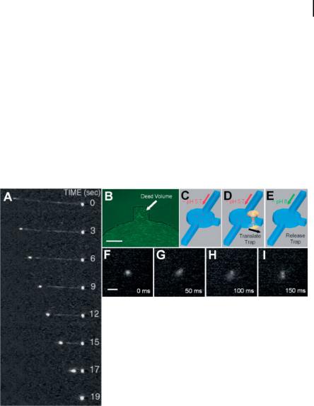
Figure 8.3. (A) Sequence of images showing the condensation of a single concatemer of k-DNA in a solution of protamine; (B) fluorescence image displaying slow flow of 1-lm fluorescent beads within the side notch of a circular microchamber (scale bar: 50 lm). (C–E) The procedure used to initiate decondensation of single DNA molecules consisted of (i) flowing condensed DNAs in a pH 5.7 solution into the circular microchamber (C), (ii) individually trapping and translating the DNA into the side notch (D), and (iii) initiating decondensation
with the optical trap after a pH 8 solution replaced the pH 5.7 solution by diffusion. (F–I) Decondensation of a single k-DNA molecule occurred over 150 ms. After t = 0, each frame was acquired with 50-ms time resolution. Slight blurring occurred because internal motions of the DNA were faster than the image acquisition rate of the camera. Reprinted with permission from Brewer et al., Science (Washington, D. C.) 286, 120, Copyright 1999 AAAS.

208 8 Single-Molecule Detection and Manipulation in Nanotechnology and Biology
lution [59, 60, 131]. In addition to understanding physical properties of the polymer, optical trapping and fluorescence visualization have been valuable towards unraveling condensation and decondensation kinetics of single DNA molecules. Packaging of DNA in cells plays a critical role in space conservation and organization, and is associated with a range of important biological functions such as gene activation and transcription. In nature, for example, k phage virus DNA with a contour length of ~17 mm is packed tightly into the phage head measuring only ~50 nm in diameter. While condensed DNA can be observed in vivo, studies in vitro provide more detailed information about the structural properties of DNA condensates. DNA molecules with micrometer-scale contour lengths have been observed in vitro to pack tightly into highly ordered toroidal, or doughnut-like, structures in the presence of several different polycations and proteins. DNA condensation is believed to be induced primarily by an electrostatic neutralization of the negatively charged DNA backbone that allows tight packing as result of a sufficient decrease in repulsive energies [132].
To probe the processes underlying DNA condensation, static visualization techniques [e.g., electron microscopy (EM) and AFM] have been used to obtain high-res- olution images of DNA condensates [132–134]. These approaches provide static images of condensed DNA that contain high information content. Little information, however, is offered on the kinetics of the condensation process, which has been implicated in determining the final structure of the DNA condensate [135]. Using dynamic light scattering, the bulk measurements on the kinetics of DNA condensation and decondensation have been collected, yet these studies were unable to visualize individual events at the single-molecule level [136]. Recently, several SMS studies have observed condensation and decondensation of single DNA molecules in solution [57, 61, 62, 137]. Balhorn and coworkers combined microfluidics and optical trapping to isolate single concatemers of k-DNA (attached to beads) in a flow of protamine, a protein known to condense DNA in sperm [61] [Fig. 8.3(A)]. Using fluorescence for visualization, dynamic changes in length of the DNA revealed information on the kinetics of condensation and decondensation of the molecule. Two studies conducted by Yoshikawa and coworkers monitored condensation and decondensation of (i) individual T4 DNA (~166 kilobase pairs) in solution using polyethylene glycol and Mg2+ [57] and (ii) single optically trapped DNA translated between condensing and decondensing environments [137]. Recently, Chiu and coworkers reported that the commonly used DNA fluorescent intercalator dye YOYO-1 can act as a condensing agent under moderately acidic pH conditions [62]. Individual YOYO-intercalated k-DNA molecules (~48.5 kbp), for example, were collapsed into toroidal structures ranging from 100 to 150 nm in diameter at pH 5.7. Using microfluidics, the solution environment could be quickly changed around an optically trapped DNA condensate [Fig. 8.3(B–E)]. Shuttering of the trap initiated conformational transitions of single, condensed YOYO-intercalated DNA molecules to an extended, random coil state that occurred over a time period of ~150 ms [Fig. 8.3(F– I)]. Interestingly, the studies performed by Balhorn and Yoshikawa observed completion of DNA condensation and decondensation on the time scale of seconds. Slower completion times can possibly be attributed to slow mass transfer in the fluidic

8.4 Applications in Single-Molecule Spectroscopy 209
designs, which required packing (or unpacking) to occur during solution exchange. For Chiu and coworkers, the solution was exchanged prior to initiating the decondensation, thus possibly allowing access to faster uncoiling dynamics. Easy visualization with SMS will allow further investigation into these biologically interesting and important processes.
8.4.2
Probing the Kinetics of Single Enzyme Molecules
To achieve single-molecule detection, researchers must tailor experimental approaches to overcome limiting factors such as noise, background, detection efficiencies, and low signals from molecules under investigation. In single-molecule enzymology, each of the limiting factors dictates the experimental direction towards elucidating catalytic activity of individual enzyme molecules. A relatively straightforward design used to overcome detection hurdles relies on using the enzyme to amplify the signal since each enzyme can catalyze the production of many fluorescent product molecules from nonfluorescent substrate molecules. Using this strategy, the first single-enzyme study was reported in 1961 by Rotman, where he incubated single b-galactosidase enzymes in aqueous microdroplets containing nonfluorescent substrates for hours until enough fluorescent product had accumulated and could be detected [138]. Unfortunately, with an incubation time of hours, better time resolution was needed to gain detailed information about catalytic rates of single enzymes. More recently, Xue and Yeung have studied single-enzyme behavior with a time resolution of minutes by injecting low concentrations of enzyme solution into micron-sized capillaries followed by incubation of the injected single lactate dehydrogenase (LDH-1) molecules in millimolar (mM) concentrations of lactate and nonfluorescent NAD+ [139]. Interestingly, the data showed catalytic rates of individual enzymes were constant over periods of ~2 hours, yet among each enzyme rates varied by up to a factor of four, which was hypothesized to be due to different long-lived conformations of each enzyme. Similarly, Dovichi and coworkers investigated the activity of single calf intestine alkaline phosphatase in capillaries and observed heterogeneity in catalytic rates, which in this case was believed to be a result of differences in chemical modification (e.g., posttranslational glycosylation) of the protein rather than different conformations of identical enzymes [140]. A fol- low-up analysis reported that highly pure enzymes have identical rates, suggesting heterogeneity is a product of chemical differences among single enzymes [141]. While these amplification techniques can provide information about static heterogeneity of enzyme catalysis, real-time detection of single-enzymatic turnover events is unattainable with characteristically low time resolutions and insufficient detection efficiencies.
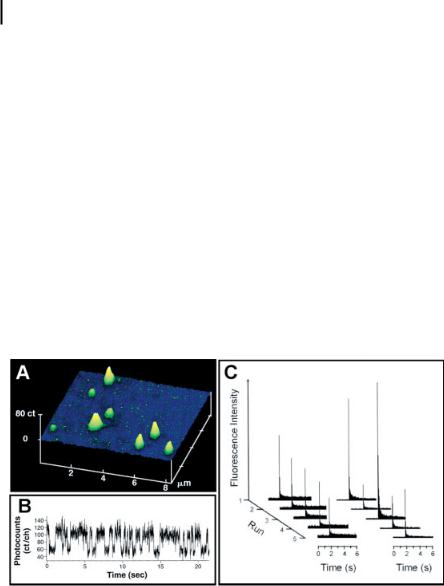
Using confocal optics, single-enzyme turnovers have been monitored in real time. Notably, Xie and coworkers detected individual turnover events from single cholesterol oxidase molecules trapped in pores of an agarose gel [Fig. 8.4(A)] containing oxygen and micromolar to millimolar concentrations of cholesterol [32]. Flavin adenine dinucleotide (FAD), a fluorescent group covalently attached to the
210 8 Single-Molecule Detection and Manipulation in Nanotechnology and Biology
enzyme, is reduced by cholesterol to a nonfluorescent form, FADH2, and then subsequently oxidized by oxygen to render the molecule fluorescent again. Each cycle of this on–off fluorescence represents a single turnover event. As configured, the experiment permitted direct study of each catalytic half-reaction. Direct monitoring of these on–off cycles [Fig. 8.4(B)] was achieved with a time resolution of ~10 ms and provided detailed information about static disorder of single enzymes as well as a new component, dynamic disorder. Detailed analysis of the on-times over durations of seconds to minutes revealed that the rate of an enzyme fluctuates over time
– a property termed “dynamic disorder” and believed to be a property dependent upon conformational changes of the enzyme. In addition to monitoring fluorescence from the enzyme, turnover events can be observed from production of single fluorophores from quenched nonfluorescent substrates. Rigler and coworkers used confocal microscopy to study catalysis of dihydrorhodamine 6G by single horseradish peroxidase molecules immobilized onto a coverslip [33]. Here, fluorescent signals rendered by the enzyme-product complex were detected with ~20-ms time resolution and used to calculate rates of single-enzyme catalysis. In addition to experimental data, theoretical discussions are also providing interesting insight into these exciting new observations [142–145].
SMS has opened the doors to new observables in single-enzyme catalysis. Interestingly, investigations into the effect of the environment surrounding the enzyme
Figure 8.4. (A) Scanning confocal image of individual, fluorescent cholesterol oxidase molecules in an agarose gel; (B) fluorescence trace of on–off cycles produced from an active cholesterol oxidase molecule Reprinted with permission from Lu et al., Science (Washington, D. C.) 282, 1877, Copyright 1998 AAAS,
(C) catalytic activity of alkaline phosphatase contained within single lipid vesicles. Every 60 s, the accumulated fluorescent products (fluorescein) formed from the nonfluorescent
substrate (fluorescein diphosphate) were probed at 488 nm and then bleached to reset the reaction clock. The left panel of (C) shows catalysis in a vesicle with a radius of 1.5 lm, and the right panel catalysis in a vesicle with a radius of 500 nm. Reprinted from Chem. Phys., 247, Chiu et al., Manipulating the biochemical nanoenvironment around single molecules contained within vesicles, 133, Copyright (1999), with permission from Elsevier.

8.4 Applications in Single-Molecule Spectroscopy 211
have received less attention. Possibly, changing surface properties or the volume surrounding an enzyme could modulate catalytic activity. In the bulk, enzymes trapped in chemically modified porous materials can exhibit significant rate enhancements as compared to enzyme in free solution [146, 147]. Furthermore, single-molecule experiments also suggest that surface properties and surface-to-volume ratios can affect activity. For instance, Tan and Yeung found that single LDH-1 molecules in a porous polycarbonate membrane showed discrete intervals of varied catalytic activity; however, when studied in fused silica supports, where adsorption of LDH-1 is not favored, rates of catalysis were constant over the same time period [148]. Another platform, designed by Zare and coworkers, investigated environmental effects by encapsulating enzymes within single vesicles meant to mimic a biological environment [149, 150]. With an optical trap immobilizing the vesicle, confocal detection monitored the catalysis of fluorescein diphosphate by alkaline phosphatase. Each fluorescence trace in Fig. 8.4(C) corresponded to an accumulation of product followed by probing and bleaching of the dye product formed by the enzyme. In a vesicle with 1.5 mm radius the catalytic rate appeared relatively homogeneous, yet for a vesicle with a 500 nm radius the incubated signal was quite variable and believed to be related to the surface-to-volume ratio of the environment surrounding the enzyme. With the exquisite detail provided by single-molecule detection, exciting potential lies in studying details of enzymatic catalysis affected by a variety of environmental factors.
8.4.3
Single-Molecule DNA Detection, Sorting, and Sequencing
The sensitivity offered by single-molecule detection has provided interesting possibilities in analytical chemistry, including DNA analysis, proteomics, sensors, and in the screening of rare molecules within a complex mixture (e.g., from combinatorial synthesis). Of these varied analytical applications, studies involving DNA molecules are the most widespread and developed. DNA molecules take center stage in many areas, such as medicine, forensics, environmental studies, and basic genetics, yet many hurdles still remain. Development of polymerase chain reaction (PCR) technology has overcome early problems with collecting enough DNA for experiments. Still, the technique requires a specific working protocol or set of PCR conditions for each DNA sample of interest and it is challenging to produce a high-quality DNA sample, so as not to amplify impurities in addition to the desired DNA sequence. With the amplified DNA, gel electrophoresis, the standard analytical tool for sizing DNA, is used as a means of sequencing, fingerprinting, creating restriction maps, and genotyping the sample. Recent innovations have improved or even eliminated these techniques in studies of single-molecule DNA and have been greatly facilitated by the appearance of new highly fluorescent dyes such as YOYO-1 (5006 increase of quantum yield upon binding to DNA) [151]. This family of dyes form stable complexes with DNA via intercalation and generate little background when unbound, thus permitting the detection of single molecules of DNA in dilute samples under a wide range of experimental conditions [151]. For example, using the enhanced sensitivity enabled by this dye as well as a known staining ratio of dye molecules to base

212 8 Single-Molecule Detection and Manipulation in Nanotechnology and Biology
pairs, it is possible to correlate the fluorescent signal detected directly with the size of the DNA fragments (tens to hundreds of kilobase pairs) present at femtomolar concentrations. With tailored microfluidic designs and by using a planar sheet of excitation laser [152], it is possible to increase volumetric sample throughput (2000 fragments per second has been demonstrated) or to sort the DNA molecules after detection with controlled changes in electroosmotic flow [153]. This method of sizing DNA fragments from the fluorescence signal of individual DNA also saves time by bypassing electrophoretic separation.
Continuous improvements are made to these techniques for single DNA analysis. Examples are: a simple modification to the geometry of the microchannel by adding a taper before the detection area to focus and create a thin sample stream that optimally flows through the tightly focused laser probe volume enhances detection efficiency by three-fold [154]; and the use of various millisecond imaging techniques to measure electrophoretic mobilities without complete separation of the DNA molecules to increase throughput [155]. By combining microfabrication and single DNA imaging, Craighead and coworkers have created entropic traps by introducing alternating regions of micrometerand nanometer-sized constrictions along the path of DNA migration driven by an applied electric field, and made the interesting observation that trapped DNA molecules escaped with a characteristic lifetime and that longer DNA molecules escaped the entropic traps faster than the shorter molecules [156]. Similar to fragment sizing in its technological requirements, single-molecule DNA restriction mapping has also been achieved [157], which yields important sequence information from the number and character of the cleavage sights and the resulting DNA fragments.
In addition to analytical assays such as DNA fragment sizing, with the complete sequencing of numerous genomes, most notably the human genome, the field of comparative genomics has evolved, and with it the need for rapid, accurate, and sensitive sequencing technology. Single-molecule DNA-sequencing techniques are still in their infancy, but proof-of-concept experiments demonstrate the possibility of an eventual viable method that could potentially sequence up to 2000 bp s–1 (greatly surpassing the fastest sequencing technique today) [158–162]. Several single-mole- cule sequencing schemes exist. To illustrate the general approach, an example of one scheme consists of: (i) fluorescent labeling or incorporation of fluorescently tagged nucleotides into the DNA, with different excitation and emission wavelengths for each of the four bases (or for at least two bases); (ii) handling of the tagged DNA molecules, which is implemented through a biotin-streptavidin bond to a microsphere, and can be controlled through suction with capillaries or using optical trapping; (iii) sequential degradation or cleavage of the DNA one base at a time with a 3¢–5¢ exonuclease; and (iv) efficient detection of each individual fluorescently labeled nucleotide as it becomes detached from the DNA after cleavage. This method will benefit from better labeling efficiency and an improved understanding of the suitable conditions required for single-base cleavage. Another example involves the simultaneous sequencing of numerous single molecules through the incorporation of dye-tagged nucleotides by DNA polymerase anchored to a coverslip with fluorescence microscopy detection [163]. Additionally, a scheme known as nanopore DNA

8.4 Applications in Single-Molecule Spectroscopy 213
sequencing utilizes the transmembrane protein a-hemolysin and electrically based detection to distinguish bases according to the distinct change in current flow [164]. Regardless of the exact detail of these single-molecule sequencing strategies, all of them face daunting challenges. Nevertheless, it seems this ambitious goal will be achieved with impressive advancements in single-molecule technologies.
8.4.4
Single-Molecule Imaging in Living Cells
The ultimate nanoscale molecular machine, perhaps, is the biological cell. The next step in understanding cellular function and behavior is to examine each component of this machine in detail as they perform their respective tasks. Proteins carry out the bulk of the work in the cell and operate within molecular networks whose functions are controlled by gene expression, energy transduction, and membrane transport. To probe these processes, single-molecule imaging of cells is often done using TIRF (see Section 8.2.3) and sometimes with epifluorescence. Remarkably, cell imaging not only determines where proteins are localized and distributed in the cell, but it can also identify associated structures and visualize how fast the proteins move, bind, and unbind within signaling pathways. Furthermore, visualizing single molecules in cells does not require the synchronization of reaction species like bulk experiments and can reveal information about rare intermediates and “memory effects” that are normally lost through ensemble averaging.
In cellular imaging, the most versatile approach is to induce expression of green fluorescent protein (GFP) in the host cell through genetic manipulation [165]. One caveat that is pertinent to single-molecule studies is the overexpression of the GFPlabeled proteins, which complicates the identification and tracking of individual molecules. Unfortunately, GFP suffers from a higher rate of photobleaching than some of the robust organic dyes, and the size of GFP (~27 kDa) can affect the diffusion rate of the tagged protein and perturb its function due to steric hindrance.
The greatest challenge in imaging single molecules in cells lies in the presence of high background noise. Presence of molecules such as flavins and NADH [55], for example, can generate high levels of autofluoresence. A number of schemes may be used to minimize background noise, including the use of excitation in the red (which also minimizes phototoxicity to the cell), time gating, and TIRF. Careful handling and culturing of cells in proper media also helps in reducing background fluorescence [166]. Some examples (Fig. 8.5) of how single-molecule imaging have been applied include (i) tracking of molecules on the cell surface [55, 166–169] as well as within the cytoplasm [55, 167, 169, 170] and the nucleus [169, 170]; (ii) the study of signal transduction [171]; (iii) visualization of viral infection of cells [172]; and (iv) measurement of disassociation kinetics between a ligand and a receptor [171]. Future advancements will rely upon both new techniques in microscopy and the development of new optical probes, such as fluorescent proteins with enhanced photostability and species that can be excited in the red [173, 174]. The capability to image molecular process at the single-molecule level will offer new levels of information about cellular processes and will increase our understanding of biological systems.
214 8 Single-Molecule Detection and Manipulation in Nanotechnology and Biology
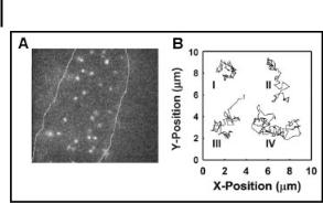
Figure 8.5. (A) Epifluorescence micrograph depicting single molecules on the surface of a cell; (B) experimentally measured single-molecule diffusional paths. Reprinted from TrAC, 22, W. E. Moerner, Optical measurements of single molecules in cells, 544, Copyright (2003, with permission from Elsevier.
8.5
Single-Molecule Detection with Bright Fluorescent Species
8.5.1
Optical Probes
In the nanoscopic single-molecular world, direct observations can be made using nonoptical techniques such as the scanning probe microscopies (SPM), patch clamp, or through the use of redox cycling. Most of these approaches, however, are unsuitable for real-time monitoring of single molecules in solution. For such studies, optical-based single-molecule detection has proved to be remarkably versatile [7, 86]. Although advancements in the hardware used to monitor single molecules have been impressive, measurements are ultimately limited by the reporter molecules, which in most cases are organic dyes. For single-molecule experiments, the dye ideally should possess the following optical characteristics: (i) excellent photostability, (ii) high quantum efficiency and absorption cross-section, (iii) fast cycling between ground and excited states to produce a high rate of photon emission, (iv) low probability of intersystem crossing from the singlet excited state to the triplet dark state,
(v) a narrow emission peak that is well separated from the absorption peak, and (vi) an excitation peak that matches well with commonly used laser wavelengths, preferably towards the red where background noise and autofluorescence from biological samples is minimal [175]. In addition to these optical properties, the dye should also contain easily modified functional groups so it can be tailored with the desired chemical functionality necessary for the end application [176].
Currently, no dye satisfies fully this set of demanding criteria, but dramatic improvements to conventional dyes (e.g., fluorescein) have been made. For improved optical properties, examples include the Alexa family of dyes from molecular probes and the family of carbocyanine dyes (CyDyes) from Amersham biosciences. For chemical functionality there is now a wide selection, which includes
8.6 Nanoscale Chemistry with Vesicles and Microdroplets 215
dyes that change fluorescence characteristics selectively when bound to biologically important ions (e.g., calcium), have diverse chemical functional groups and coupling chemistries, and are compatible with long-term cell culture to report particular cellular functions or states [177]. One particularly powerful reporter is the family of GFPs, owing to the ease by which these fluorescent reporters can be genetically manipulated [25, 176, 178]. In this case, the marriage of genetics with a good fluorescent reporter and high-sensitivity optical imaging has provided unprecedented insight into cellular function.
8.5.2
Quantum Dots
Advances in inorganic crystal generation have enabled nanoscopic particles to be generated for use as molecular probes. Semiconductor nanocrystals, more commonly called quantum dots (QDs), have unique optical characteristics making them behave neither as small molecules nor as bulk solids. Composed usually of a CdSe core with a CdS or ZnS shell, these nanoparticles typically range in size from 2 to 10 nm [179]. QDs have an advantage over organic dyes in that they can be tailored to obtain the desired florescent properties: high quantum yield, high photostability, high extinction coefficient, and narrow emission spectra, but with a broad excitation band (multiple reporters can be excited at a single wavelength), and long fluorescence lifetime [179–182]. These properties make emissions intense and ideal for sin- gle-molecule studies; however, synthetic shells added to increase solubility and biocompatibility can make the particles bulky in comparison to other probes. The major emerging area of QDs is in single-molecule bioconjugate work for in vitro and in vivo studies of biological systems, which can be performed in some cases without interrupting cellular processes [183–185]. Unfortunately, unlike GFPs, QDs can be attached to proteins only after they are expressed.
8.6
Nanoscale Chemistry with Vesicles and Microdroplets
While the area of nanoscale science has experienced tremendous growth in both the fabrication of nanostructures and the imaging of such nanoscale objects, few if any experiments have demonstrated the ability to control chemical reactions in the nanoscale, confined within femtoliter (10–15 L) volumes of solution. This capability is especially pertinent with advancements in single-molecule detection. Although we can detect single molecules, most of these studies rely on the use of bulk solutions that must contain sufficiently high concentrations of the molecules of interest so a single molecule can be isolated easily [186, 187]. To manipulate chemically and selectively only one or a small number of molecules at a time, a strategy is needed to localize, confine, and chemically transform in solution the selected molecules of interest.
Two approaches have been pursued to control chemical transformations of molecules within ultrasmall volumes (femtoliters or less). Reported by Zare and cowork-
216 8 Single-Molecule Detection and Manipulation in Nanotechnology and Biology
ers, the first approach relies on the use of lipid vesicles, in which the molecules of interest are first encapsulated within the vesicle [149]. Individual vesicles can then be mechanically manipulated (e.g., with optical trap or micropipettes). To initiate a chemical transformation at this small length scale, two vesicles each containing different types of reactive molecules are brought into contact and fused via application of a short (ms) and intense (kV cm–1) electric field through a pair of carbon-fiber microelectrodes [150, 188, 189]. In addition to the use of an electric field, the fusion of liposomes can be accomplished on an individual basis using a focused laser beam and on the bulk scale using chemicals [190]. A second method to achieve controlled nanoscale chemical transformations is to use aqueous microand nanodroplets that are dispersed in an immiscible medium [191]. These droplets can be generated on chip in a microfluidic system with excellent control, and the molecules of interest can be encapsulated within the droplets during their formation. The direct mechanical manipulation of such droplets, however, is nontrivial and is best achieved through the use of vortex trap [110, 117]. Like vesicles, these individual droplets can be fused together either spontaneously or with a small applied force (Fig. 8.6) so their respective contents will mix and a reaction can be initiated [191]. The advantage to using lipid vesicles lies in their biomimetic nature and, as demonstrated by Orwar and coworkers, the remarkable range of topology and shape that these liposomes exhibit [192–194]. The usefulness of the droplet platform is based on the ease and control with which droplets can be produced in a microfluidic platform, as well as on the wide range of interesting interfacial phenomena that may be studied and exploited in using droplets. One particular useful example is the possibility to concentrate molecules within individual aqueous droplets to very high levels [195], which offers new possibilities in understanding spatially confined single-molecule reactions and the effects of macromolecular crowding.
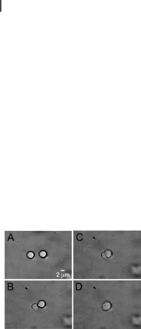
Figure 8.6. Sequence of images showing the directed fusion of two aqueous microdroplets in oil. The droplets measured ~4 lm in diameter, corresponding to a volume of 2010–13 L. Reprinted from TrAC, 22, D. T. Chiu, Microand nano-scale chemical analysis of individual sub-cellular compartments, 528, Copyright (2003), with permission from Elsevier.

References 217
8.7
Perspectives
Single-molecule studies and technologies are poised to offer both fundamental understandings of the molecular and nanoscopic world and to provide new tools that probe the inner workings of individual living cells. Over the past years, we have witnessed the increased application of established single-molecule methods in biology, such as AFM and high-sensitivity fluorescence microscopy, as well as the birth of new techniques that were made possible by advances in optics and instrumentation. The rapid pace with which this area has progressed will surely continue in the coming years to offer us yet more striking views of the nanoscale machinery and molecular engines that make biology work.
Acknowledgments
C.L.K. thanks the National Science Foundation for a graduate research fellowship. This work was supported by the National Institutes of Health (R01 GM65293) and the Keck Foundation.
References
1 W. Ho, Single-molecule chemistry, J. Chem. Phys. 2002, 117, 11033–11061.
2A. Ikai, STM and AFM of bio/organic molecules and structures, Surf. Sci. Rep. 1996, 26, 263–332.
3M. A. Poggi, L. A. Bottomley, P. T. Lillehei, Scanning probe microscopy, Anal. Chem. 2002, 74, 2851–2862.
4P. T. Lillehei, L. A. Bottomley, Scanning probe microscopy, Anal. Chem. 2000, 72, 189R–196R.
5L. A. Bottomley, Scanning probe microscopy,
Anal. Chem. 1998, 70, 425R–475R.
6S. Nie, R. N. Zare, Optical detection of single molecules, Ann. Rev. Biophys. Biomol. Struct.
1997, 26, 567–596.
7X. S. Xie, J. K. Trautman, Optical studies of single molecules at room temperature, Annu. Rev. Phys. Chem. 1998, 49, 441–480.
8S. Weiss, Fluorescence spectroscopy of single biomolecules, Science (Washington, D. C.)
1999, 283, 1676–1683.
9W. E. Moerner, A dozen years of single-mole- cule spectroscopy in physics, chemistry, and biophysics, J. Phys. Chem. B 2002, 106, 910–927.
10C. Bustamante, J. C. Macosko, G. J. L. Wuite, Grabbing the cat by the tail: manipulating molecules one by one, Nat. Rev. Mol. Cell. Biol. 2000, 1, 130.
11K. Svoboda, S. M. Block, Biological applications of optical forces, Ann. Rev. Biophys. Biomol. Struct. 1994, 23, 247–285.
12A. D. Mehta, M. Rief, J. A. Spudich,
D.A. Smith, R. M. Simmons, Single-mole- cule biomechanics with optical methods,
Science (Washington, D. C.) 1999, 283, 1689–1695.
13C. L. Kuyper, D. T. Chiu, Optical trapping: a versatile technique for biomanipulation,
Appl. Spectrosc. 2002, 56, 300A–312A.
14M. Ludwig, M. Rief, L. Schmidt, H. Li,
F.Oesterhelt, M. Gautel, H. E. Gaub, AFM, a tool for single-molecule experiments,
Appl. Phys. A 1999, 68, 173–176.
15J. K. Trautman, J. J. Macklin, L. E. Brus,
E.Betzig, Near-field spectroscopy of single molecules at room temperature, Nature (London) 1994, 369, 40–42.
16S. Xie, R. C. Dunn, Probing single molecule dynamics, Science (Washington, D. C.) 1994,
265, 361–364.

218 8 Single-Molecule Detection and Manipulation in Nanotechnology and Biology
17W. Denk, J. H. Strickler, W. W. Webb, 2-photon laser scanning fluorescence microscopy, Science (Washington, D. C.) 1990, 248, 73–76.
18T. Plakhotnik, D. Walser, M. Pirotta, A. Renn,
U.P. Wild, Nonlinear spectroscopy on a single quantum system: two-photon absorption of a single molecule, Science (Washington,
D.C.) 1996, 271, 1703.
19E. J. Sanchez, L. Novotny, G. R. Holtom,
S.Xie, Room-temperature fluorescence imaging and spectroscopy of single molecules by two-photon excitation, J. Phys. Chem. A 1997, 101, 7019–7023.
20T. Wilson, Confocal Microscopy, Academic Press, San Diego, 1990.
21J. B. Pawley, Handbook of Biological Confocal Microscopy, Plenum Press, New York, 1995.
22M. Eigen, R. Rigler, Sorting single molecules: application to diagnostics and evolutionary biotechnology, Proc. Natl. Acad. Sci. U. S. A.
1994, 91, 5740–5747.
23P. Schwille, U. Haupts, S. Maiti, W. W. Webb, Molecular dynamics in living cells observed by fluorescence correlation spectroscopy with oneand two-photon excitation, Biophys. J. 1999, 77, 2251–2265.
24M. J. Wirth, D. Swinton, Single-molecule study of an adsorbed oligonucleotide undergoing both lateral diffusion and strong adsorption, J. Phys. Chem. B 2001, 105, 1472–1477.
25A. Zumbusch, Single-molecule spectroscopy of the green fluorescent protein, Single Mol. 2001, 2, 287–288.
26M. Boehmer, J. Enderlein, Fluorescence spectroscopy of single molecules under ambient conditions: methodology and technology,
Chem. Phys. Chem. 2003, 4, 792–808.
27M. Diez, M. Boersch, B. Zimmermann,
P.Turina, S. D. Dunn, P. Graeber, Binding of the b-subunit in the ATP synthase from
Escherichia coli, Biochemistry 2004, 43, 1054–1064.
28S. A. Sanchez, J. E. Brunet, D. M. Jameson,
R.Lagos, O. Monasterio, Tubulin equilibrium unfolding followed by time-resolved fluorescence and fluorescence correlation spectroscopy, Protein Sci. 2004, 13, 81–88.
29A. Schenk, S. Ivanchenko, C. Roecker,
J.Wiedenmann, G. U. Nienhaus, Photodynamics of red fluorescent proteins studied by fluorescence correlation spectroscopy,
Biophys. J. 2004, 86, 384–394.
30S. Nie, D. T. Chiu, R. N. Zare, Probing individual molecules with confocal fluorescence microscopy, Science (Washington, D. C.) 1994,
266, 1018–1021.
31S. Nie, D. T. Chiu, R. N. Zare, Real-time detection of single molecules in solution by confocal fluorescence microscopy,
Anal. Chem. 1995, 67, 2849–2857.
32H. P. Lu, L. Xun, X. S. Xie, Single-molecule enzymatic dynamics, Science (Washington,
D.C.) 1998, 282, 1877–1882.
33L. Edman, Z. Foeldes-Papp, S. Wennmalm,
R.Rigler, The fluctuating enzyme: a single molecule approach, Chem. Phys. 1999, 247, 11–22.
34S. Wennmalm, L. Edman, R. Rigler, Conformational fluctuations in single DNA molecules, Proc. Natl. Acad. Sci. U. S. A. 1997, 94, 10641–10646.
35D. M. Warshaw, E. Hayes, D. Gaffney, A.-M. Lauzon, J. Wu, G. Kennedy, K. Trybus,
S.Lowey, C. Berger, Myosin conformational states determined by single fluorophore polarization, Proc. Natl. Acad. Sci. U. S. A.
1998, 95, 8034–8039.
36T. Ha, A. Y. Ting, J. Liang, W. B. Caldwell,
A.A. Deniz, D. S. Chemla, P. G. Schultz,
S. Weiss, Single-molecule fluorescence spectroscopy of enzyme conformational dynamics and cleavage mechanism, Proc. Natl. Acad.
Sci. U. S. A. 1999, 96, 893–898.
37H. Yang, G. Luo, P. Karnchanaphanurach, T.-M. Louie, I. Rech, S. Cova, L. Xun, X. S. Xie, Protein conformational dynamics probed by single-molecule electron transfer, Science (Washington, D. C.) 2003, 302, 262–266.
38X. Zhuang, H. Kim, M. J. B. Pereira,
H.P. Babcock, N. G. Walter, S. Chu, Correlating structural dynamics and function in single ribozyme molecules, Science (Washington,
D.C.) 2002, 296, 1473–1476.
39E. Tan, T. J. Wilson, M. K. Nahas, R. M. Clegg,
D.M. J. Lilley, T. Ha, A four-way junction accelerates hairpin ribozyme folding via a discrete intermediate, Proc. Natl. Acad. Sci. U. S.
A.2003, 100, 9308–9313.

40K. Weninger, M. E. Bowen, S. Chu,
A.T. Brunger, Single-molecule studies of snare complex assembly reveal parallel and antiparallel configurations, Proc. Natl. Acad. Sci. U. S. A. 2003, 100, 14800–14805.
41C. Hofmann, T. J. Aartsma, H. Michel,
J.Koehler, Direct observation of tiers in the energy landscape of a chromoprotein: a sin- gle-molecule study, Proc. Natl. Acad. Sci. U. S.
A.2003, 100, 15534–15538.
42E. L. Chandler, A. L. Smith, L. M. Burden,
J.J. Kasianowicz, D. L. Burden, Membrane surface dynamics of DNA-threaded nanopores revealed by simultaneous single-mole- cule optical and ensemble electrical recording, Langmuir 2004, 20, 898–905.
43T. Ha, T. A. Laurence, D. S. Chemla, S. Weiss, Polarization spectroscopy of single fluorescent molecules, J. Phys. Chem. B 1999, 103, 6839–6850.
44C. G. Hubner, A. Renn, I. Renge, U. P. Wild, Direct observation of the triplet lifetime quenching of single dye molecules by molecular oxygen, J. Chem. Phys. 2001, 115, 9619–9622.
45B. Bowen, N. Woodbury, Single-molecule fluorescence lifetime and anisotropy measurements of the red fluorescent protein, DsRed, in solution, Photochem. Photobiol.
2003, 77, 362–369.
46J. Hernando, M. van der Schaaf,
E.M. H. P. van Dijk, M. Sauer,
M. F. Garcia-Parajo, N. F. van Hulst, Excitonic behavior of rhodamine dimers: a single-molecule study, J. Phys. Chem. A 2003, 107, 43–52.
47D. S. Ko, Photobleaching time distribution of a single tetramethylrhodamine molecule in agarose gel, J. Chem. Phys. 2004, 120, 2530–2531.
48L. A. Deschenes, D. A. Vanden Bout, Singlemolecule studies of heterogeneous dynamics in polymer melts near the glass transition,
Science (Washington, D. C.) 2001, 292, 255–258.
49R. A. L. Vallee, M. Cotlet, J. Hofkens,
F. C. De Schryver, K. Muellen, Spatially heterogeneous dynamics in polymer glasses at room temperature probed by single molecule lifetime fluctuations, Macromolecules 2003, 36, 7752–7758.
References 219
50X. Tan, P. Nalbant, A. Toutchkine, D. Hu,
E.R. Vorpagel, K. M. Hahn, H. P. Lu, Singlemolecule study of protein-protein interaction dynamics in a cell signaling system, J. Phys. Chem. B 2004, 108, 737–744.
51M. Unger, E. Kartalov, C.-S. Chiu, H. A. Lester,
S.R. Quake, Single-molecule fluorescence observed with mercury lamp illumination,
BioTechniques 1999, 27, 1008–1014.
52J. S. Kuo, C. L. Kuyper, P. B. Allen, D. T. Chiu, High power LED as excitation source for fluorescence applications, Electrophoresis 2004 in press.
53K. S. McCain, D. C. Hanley, J. M. Harris, Single-molecule fluorescence trajectories for investigating molecular transport in thin silica sol-gel films, Anal. Chem. 2003, 75, 4351–4359.
54M. Tokunaga, K. Kitamura, K. Saito,
A.H. Iwane, T. Yanagida, Single molecule imaging of fluorophores and enzymic reactions achieved by objective-type total internal reflection fluorescence microscopy, Biochem.
Biophys. Res. Commun. 1997, 235, 47–53.
55W. E. Moerner, Optical measurements of single molecules in cells, Trends Anal. Chem. 2003, 22, 544–548.
56T. T. Perkins, D. E. Smith, S. Chu, Direct observation of tube-like motion of a single polymer chain, Science (Washington, D. C.)
1994, 264, 819–822.
57K. Yoshikawa, Y. Matsuzawa, Nucleation and growth in single DNA molecules, J. Am. Chem. Soc. 1996, 118, 929–930.
58D. T. Chiu, R. N. Zare, Biased diffusion, optical trapping, and manipulation of single molecules in solution, J. Am. Chem. Soc. 1996, 118, 6512–6513.
59S. R. Quake, H. Babcock, S. Chu,
The dynamics of partially extended single molecules of DNA, Nature (London) 1997, 388, 151–154.
60Y. Arai, R. Yasuda, K.-I. Akashi, Y. Harada,
H.Miyata, K. Kinosita, H. Itoh, Tying a molecular knot with optical tweezers, Nature (London) 1999, 399, 446–448.
61L. R. Brewer, M. Corzett, R. Balhorn, Prota- mine-induced condensation and decondensation of the same DNA molecule, Science (Washington, D. C.) 1999, 286, 120–123.

220 8 Single-Molecule Detection and Manipulation in Nanotechnology and Biology
62C. L. Kuyper, G. P. Brewood, D. T. Chiu, Initiating conformation transitions of individual YOYO-intercalated DNA molecules with optical trapping, Nano Lett. 2003, 3, 1387–1389.
63I. Sase, H. Miyata, S. i. Ishiwata, K. Kinosita, Jr., Axial rotation of sliding actin filaments revealed by single-fluorophore imaging, Proc. Natl. Acad. Sci. U. S. A. 1997, 94, 5646–5650.
64T. Sakamoto, I. Amitani, E. Yokota, T. Ando, Direct observation of processive movement by individual myosin v molecules, Biochem. Biophys. Res. Commun. 2000, 272, 586–590.
65A. B. Asenjo, N. Krohn, H. Sosa, Configuration of the two kinesin motor domains during ATP hydrolysis, Nat. Struct. Bio. 2003, 10, 836–842.
66H. Hess, C. M. Matzke, R. K. Doot,
J.Clemmens, G. D. Bachand, B. C. Bunker,
V.Vogel, Molecular shuttles operating undercover: a new photolithographic approach for the fabrication of structured surfaces supporting directed motility, Nano Lett. 2003, 3, 1651–1655.
67K. Adachi, R. Yasuda, H. Noji, H. Itoh,
Y.Harada, M. Yoshida, K. Kinosita, Jr., Stepping rotation of F1-ATPase visualized through angle-resolved single-fluorophore imaging, Proc. Natl. Acad. Sci. U. S. A. 2000,
97, 7243–7247.
68A. M. van Oijen, P. C. Blainey, D. J. Crampton,
C.C. Richardson, T. Ellenberger, X. S. Xie, Single-molecule kinetics of k-exonuclease reveal base dependence and dynamic disorder, Science (Washington, D. C.) 2003, 301, 1235–1238.
69T. Schmidt, G. J. Schutz, W. Baumgartner,
H.J. Gruber, H. Schindler, Imaging of single molecule diffusion, Proc. Natl. Acad. Sci. U.
S.A. 1996, 93, 2926–2929.
70P. C. Ke, C. A. Naumann, Hindered diffusion in polymer-tethered phospholipid monolayers at the air-water interface: a single-molecule fluorescence imaging study, Langmuir 2001, 17, 5076–5081.
71C. Seebacher, C. Hellriegel, F.-W. Deeg,
C.Braeuchle, S. Altmaier, P. Behrens,
K. Muellen, Observation of translational diffusion of single terrylenediimide molecules in a mesostructured molecular sieve, J. Phys. Chem. B 2002, 106, 5591–5595.
72D. Axelrod, Total internal-reflection fluorescence microscopy, Metods. Cell Biol. 1989, 30, 245–270.
73W. P. Ambrose, P. M. Goodwin, J. H. Jett,
A.Van Orden, J. H. Werner, R. A. Keller, Single-molecule fluorescence spectroscopy at ambient temperature, Chem. Rev. 1999, 99, 2929–2956.
74M. F. Paige, E. J. Bjerneld, W. E. Moerner, A comparison of through-the-objective total internal reflection microscopy and epi-fluo-
rescence microscopy for single-molecule fluorescence imaging, Single Mol. 2001, 2, 191–201.
75R. M. Dickson, D. J. Norris, W. E. Moerner, Simultaneous imaging of individual molecules aligned both parallel and perpendicular to the optic axis, Phys. Rev. Lett. 1998, 81, 5322–5325.
76A. Bartko, R. M. Dickson, Imaging threedimensional single molecule orientations,
J.Phys. Chem. B 1999, 103, 11237–11241.
77S. H. Kang, M. R. Shortreed, E. S. Yeung, Real-time dynamics of single-DNA molecules undergoing adsorption and desorption at liquid-solid interfaces, Anal. Chem. 2001, 73, 1091–1099.
78M. A. Osborne, C. L. Barnes,
S.Balasubramanian, D. Klenerman, Probing DNA surface attachment and local environment using single-molecule spectroscopy,
J.Phys. Chem. B 2001, 105, 3120–3126.
79R. Yamasaki, M. Hoshino, T. Wazawa, Y. Ishii,
T.Yanagida, Y. Kawata, T. Higurashi, K. Sakai,
J.Nagai, Y. Goto, Single molecular observation of the interaction of GroEL with substrate proteins, J. Mol. Biol. 1999, 292, 965–972.
80R. D. Vale, T. Funatsu, D. W. Pierce,
L.Romberg, Y. Harada, T. Yanagida, Direct observation of single kinesin molecules moving along microtubules, Nature (London) 1996, 380, 451–453.
81A. Seitz, H. Kojima, K. Oiwa,
E.-M. Mandelkow, Y.-H. Song, E. Mandelkow, Single-molecule investigation of the interference between kinesin, tau and map2c, EMBO
J.2002, 21, 4896–4905.
82T. Funatsu, Y. Harada, M. Tokunaga, K. Saito,
T.Yanagida, Imaging of single fluorescent molecules and individual ATP turnovers by single myosin molecules in aqueous solution,
Nature (London) 1995, 374, 555–559.

83 K. Oiwa, J. F. Eccleston, M. Anson, M. Kikumoto, C. T. Davis, G. P. Reid,
M. A. Ferenczi, J. E. T. Corrie, A. Yamada, H. Nakayama, D. R. Trentham, Comparative single-molecule and ensemble myosin enzymology: sulfoindocyanine ATP and ADP derivatives, Biophys. J. 2000, 78, 3048–3071.
84Y. Harada, T. Funatsu, K. Murakami,
Y.Nonoyama, A. Ishihama, T. Yanagida, Single-molecule imaging of RNA polymeraseDNA interactions in real-time, Biophys. J. 1999, 76, 709–715.
85F. Hashimoto, S. Tsukahara, H. Watarai, Lateral diffusion dynamics for single molecules of fluorescent cyanine dye at the free and surfactant-modified dodecane-water interface, Langmuir 2003, 19, 4197–4204.
86S. Nie, S. R. Emory, Probing single molecules and single nanoparticles by surface-enhanced Raman scattering, Science (Washington, D. C.)
1997, 275, 1102–1106.
87K. Kneipp, Y. Wang, H. Kneipp,
L.T. Perelman, I. Itzkan, R. R. Dasari,
M.S. Feld, Single-molecule detection using surface-enhanced Raman scattering (SERS),
Phys. Rev. Lett. 1997, 78, 1667–1670.
88J. Jiang, K. Bosnick, M. Maillard, L. Brus, Single-molecule Raman spectroscopy at the junctions of large Ag nanocrystals, J. Phys. Chem. B 2003, 107, 9964–9972.
89C. J. L. Constantino, T. Lemma, P. A. Antunes,
R.Aroca, Single-molecule detection using surface-enhanced resonance Raman scattering and Langmuir-Blodgett monolayers, Anal. Chem. 2001, 73, 3674–3678.
90T. Lemma, R. F. Aroca, Single-molecule sur- face-enhanced resonance Raman scattering on colloidal silver and Langmuir-Blodgett monolayers coated with silver overlayers,
J.Raman Spectrosc. 2002, 33, 197–201.
91P. Goulet, N. Pieczonka, R. Aroca, Singlemolecule SERRS of mixed perylene Lang- muir-Blodgett monolayers on novel metal island substrates, Can. J. Anal. Sci. Spectrosc.
2003, 48, 146–152.
92Z. Wang, S. Pan, T. D. Krauss, H. Du,
L.J. Rothberg, The structural basis for giant enhancement enabling single-molecule Raman scattering, Proc. Natl. Acad. Sci. U. S.
A.2003, 100, 8638–8643.
References 221
93A. Weiss, G. Haran, Time-dependent singlemolecule Raman scattering as a probe of surface dynamics, J. Phys. Chem. B 2001, 105, 12348–12354.
94J. M. North, L. J. van de Burgt, N. S. Dalal, A Raman study of the single molecule magnet Mn12-acetate and analogs, Solid State Commun. 2002, 123, 75–79.
95J. M. North, N. S. Dalal, Raman and infrared
modes of the single molecule magnet Fe8Br8 and analogs, J. Appl. Phys. 2003, 93, 7092–7094.
96H. Xu, E. J. Bjerneld, M. Kaell, L. Borjesson, Spectroscopy of single hemoglobin molecules by surface enhanced Raman scattering, Phys. Rev. Lett. 1999, 83, 4357–4360.
97A. R. Bizzarri, S. Cannistraro, Surfaceenhanced resonance Raman spectroscopy signals from single myoglobin molecules, Appl. Spectrosc. 2002, 56, 1531–1537.
98E. J. Bjerneld, Z. Foeldes-Papp, M. Kaell,
R.Rigler, Single-molecule surface-enhanced Raman and fluorescence correlation spectroscopy of horseradish peroxidase, J. Phys. Chem. B 2002, 106, 1213–1218.
99S. Habuchi, M. Cotlet, R. Gronheid, G. Dirix,
J.Michiels, J. Vanderleyden, F. C. De Schryver,
J.Hofkens, Single-molecule surface enhanced resonance Raman spectroscopy of the enhanced green fluorescent protein,
J.Am. Chem. Soc. 2003, 125, 8446–8447.
100C. Zander, Single-molecule detection in solution: a new tool for analytical chemistry,
Fresenius J. Anal. Chem. 2000, 366, 745–751.
101K. Kneipp, H. Kneipp, I. Itzkan, R. R. Dasari,
M.S. Feld, Surface-enhanced Raman scattering and biophysics, J. Phys. Condens. Matter
2002, 14, R597–R624.
102R. M. Dickson, A. B. Cubitt, R. Y. Tsien,
W.E. Moerner, On/off blinking and switching behavior of single molecules of green fluorescent protein, Nature (London) 1997, 388, 355–358.
103A. Ashkin, J. M. Dziedzic, J. E. Bjorkholm,
S.Chu, Observation of a single-beam gradient force optical trap for dielectric particles,
Opt. Lett. 1986, 11, 288–290.
104A. Ashkin, History of optical trapping and manipulation of small-neutral particle, atoms, and molecules, IEEE J. Select. Topics Quantum Electr. 2000, 6, 841–856.

222 8 Single-Molecule Detection and Manipulation in Nanotechnology and Biology
105M. D. Wang, Manipulation of single molecules in biology, Curr. Opin. Biotechnol. 1999,
10, 81–86.
106G. J. Sonek, W. Wang, Theory of optical trapping forces: a review, Rev. Laser Eng. 1994, 24, 3–11.
107A. Ashkin, Forces of a single beam gradient laser trap on a dielectric sphere in the ray optics regime, Biophys. J. 1992, 61, 569–582.
108M. A. Clifford, J. Arlt, J. Courtial, K. Dholakia, High-order Laguerre-Gaussian laser modes for studies of cold atoms, Opt. Commun. 1998, 156, 300–306.
109H. He, N. R. Heckenberg, H. RubinszteinDunlop, Optical particle trapping with high- er-order doughnut beams produced using high efficiency computer generated holograms, J. Mod. Opt. 1995, 42, 217–223.
110D. G. Grier, A revolution in optical manipulation, Nature (London) 2003, 424, 810–816.
111K. T. Gahagan, G. A. Swartzlander, Jr., Optical trapping of particles, Opt. Lett. 1996, 21, 827–829.
112J. Arlt, K. Dholakia, L. Allen, M. J. Padgett, The production of multiringed LaguerreGaussian modes by computer-generated holograms, J. Mod. Opt. 1998, 45, 1231–1237.
113V. Y. Bazhenov, M. S. Soskin, M. V. Vasnetsov, Screw dislocations in light wavefronts, J. Mod. Opt. 1992, 39, 985–990.
114D. Ganic, X. Gan, M. Gu, M. Hain,
S.Somalingam, S. Stankovic, T. Tschudi, Generation of doughnut laser beams by use of a liquid-crystal cell with a conversion efficiency near 100%, Opt. Lett. 2002, 27, 1351–1353.
115C. Mio, T. Gong, A. Terray, D. W. M. Marr, Design of a scanning laser optical trap for multiparticle manipulation, Rev. Sci. Instrum. 2000, 71, 2196–2200.
116M. P. MacDonald, L. Paterson, W. Sibbett,
K.Dholakia, P. E. Bryant, Trapping and manipulation of low-index particles in a twodimensional interferometric optical trap, Opt. Lett. 2001, 26, 863–865.
117K. T. Gahagan, G. A. Swartzlander, Jr., Simultaneous trapping of low-index and high-index microparticles observed with an optical-vortex trap, J. Opt. Soc. Am. B Opt. Phys. 1999, 16, 533–537.
118K. T. Gahagan, G. A. Swartzlander, Jr., Trapping of low-index microparticles in an optical vortex, J. Opt. Soc. Am. B Opt. Phys.
1998, 15, 524–534.
119N. B. Simpson, L. Allen, M. J. Padgett, Optical tweezers and optical spanners with Laguerre-Gaussian modes, J. Mod. Opt. 1996, 43, 2485–2491.
120N. B. Simpson, D. McGloin, K. Dholakia,
L.Allen, M. J. Padgett, Optical tweezers with increased axial trapping efficiency, J. Mod. Opt. 1998, 45, 1943–1949.
121A. T. O’Neil, M. J. Padgett, Axial and lateral trapping efficiency of Laguerre-Gaussian modes in inverted optical tweezers, Opt. Commun. 2001, 193, 45–50.
122H. He, M. E. J. Friese, N. R. Heckenberg,
H.Rubinsztein-Dunlop, Direct observation of transfer of angular momentum to absorptive particles from a laser beam with a phase singularity, Phys. Rev. Lett. 1995, 75, 826–829.
123V. Garces-Chavez, K. Volke-Sepulveda,
S.Chavez-Cerda, W. Sibbett, K. Dholakia, Transfer of orbital angular momentum to an optically trapped low-index particle, Phys. Rev. A Atom. Mol. Opt. Phys. 2002, 66, 063402/ 063401–063402/063408.
124M. E. J. Friese, T. A. Nieminen,
N.R. Heckenberg, H. Rubinsztein-Dunlop, Optical alignment and spinning of lasertrapped microscopic particles, Nature (London) 1998, 395, 621.
125L. Paterson, M. P. MacDonald, J. Arlt,
W.Sibbett, P. E. Bryant, K. Dholakia, Controlled rotation of optically trapped microscopic particles, Science (Washington, D. C.)
2001, 292, 912–914.
126J. Liesener, M. Reicherter, T. Haist,
H.J. Tiziani, Multi-functional optical tweezers using computer-generated holograms,
Opt. Commun. 2000, 185, 77–82.
127J. E. Curtis, B. A. Koss, D. G. Grier, Dynamic holographic optical tweezers, Opt. Commun. 2002, 207, 169–175.
128E. R. Dufresne, G. C. Spalding, M. T. Dearing,
S.A. Sheets, D. G. Grier, Computer-generated holographic optical tweezer arrays, Rev. Sci. Instrum. 2001, 72, 1810–1816.
129V. R. Daria, P. J. Rodrigo, J. Gluckstad, Dynamic array of dark optical traps, Appl. Phys. Lett. 2004, 84, 323–325.
|
|
|
References |
223 |
|
|
|
|
|
130 |
P. Korda, G. C. Spalding, E. R. Dufresne, |
143 |
H. Qian, E. L. Elson, Single-molecule enzy- |
|
|
D. G. Grier, Nanofabrication with holographic |
|
mology: stochastic Michaelis–Menten |
|
|
optical tweezers, Rev. Sci. Instrum. 2002, 73, |
|
kinetics, Biophys. Chem. 2002, 101–102, |
|
|
1956–1957. |
|
565–576. |
|
131 T. T. Perkins, S. R. Quake, D. E. Smith, |
144 |
L. Edman, R. Rigler, Memory landscapes of |
||
|
S. Chu, Relaxation of a single DNA molecule |
|
single-enzyme molecules, Proc. Natl. Acad. |
|
|
observed by optical microscopy, Science |
|
Sci. U. S. A. 2000, 97, 8266–8271. |
|
|
(Washington, D. C.) 1994, 264, 822–826. |
145 |
G. K. Schenter, H. P. Lu, X. S. Xie, Statistical |
|
132 |
V. A. Bloomfield, DNA condensation, |
|
analyses and theoretical models of single- |
|
|
Curr. Opin. Struct. Biol. 1996, 6, 334–341. |
|
molecule enzymatic dynamics, J. Phys. Chem. |
|
133 |
N. V. Hud, K. H. Downing, R. Balhorn, |
|
A 1999, 103, 10477–10488. |
|
|
A constant radius of curvature model for the |
146 |
C. Lei, Y. Shin, J. Liu, E. J. Ackerman, Entrap- |
|
|
organization of DNA in toroidal condensates, |
|
ping enzyme in a functionalized nanoporous |
|
|
Proc. Natl. Acad. Sci. U. S. A. 1995, 92, |
|
support, J. Am. Chem. Soc. 2002, 124, |
|
|
3581–3585. |
|
11242–11243. |
|
134 |
D. Liu, C. Wang, J. Li, Z. Lin, T. Zukun, |
147 |
J. Drott, L. Rosengren, K. Lindstrom, |
|
|
C. Bai, Atomic force microscopy analysis of |
|
T. Laurell, Pore morphology influence on cat- |
|
|
intermediates in cobalt hexamine-induced |
|
alytic turn-over for enzyme activated porous |
|
|
DNA condensation, J. Biomol. Struct. Dyn. |
|
silicon matrices, Thin Solid Films 1998, 330, |
|
|
2000, 18, 1–9. |
|
161–166. |
|
135 |
N. V. Hud, F. Milanovich, R. Balhorn, A con- |
148 W. Tan, E. S. Yeung, Monitoring the reactions |
||
|
stant radius of curvature model for the orga- |
|
of single enzyme molecules and single metal |
|
|
nization of DNA in toroidal condensates, |
|
ions, Anal. Chem. 1997, 69, 4242–4248. |
|
|
Proc. Natl. Acad. Sci. U. S. A. 1995, 92, |
149 D. T. Chiu, C. F. Wilson, A. Karlsson, |
||
|
3581–3585. |
|
A. Danielsson, A. Lundqvist, A. Stromberg, |
|
136 |
D. Porschke, Dynamics of DNA condensa- |
|
F. Ryttse, M. Davidson, S. Nordholm, |
|
|
tion, Biochemistry 1984, 23, 4821–4828. |
|
O. Orwar, R. N. Zare, Manipulating the bio- |
|
137 |
Y. Yoshikawa, S. M. Nomura, T. Kanbe, |
|
chemical nanoenvironment around single |
|
|
K. Yoshikawa, Controlling the folding/unfold- |
|
molecules contained within vesicles, Chem. |
|
|
ing transition of the DNA-histone H1 com- |
|
Phys. 1999, 247, 133–139. |
|
|
plex by direct optical manipulation, Chem. |
150 D. T. Chiu, C. F. Wilson, F. Ryttsen, |
||
|
Phys. Lett. 2000, 330, 77–82. |
|
A. Stromberg, C. Farre, A. Karlsson, |
|
138 |
B. Rotman, Measurement of activity of single |
|
S. Nordholm, A. Gaggar, B. P. Modi, |
|
|
molecules of b-D-galactosidase, Proc. Natl. |
|
A. Moscho, R. A. Garza-Lopez, O. Orwar, |
|
|
Acad. Sci. U. S. A. 1961, 47, 1981–1991. |
|
R. N. Zare, Chemical transformations in indi- |
|
139 |
Q. Xue, E. S. Yeung, Differences in the chem- |
|
vidual ultrasmall biomimetic containers, |
|
|
ical reactivity of individual molecules of an |
|
Science (Washington, D. C.) 1999, 283, |
|
|
enzyme, Nature (London) 1995, 373, 681–684. |
|
1892–1895. |
|
140 |
D. B. Craig, E. Arriaga, J. C. Y. Wong, H. Lu, |
151 S. Gurrieri, K. S. Wells, I. D. Johnson, |
||
|
N. J. Dovichi, Studies on single alkaline phos- |
|
C. Bustamante, Direct visualization of indi- |
|
|
phatase molecules: Reaction rate and activa- |
|
vidual DNA molecules by fluorescence mi- |
|
|
tion energy of a reaction catalyzed by a single |
|
croscopy: characterization of the factors |
|
|
molecule and the effect of thermal denatura- |
|
affecting signal/background and optimization |
|
|
tion: the death of an enzyme, J. Am. Chem. |
|
of imaging conditions using YOYO, Anal. |
|
|
Soc. 1996, 118, 5245–5253. |
|
Biochem. 1997, 249, 44–53. |
|
141 |
R. Polakowski, D. B. Craig, A. Skelley, |
152 A. Van Orden, R. A. Keller, W. P. Ambrose, |
||
|
N. J. Dovichi, Single molecules of highly pur- |
|
High-throughput flow cytometric DNA frag- |
|
|
ified bacterial alkaline phosphatase have iden- |
|
ment sizing, Anal. Chem. 2000, 72, 37–41. |
|
|
tical activity, J. Am. Chem. Soc. 2000, 122, |
153 H. P. Chou, C. Spence, A. Scherer, S. Quake, |
||
|
4853–4855. |
|
A microfabricated device for sizing and sort- |
|
142 |
D. Astumian, Protein conformational fluctua- |
|
ing DNA molecules, Proc. Natl. Acad. Sci. U. |
|
|
tions and free-energy transduction, Appl. |
|
S. A. 1999, 96, 11–13. |
|
|
Phys. A 2002, 75, 193–206. |
|
|
|

224 8 Single-Molecule Detection and Manipulation in Nanotechnology and Biology
154B. B. Haab, R. A. Mathies, Single-molecule detection of DNA separations in microfabricated capillary electrophoresis chips employing focused molecular streams, Anal. Chem. 1999, 71, 5137–5145.
155M. R. Shortreed, H. Li, W.-H. Huang,
E.S. Yeung, High-throughput single-mole- cule DNA screening based on electrophoresis,
Anal. Chem. 2000, 72, 2879–2885.
156J. Han, S. W. Turner, H. G. Craighead, Entropic trapping and escape of long DNA molecules at submicron size constriction, Phys. Rev. Lett. 1999, 83, 1688–1691.
157B. Schafer, H. Gemeinhardt, V. Uhl,
K.O. Greulich, Single-molecule DNA restriction analysis in the light microscope, Single Mol. 2000, 1, 33–40.
158J. H. Werner, H. Cai, J. H. Jett,
L.Reha-Krantz, R. A. Keller, P. M. Goodwin, Progress towards single-molecule DNA sequencing: a one color demonstration,
J.Biotechnol. 2003, 102, 1–14.
159M. Sauer, B. Angerer, W. Ankenbauer,
Z.Foldes-Papp, F. Gobel, K. T. Han, R. Rigler,
A.Schulz, J. Wolfrum, C. Zander, Single-mol- ecule DNA sequencing in submicrometer channels: state of the art and future prospects, J. Biotechnol. 2001, 86, 181–201.
160M. Sauer, B. Angerer, K. T. Han, C. Zander, Detection and identification of single dyelabeled mononucleotide molecules released from an optical fiber in a microcapillary: First steps towards a new single-molecule DNA sequencing technique, PCCP Phys. Chem. Chem. Phys. 1999, 1, 2471–2477.
161M. A. Augustin, W. Ankenbauer, B. Angerer, Progress towards single-molecule sequencing: enzymatic synthesis of nucleotide-speci- fically labeled DNA, J. Biotechnol. 2001, 86, 289–301.
162Z. Foldes-Papp, B. Angerer, W. Ankenbauer,
R.Rigler, Fluorescent high-density labeling of DNA: error-free substitution for a normal nucleotide, J. Biotechnol. 2001, 86, 237–253.
163I. Braslavsky, B. Hebert, E. Kartalov,
S.R. Quake, Sequence information can be obtained from single DNA molecules, Proc. Natl. Acad. Sci. U. S. A. 2003, 100, 3960–3964.
164M. Akeson, D. Branton, J. J. Kasianowicz,
E.Brandin, D. W. Deamer, Microsecond timescale discrimination among polycytidylic acid, polyadenylic acid, and polyuridylic acid as homopolymers or as segments within single RNA molecules, Biophys. J. 1999, 77, 3227–3233.
165R. Brock, G. Vamosi, G. Vereb, T. M. Jovin, Rapid characterization of green fluorescent protein fusion proteins on the molecular and cellular level by fluorescence correlation microscopy, Proc. Natl. Acad. Sci. U. S. A. 1999,
96, 10123–10128.
166R. Iino, I. Koyama, A. Kusumi, Single-mole- cule imaging of green fluorescent proteins in living cells: E-cadherin forms oligomers on the free cell surface, Biophys. J. 2001, 80, 2667–2677.
167G. I. Mashanov, D. Tacon, A. E. Knight,
M.Peckham, J. E. Molloy, Visualizing single molecules inside living cells using total internal reflection fluorescence microscopy, Methods 2003, 29, 142–152.
168M. Vrljic, S. Y. Nishimura, S. Brasselet,
W.E. Moerner, H. M. McConnell, Translational diffusion of individual class II MHC membrane proteins in cells, Biophys. J. 2002, 83, 2681–2692.
169T. A. Byassee, W. C. W. Chan, S. M. Nie, Probing single molecules in single living cells, Anal. Chem. 2000, 72, 5606–5611.
170U. Kubitscheck, Single protein molecules visualized and tracked in the interior of eukaryotic cells, Single Mol. 2002, 3, 267–274.
171Y. Sako, T. Uyemura, Total internal reflection fluorescence microscopy for single-molecule imaging in living cells, Cell Struct. Func. 2002, 27, 357–365.
172G. Seisenberger, M. U. Ried, T. Endress,
H.Buning, M. Hallek, C. Brauchle, Real-time single-molecule imaging of the infection pathway of an adeno-associated virus, Science (Washington, D. C.) 2001, 294, 1929–1932.
173R. E. Campbell, O. Tour, A. E. Palmer,
P.A. Steinbach, G. S. Baird, D. A. Zacharias,
R.Y. Tsien, A monomeric red fluorescent protein, Proc. Natl. Acad. Sci. U. S. A. 2002,
99, 7877–7882.
|
|
|
References |
225 |
|
|
|
|
|
174 |
N. G. Gurskaya, A. F. Fradkov, A. Terskikh, |
185 |
X. Gao, S. Nie, Molecular profiling of single |
|
|
M. V. Matz, Y. A. Labas, V. I. Martynov, |
|
cells and tissue specimens with quantum |
|
|
Y. G. Yanushevich, K. A. Lukyanov, |
|
dots, Trends Biotech. 2003, 21, 371–373. |
|
|
S. A. Lukyanov, GFP-like chromoproteins as a |
186 |
M. D. Barnes, K. C. Ng, W. B. Whitten, |
|
|
source of far-red fluorescent proteins, FEBS |
|
J. M. Ramsey, Detection of single rhodamine |
|
|
Lett. 2001, 507, 16–20. |
|
6G molecules in levitated microdroplets, |
|
175 |
A. N. Kapanidis, S. Weiss, Fluorescent probes |
|
Anal. Chem. 1993, 65, 2360–2365. |
|
|
and bioconjugation chemistries for single- |
187 |
M. D. Barnes, N. Lermer, C. Y. Kung, |
|
|
molecule fluorescence analysis of biomole- |
|
W. B. Whitten, J. M. Ramsey, S. C. Hill, Real- |
|
|
cules, J. Chem. Phys. 2002, 117, 10953–10964. |
|
time observation of single-molecule fluores- |
|
176 |
J. Zhang, R. E. Campbell, A. Y. Ting, |
|
cence in microdroplet streams, Opt. Lett. |
|
|
R. Y. Tsien, Creating new fluorescent probes |
|
1997, 22, 1265–1267. |
|
|
for cell biology, Nat. Rev. Mol. Cell. Biol. 2002, |
188 |
C. F. Wilson, G. J. Simpson, D. T. Chiu, |
|
|
3, 906–918. |
|
A. Stroemberg, O. Orwar, N. Rodriguez, |
|
177 |
K. A. Willets, O. Ostroverkhova, M. He, |
|
R. N. Zare, Nanoengineered structures for |
|
|
R. J. Twieg, W. E. Moerner, Novel fluoro- |
|
holding and manipulating liposomes and |
|
|
phores for single-molecule imaging, J. Am. |
|
cells, Anal. Chem. 2001, 73, 787–791. |
|
|
Chem. Soc. 2003, 125, 1174–1175. |
189 |
A. Stromberg, F. Ryttsen, D. T. Chiu, |
|
178 |
R. Tsien, Rosy dawn for fluorescent proteins, |
|
M. Davidson, P. S. Eriksson, C. F. Wilson, |
|
|
Nat. Biotech. 1999, 17, 956–957. |
|
O. Orwar, R. N. Zare, Manipulating the |
|
179 |
M. Bruchez, Jr., M. Moronne, P. Gin, |
|
genetic identity and biochemical surface |
|
|
S. Weiss, A. P. Alivisatos, Semiconductor |
|
properties of individual cells with electric- |
|
|
nanocrystals as fluorescent biological labels, |
|
field-induced fusion, Proc. Natl. Acad. Sci. U. |
|
|
Science (Washington, D. C.) 1998, 281, |
|
S. A. 2000, 97, 7–11. |
|
|
2013–2016. |
190 |
S. Kulin, R. Kishore, K. Helmerson, |
|
180 |
X. Michalet, F. Pinaud, T. D. Lacoste, |
|
L. Locascio, Optical manipulation and fusion |
|
|
M. Dahan, M. P. Bruchez, A. P. Alivisatos, |
|
of liposomes as microreactors, Langmuir |
|
|
S. Weiss, Properties of fluorescent semicon- |
|
2003, 19, 8206–8210. |
|
|
ductor nanocrystals and their application to |
191 |
D. T. Chiu, Microand nano-scale chemical |
|
|
biological labeling, Single Mol. 2001, 2, |
|
analysis of individual sub-cellular compart- |
|
|
261–276. |
|
ments, Trends Anal. Chem. 2003, 22, 528–536. |
|
181 |
Y. Ebenstein, T. Mokari, U. Banin, Fluores- |
192 |
A. Karlsson, M. Karlsson, R. Karlsson, K. Sott, |
|
|
cence quantum yield of CdSe/ZnS nanocrys- |
|
A. Lundqvist, M. Tokarz, O. Orwar, Nanoflui- |
|
|
tals investigated by correlated atomic-force |
|
dic networks based on surfactant membrane |
|
|
and single-particle fluorescence microscopy, |
|
technology, Anal. Chem. 2003, 75, 2529–2537. |
|
|
Appl. Phys. Lett. 2002, 80, 4033–4035. |
193 |
M. Karlsson, K. Sott, M. Davidson, A.-S. Cans, |
|
182 |
B. O. Dabbousi, J. Rodriguez-Viejo, |
|
P. Linderholm, D. Chiu, O. Orwar, Formation |
|
|
F. V. Mikulec, J. R. Heine, H. Mattoussi, |
|
of geometrically complex lipid nanotube-vesi- |
|
|
R. Ober, K. F. Jensen, M. G. Bawendi, |
|
cle networks of higher-order topologies, Proc. |
|
|
(CdSe).ZnS core-shell quantum dots: synthe- |
|
Natl. Acad. Sci. U. S. A. 2002, 99, |
|
|
sis and optical and structural characterization |
|
11573–11578. |
|
|
of a size series of highly luminescent materi- |
194 |
A. Karlsson, R. Karlsson, M. Karlsson, |
|
|
als, J. Phys. Chem. B 1997, 101, 9463–9475. |
|
A.-S. Cans, A. Stromberg, F. Ryttsen, |
|
183 |
W. C. W. Chan, S. Nile, Quantum dot biocon- |
|
O. Orwar, Molecular engineering: networks |
|
|
jugates for ultrasensitive nonisotopic detec- |
|
of nanotubes and containers, Nature (London) |
|
|
tion, Science (Washington, D. C.) 1998, 281, |
|
2001, 409, 150–152. |
|
|
2016–2018. |
195 |
M. He, C. Sun, D. T. Chiu, Concentrating |
|
184 |
M. Han, X. Gao, J. Z. Su, S. Nie, Quantum- |
|
solutes and nanoparticles within individual |
|
|
dot-tagged microbeads for multiplexed optical |
|
aqueous microdroplets, Anal. Chem. 2004, 76, |
|
|
coding of biomolecules, Nat. Biotech. 2001, |
|
1222–1227. |
|
|
19, 631–635. |
|
|
|