Latha M. Santhakumaran, Alex Chen, C. K. S. Pillai, Thresia Thomas, Huixin He, and T. J. Thomas
Abstract
Gene therapy relies on the efficient transport of oligonucleotides and plasmid DNA through the cell membrane by mechanisms that are not well defined at present. Research on viral and nonviral gene delivery vehicles is taking place at an accelerated pace in academic and industrial settings to utilize the full potential of the knowledge gained through the human genome sequencing effort. Viral vectors are beset by toxic side effects on the immune system. Nonviral gene delivery vehicles can be devoid of this adverse effect; however, most nonviral gene delivery vehicles are not sufficiently effective for use in clinical settings. A major stumbling block in the development of efficient gene delivery vehicles, including the oligonucleotide delivery vehicles, is our lack of understanding of the physical properties of DNA in the presence of delivery vehicles. Several investigations, including those originating from our laboratories, have shown that DNA nanoparticle formation is a critical first stage in the uptake of oligonucleotides and plasmid DNA in cancer cells. In this chapter, we describe the agents that provoke DNA nanoparticle formation, physical characterization of DNA nanoparticles, mechanism(s) involved in nanoparticle formation, and gene therapy applications.
10.1
Introduction
The identification of disease-related genes and their complete nucleotide sequence through the human genome project provides us with a remarkable opportunity to combat a large number of diseases with designer genes in the form of either therapeutic oligonucleotides or plasmids carrying gene sequences. Gene therapy relies on the efficient transport of DNA/oligonucleotides (ODNs) through the cell membrane. This process is very inefficient, and the underlying mechanism or mechanisms are not clear at present [1, 2]. Interestingly, viruses have mastered the technique of penetrating through the cell membrane because of their nanoparticulate structure and/ or the nature of the proteins on the viral envelope [3, 4]. Therefore, different types of viruses, including retroviruses, adenoviruses, and adeno-associated viruses (AAV),
Nanofabrication Towards Biomedical Applications. C. S. S. R. Kumar, J. Hormes, C. Leuschner (Eds.) Copyright 2005 WILEY-VCH Verlag GmbH & Co. KGaA, Weinheim
ISBN 3-527-31115-7
254 10 Nanotechnology in Nonviral Gene Delivery
have been used as gene delivery vehicles by many investigators [5]. In addition to their ability to transfer the genes, viral particles interfere with the immune system by triggering the production of antibodies and other proteins [6, 7]. In many cases, the production of undesired proteins has triggered serious side effects, including deaths in clinical trials [7]. Therefore, several academic and industrial laboratories are involved in the development of nonviral gene delivery vehicles.
Development of nonviral gene delivery vehicles involves the interaction of negatively charged DNA phosphate groups with agents that carry multiple positively charged groups and/or polymeric chains. This process, known as DNA condensation, has been the subject of intense research during the past three decades [1, 8–12]. In many cases, the cationic molecules are derived from the chemical structures of the natural polyamines putrescine (H2N(CH2)4NH2), spermidine (H2N(CH2)3NH(CH2)4NH2), and spermine (H2N(CH2)3NH(CH2)4NH(CH2)3NH2). Under physiological ionic and pH conditions, these molecules are positively charged, and hence a dominant force in their interaction with DNA is electrostatic, although site-specific interactions have also been implicated as playing a secondary role [13–15]. An inorganic cation, cobalt hexamine (Co(NH3)63+) has also been used by many investigators to study the mechanism of DNA condensation [9, 14–16]. In general, condensation results in morphologically distinct DNA nanoparticles, as evidenced by electron and atomic force microscopic techniques [17, 18]. However, DNA complexes formed with these small cationic molecules are labile to dissociation under physiological ionic conditions. Therefore, higher-valency polyamines and derivatives of spermidine and spermine have been synthesized as gene delivery vehicles [1, 19]. The synthesized polycations condense DNA into nanoparticles and facilitate its cellular uptake. The cationic polyamines and polymers can provoke the condensation of large DNA molecules into compact particles, permitting the incorporation of gene-regulatory regions [20]. Nonviral vectors are also flexible to incorporate functional groups so that cell-specific targeting and nuclear localization can be facilitated [21]. Stable gene delivery vehicles that are nontoxic and biodegradable with the ability to protect DNA from degradation are of utmost interest [20–22]. In addition, these vehicles may facilitate cellular uptake through membrane receptors and allow endosomal release of the DNA [23–26].
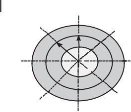
The first step in the cellular transport of DNA nanoparticles is the electrostatic interaction between the carrier/DNA complex and the anionic plasma membrane [1, 27, 28]. Complex formation between DNA and cationic molecules results in positively charged nanoparticles, as measured by f potential. A positively charged structure facilitates adherence to the negatively charged cell surface receptors and endocytosis. The amount of negative charges on the cell surface and the size of the carrier/DNA complex appear to be the determinants of successful gene delivery [29]. Depending on the size of the carrier/DNA complex, receptor-mediated endocytosis, pinocytosis, or phagocytosis may occur. In the cytoplasm, endosomes are destabilized, leading to the release of the DNA. A schematic diagram of DNA uptake in cells is shown in Fig. 10.1 [1]. The common agents used for DNA condensation and gene delivery applications are listed below.
10.2 Agents That Provoke DNA Nanoparticle Formation 255
Figure 10.1. DNA uptake by mammalian cells. DNA is compacted in the presence of polycations into ordered structures such as toroids, rods, and spheroids. These particles interact with the anionic proteoglycans at the cell surface and are transported by endocytosis. The cationic agents accumulate in the acidic vesicles and raise the pH of the endosomes, inhibiting the degradation of DNA by lysosomal
10.2
enzymes. They also sustain a protein influx, which destabilizes the endosome and releases DNA. The DNA is then translocated to the nucleus either through the nuclear pore or with the aid of nuclear localization signals, and decondenses after separation from the cationic delivery vehicle. (Reproduced with permission from Ref. [1].)
Agents That Provoke DNA Nanoparticle Formation
10.2.1
Polyamines
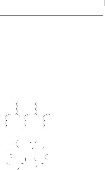
The polyamines spermidine and spermine and their synthetic analogues are excellent promoters of DNA nanoparticle formation [8, 9, 14, 18, 30–33]. The ability of polyamines and their analogues (Fig. 10.2) to compact DNA has been studied by several investigators as a model system to understand the mechanism of DNA compaction in phage head [8, 9]. Bloomfield and colleagues [8, 9, 13, 14, 32] have used the counterion condensation theory developed by Manning [34] and Record et al. [35] to calculate DNA charge neutralization in the presence of spermidine and spermine, and found that DNA collapse occurs at >89% charge neutralization. Earlier work considers cationic polyamines as point charges without any defined structure. However, two trivalent cations, cobalt hexamine3+ and spermidine3+, differ in their ability to condense DNA, suggesting the importance of site-specific interactions in addition to the overriding electrostatic interactions [14]. Recent studies with a series of isovalent spermine ho-
256 10 Nanotechnology in Nonviral Gene Delivery
mologues further show the importance of polyamine structure in DNA condensation (Fig. 10.3) [30]. There are also significant differences in the relative affinity of these homologues to DNA, as measured by the ethidium bromide displacement assay (Tab. 10.1). Although tetravalent polyamines are not very efficient in DNA transport in cells, higher-valency analogues, including the hexamines facilitated the transport of a 37nucleotide (nt) oligonucleotide in breast cancer cells (Fig. 10.4) [19]. The hexamines can condense a plasmid DNA to toroidal nanoparticles, as measured by atomic force microscopy (AFM) (Fig. 10.5; Tab. 10.2).
3-2-3:
NH2
NH
NH2
NH
3-3-3:
NH2
NH
NH
NH2
3-4-3:
NH2
NH
NH
NH2
3-5-3:
NH2
NH
NH
NH2
3-6-3:
NH2
NH
NH
NH2
3-7-3:
NH2
NH
NH
NH2
3-8-3:
NH2
NH
NH
NH2
3-9-3:
NH2
NH
NH
NH2
3-10-3:
NH2
NH
NH
NH2
3-11-3:
NH2
NH
NH
NH2
3-12-3:
NH2
NH
NH
NH2
3-3-3-3:
NH2
NH
NH
NH
NH2
3-3-3-3-3:
NH2
NH
NH
NH
NH2
NH2
3-4-3-4-3:
NH2
NH
NH
NH
NH
NH2
3-3-4-3-3:
NH2
NH
NH
NH
NH
NH2
Figure 10.2. Chemical structures of the natural polyamine, spermine (3–4-3) and its analogues. The polyamine analogues are abbre-
viated by a number system that represents the number of methylene bridging groups between the primary and secondary amino groups.
Figure 10.3. Typical plots of the relative intensity of scattered light at 90 plotted against the concentrations of spermine (O), 3–10–3 (~), 3–11–3 (d, and 3–12–3 (h). The k-DNA
solution had a concentration of 1.5 lM DNA phosphate, dissolved in 10 mM Na cacodylate buffer, pH 7.4. (Reproduced with permission from Ref. [30].)
10.2 Agents That Provoke DNA Nanoparticle Formation 257
Uptake, cpm
LabelledTFO P]- 32 [
50000
3-3-3-3-3
40000
30000
20000
10000
0
0
5
10
15
20
25
30
60000
3-4-3-4-3
50000
40000
30000
20000
10000
0 0
5
10
15
20
25
30
50000
3-3-4-3-3
40000
30000
20000
10000
0
0
5
10
15
20
25
30
Time, h
Figure 10.4. Time-course of uptake of a 37-nt triplex-forming oligonucleotide (TFO) in the presence of hexamines, 3–3-3–3-3, 3–4-3–4-3 and 3–3-4–3-3. MCF-7 breast cancer cells
(5 x 10⁵ per well) were plated in six well plates and allowed to adhere for 24 h. Triplicate wells were treated with a 250,000 cpm level of 32P-labeled TFO in 0.5 ml prewarmed (37 C)
medium. Cells were harvested at the indicated time points. Cell lysate was prepared and radioactivity determined by scintillation counting.
The concentrations of hexamines used in these experiments were: 0 (O), 0.25 (r), 0.5 (j),
1 (,), and 2.5 lM (d). Data shown are mean
– SD from triplicate experiments. (Reproduced with permission from Ref. [19].)
258 10 Nanotechnology in Nonviral Gene Delivery
A B
C D
Figure 10.5. Atomic force microscopy images showing the toroid structures of pGL3 plasmid DNA formed by incubation with 25 lM spermine (A), 5 lM 3–3-3–3 (B), 2 lM 3–4-3–4-3 (C), and the partly formed toroids observed in the presence of 2 lM 3–4-3–4-3 (D). D provides
evidence for spooling of DNA to form toroids. Scale bar is 200 nm. (Reproduced with permission from Nucleic Acids Research 2004, 32, 127–134. Copyright 2004 Oxford University Press Ref. [33].)
Table 10.1 Relative binding constants of polyamine analogues for calf thymus DNA, as measured by the ethidium bromide competition method.
Polyamine homologue
Relative binding constanta
3–2-3
0.4
3–3-3
0.6
3–4-3 (spermine)
1.0
3–5-3
1.3
3–6-3
1.1
3–7-3
1.0
3–8-3
0.7
3–9-3
0.5
3–10–3
0.5
3–11–3
0.4
aThe binding constants were calculated as the reciprocal of the 50% concentration of polyamine homologues required to displace 50% ethidium bromide bound to k-DNA. The reproducibility with these results was within 3% in repeated measurements. The results are normalized with respect to spermine. (Reproduced with permission from Ref. [30].)
10.2 Agents That Provoke DNA Nanoparticle Formation 259
Table 10.2 AFM measurement of the outer diameter and height of toroidal DNA nanoparticles formed in the presence of polyamine analogues.
Polyamine
Outer diameter
Mean height
(nm)
(nm)
3–4-3 (Spermine)
191
– 12a
2.61 – 0.77b
3–3-3–3
168
– 5.4
3–3-3–3-3
117
– 8.8
3–4-3–4-3
118
– 10.8
a Mean – S.E.M. of 5–7 toroids measured in each case.
bThe toroid height given here is the average value for 28 toroids, formed in the presence of polyamines shown in column 1 of this table.
(Reproduced with permission from Ref. [33].)
10.2.2
Cationic Lipids
A group of cationic molecules that has attracted much attention in preclinical and clinical gene therapy trials is the cationic lipids [26, 27, 36, 37]. These molecules possess a hydrophobic group and a polar group (Fig. 10.6). A large number of lipids have been developed to transfect DNA; the best known are DOTAP (N-1 (-2,3-dio- leoyloxy) propyl)-N, N, N-trimethylammoniumethyl sulfate), DOTMA (N-1- (2,3-dio- leoyloxy) propyl)-N, N, N-trimethylammonium chloride), DOGS (dioctadecylamido- glycylspermine-4-trifluoroacetic acid), and DOSPA (2,3-dioleoyloxy-N-[2-(spermine- carboxamido)-ethyl]N,N-dimethyl-propan-1-aminium trifluoroacetate). These are available as commercial DNA transfection agents. These agents can form DNA nanoparticles by electrostatic interactions between the positively charged polar head group of the lipid and the negatively charged phosphate group of DNA [36]. Increasing the number of amino groups per molecule and the distance between amino group and hydrophobic group favors the transfection efficiency of cationic lipids. Nanoparticle formation by complexation with lipids not only improves the transport of DNA through the cellular barriers, but also protects it from enzymatic degradation in cell culture media [38–41]. The size of the particles ranges from 50 to 1000 nm. The large particles of liposome/DNA complexes are found to transfect cells in vitro. Liposome DNA complexes are taken up by endocytosis, and after internalization DNA is released from the endocytic vehicle, leaving behind the liposome [42]. However, the transfection efficiency of liposome/DNA complex is relatively low in vivo. In addition, many available cationic lipids are reported to be toxic [43]. Studies in mice and macaques have shown that exposure to high doses or repeated doses results in microscopic pathology and gross lung pathology.
260 10 Nanotechnology in Nonviral Gene Delivery
O N+
O
DOTMA O
O N+
O
+
NH3
DOTAP O
+
NH2
O
N
NH
O NH2+
DOGS
NH3+
NH3+
NH2+
O
O
N +
NH
NH2+
O
NH3+
DOSPA
Figure 10.6. Chemical structures of commonly used cationic lipids. The abbreviations are defined in the text.
10.2.3
Polyethylenimine
Polyethylenimine (PEI) is a group of synthetic polymers that are known to be efficient in the transport of oligonucleotides and plasmid DNA in a variety of cell types and animal models [44]. Linear and branched PEI (Fig. 10.7) can induce the condensation of DNA to nanoparticles. The linear and branched nature of PEI as well as its molecular weight play important roles in DNA condensation and transfection effi-
10.2 Agents That Provoke DNA Nanoparticle Formation 261
ciency [45]. Branched PEI contains primary, secondary, and tertiary amino groups, and acts as a proton sponge at the endosomal pH [1, 23]. The buffering capacity of PEI is believed to contribute to its ability to deliver DNA within cells without degradation. Unlike other polymers, PEI possesses intrinsic endosomolytic activity and does not need any endosomolytic agent to escape from endosome [23, 45, 46]. A confocal microscopic investigation shows that PEI DNA transport occurs without the dissociation of the complex [47]. Poor solubility of the DNA/PEI complex at physiological pH and toxicity of PEI in animal model studies are the major limitations in using this polymer as a gene carrier. Derivatization of PEI with poly(ethylene glycol) (PEG) is reported to increase the solubility and reduce the cytotoxicity of the PEI [47–50]. PEGylation improves the stability of nanoparticles and increases their in vivo circulation time. A terpolymer of polylysine, PEG, and PEI has also been investigated as an agent for DNA nanoparticle formation [51].
NH
NH
NH
]n
[ NH
NH
NH
NH
Polyethylenimine (linear)
N
NH
[ NH
N
N
]n
N
NH
NH
NH
NH
N
NH
NH
NH2
Polyethylenimine (branched)
O
[
O ]
O
n
N
NH
]n
[ NH
NH
N
NH
O [
O
]
O
n
PEGylated Polyethylenimine
Figure 10.7. Chemical structures of linear, branched, and polyethylene glycol (PEG) derivatized polyethylenimines.
26210 Nanotechnology in Nonviral Gene Delivery
10.2.4
Dendrimers
Polyamidoamine (PAMAM) and polypropylenimine (PPI, Fig. 10.8) dendrimers have been studied for their ability to provoke DNA nanoparticle formation and facilitate DNA transport [52–57]. Tomalia et al. [52] reported the first preparation of an
entire series of dendrimers, possessing trigonal, 1- > 2 N-based, branching centers. Compared to linear and branched polymers, the monodispersity and
H N
2
NH
N
2
N
H N
2
NH
2
NH
2
H N
NH
2
N
2
N
N
NH2
H N
N
2
N
N
NH
NH
2
2
NH
2
G - 1
G - 2
H
N
H
N
2
H N
2
2
H
N
N
2
H N
N
NH
2
N
N
N
NH
N
2
N
H N
NH
2
N
2
N
H
N
N
2
N
H
N
NH
2
N
NH2
N
2
NH
NH
2
2
H N
NH
2
2
H
N
NH
NH
NH2
2
2
H N
2
NH
2
NH
2
N
2
H
N
N
2
NH
N
N
2
H
N
N
NH
2
2
N
H
N
N
N
N
NH
2
N
2
N
N
NH
2
N
N
N
NH
H N
2
2
NH
H N
N
N
2
2
N
N
H N
N
N
2
N
N
N
H
N
NH
2
N
N
2
H
N
N
NH
2
2
N
N
NH2
H
N
N
2
NH
2
H
N
2
H
2
N
NH
NH
2
NH
NH
2
2
2
G - 3
G - 4
H N NH2 NH2
NH
2
NH
H
N
2NH
2NH
2
H N
2
2
NH2 NH
H
N
2
2
2
NH
2
H
N
2
N
N
N
N
NH
2
H
N
2
N
NH
H
N
N
2
NH
2
N
H
N
N
N
N
2
NH
2
2
N
N
N
H N
N
NH
2
2
N
N
H
N
N
NH2
N
2
N
N
NH
N
2
N
N
H N
NH
2
2
H
N
N
N
2
N
N
N
NH
2
H
N
N
2
NH
H N
N
N
N
N
2
NH
2
N
2
H
N
N
NH
N
2
2
N
N
N
H
N
2
N
NH
H
N
N
2
2
N
NH
H N
N
N
2
N
N
N
2
N
NH
2
H
N
N
2
N
N
N
NH
2
H N
N
2
N
N
N
NH
H
N
N
2
NH
2
N
H
N
N
N
2
2
N
N
N
NH2
H
2
NH
H N
NH
2
2
H
N
H
N
2
NH
2
H
N
2
NH
2
2
H
N
NH
NH
NH
NH
2
2
2
2
2
2
G - 5
Figure 10.8. Chemical structures of five generations of polypropylenimine dendrimers. The complexity of the dendrimers is represented by generation numbers (G1 to G5).
10.2 Agents That Provoke DNA Nanoparticle Formation 263
Figure 10.9. Cellular uptake of a 32P-labeled 31-nt oligonucleotide (ODN) in the presence of polypropylenimine generation 4 (G-4) dendrimer. Different cell lines (MCF-7 and SK-BR-3 breast cancer, LNCaP prostate cancer, and SK-OV-3 ovarian cancer) were treated with a 250,000 cpm level (0.4 nM) of 32P-labeled ODN after complexing with G-4 dendrimer in six-well plates. At the indicated times, the medium was removed, cells washed three times
with cold phosphate buffered saline, lysed, and cell-associated radioactivity quantified by scintillation counting. Control indicates the use of labeled ODN in the absence of dendrimer.
The percentage increase in uptake is calculated with reference to the control group. Results presented are the mean of three separate triplicate measurements. Error bars indicate S.E.M. (Reproduced with permission from Ref. [56].)
264 10 Nanotechnology in Nonviral Gene Delivery
4 h
6 h
8 h
24 h
48 h
C
+
+
+
+
+
Figure 10.10. Stabilization of DNA by nanoparticle formation with polypropylenimine G-4 dendrimer. MDA-MB-231 breast cancer cells were treated with a 31-nt 32P-labeled ODN alone or 32P-labeled ODN/G-4 dendrimer complex for the indicated time points. Cells were washed with phosphate buffered saline and DNase I (300 units ml–1) to remove cell sur- face-associated ODN. 32P-labeled ODN was then extracted from cells and characterized by 20% polyacrylamide gel electrophoresis. Lanes
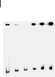
2, 4, 6, 8, and 10 show 32P-labeled ODN extracted from cells treated with 32P-labeled ODN alone. Lanes 3, 5, 7, 9, and 11 show 32P-labeled ODN extracted from cells treated with 32P-labeled ODN/G-4 complex. Lane 1 (C) is 32P-labeled ODN in sterile water, used as a marker of the intact 32P-labeled ODN migration in the gel. ODN/G-4 nanoparticles had a remarkable stability within the cells. (Reproduced with permission from Ref. [56].)
controllable surface functionality make the dendrimers an interesting class of gene delivery vehicles. Haensler and Sz.ka [53] were the first to show that PAMAM dendrimers are efficient gene transfer agents for a variety of cultured mammalian cells. Generations 2 and 3 of PPI dendrimers have been shown to be effective in transfecting a plasmid DNA in A431 cells [54]. We have found that all five generations of PPI dendrimers can facilitate the transport of a 31-nt triplex-forming oligonucleotide in breast, prostate, and ovarian cancer cell lines (Fig. 10.9) [56]. Nanoparticles formed by the oligonucleotide have hydrodynamic radii ranging from 100 to 200 nm (Tab. 10.3). The oligonucleotide nanoparticles are protected from enzymatic digestion within the cell for up to 48 h of treatment (Fig. 10.10). Synchrotron X-ray diffraction studies show that the complex formed between generation 4 dendrimer and DNA is in the liquid crystalline state [58], similar to the liquid crystalline textures observed with polyamine/DNA complexes [59].
10.2 Agents That Provoke DNA Nanoparticle Formation 265
Table 10.3 Hydrodynamic radius of the nanoparticles formed from a 31-nt oligonucleotide in the presence of polypropylenimine dendrimers.a
Concentration
Hydrodynamic radius (nm)
of dendrimer (lM)
G-1
G-2
G-3
G-4
G-5
1
–
–b
240–11
172–1
196–2
2.5
–
–
181–12
143–10
160–8
5
279–9
193–16
166–1
156 –1
163–8
10
235–4
233–15
137–9
137–3
158–4
aAll measurements were done using dynamic laser light scattering equipment. Measurements were done in a buffer containing approximately physiological concentration of cations (120 mM KCl, 10 mM NaCl, 2 mM MgCl2, and 0.1 mM CaCl2) at 22 C. The concentration of oligonucleotide used in these experiments was 0.1 lM oligomer.
bDashes indicate that no particles were formed. (Reproduced with permission from Ref. [56].)
10.2.5
Proteins and Polypeptides
DNA nanoparticle formation within the cell is mainly achieved by the interaction of DNA with cationic proteins or peptides, such as histones and protamines. DNA in sperm and certain viruses is condensed by protamine, which contains arginine-rich
NH2
NH2
O
O
O
[NH
NH
NH
NH
NH
]
n
O
O
NH2
NH2
NH2
Poly-L-lysine (linear)
Lys
Lys
Lys
Lys
Lys
Lys
Lys
Lys
[
Lys
Lys
Lys
Lys
Lys ]
Lys
Lys
n
Lys
Lys
Lys
Lys
Lys
Lys
Lys
Lys
Lys
Lys
Lys
Lys
Lys
Lys
Lys
Poly-L-lysine (branched)
Figure 10.11. Chemical structures of linear and branched poly-l-lysine.
266 10 Nanotechnology in Nonviral Gene Delivery
domains that can bind with the phosphodiester backbone of DNA. Protamine-medi- ated DNA condensation causes adjacent arginine residues to interlock both strands of the DNA helix, making it transcriptionally inactive [60, 61]. There have been several studies using protamine and related proteins to induce and stabilize DNA nanoparticles for gene delivery.
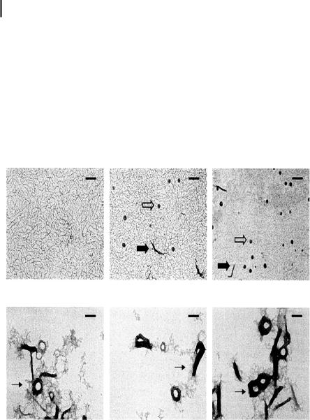
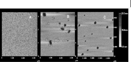
The cationic polypeptide, poly-l-lysine (PLL) (Fig. 10.11) contains protonated amine groups, which can interact with DNA and provoke nanoparticle formation [17, 18, 62, 63]. The size of the nanoparticles thus formed depends on the nature of the PLL (linear versus branched as well as ionic conditions of the medium) (Fig. 10.12) [17]. Although PLL is not a very efficient agent for transfecting DNA in
A B C
D E F
Figure 10.12. Electron micrographs of the structures of condensed DNA complexes prepared at different ionic strengths and poly-l-lysine/DNA ratios. Electron micrographs of 0.04% uranyl acetate-stained DNA complexes were prepared using plasmids pCMVLuc and GalPLL256 [17]. Samples were stained within 30 min after their preparation.
(A–C) DNA samples prepared at 1 M NaCl with GalPLL256. (D–F) DNA samples prepared at
0 M NaCl with GalPLL256. r values (poly-l-ly- sine to DNA ratio) are as follows: (A, D) 0.25; (B, E) 0.5; (C, F) 0.75. There was no adjustment of salt concentration before processing samples. Open arrows, spherical complexes; solid arrows, rod-like complexes (major diameters of ~100 nm); thin arrows, aggregated structure including large rods and toroids complexes. The bars in all panels represent 100 nm. (Reproduced with permission from Ref. [17].)
10.3 Characterization of DNA Nanoparticles 267
cells, conjugation to a receptor enhances its ability as a gene delivery vehicle [64–67]. Complex formation between PLL and DNA can also protect the DNA from enzymatic degradation within the cellular environment [68].
Low-molecular-weight peptides are also reported to condense DNA to nanoparticles, but the in vivo efficiency of these complexes for gene delivery is poor because of the low affinity of the peptides for DNA. To improve the DNA binding and transfection efficiency, stable cross-links were introduced to the condensates using bifunctional agents such as glutaraldehyde or sulfhydryl groups [9, 69–71]. Short peptides possessing multiple cysteine residues form interpeptide disulfide bonds when bound to DNA and after internalization. The half-life of DNA in mouse liver is extended by a formulation of sulfhydryl-linked gene delivery system [72].
10.2.6
Polymers
Chitosan is a natural polymer consisting of two subunits, d-glucosamine and N- acetyl-d-glucosamine linked together by glycosidic bonds (Fig. 10.13). It is comparatively nontoxic and has a high transfection efficiency after nanoparticle formation [73–77]. Chemical modification is feasible in this system because of the availability of a large number of surface functional groups. Chitosan and its derivatives were found to be promising vectors for gene delivery [73].
CH2OH
CH2OH
CH2OH
CH2OH
O
O
O
OH
OH
O
[
O
O
OH
O
OH
O
NH2
NH2
NH2
NH2
Figure 10.13.
Chemical structure of chitosan.
]
n
Neutral polymers such as PEG and poly(ethylene oxide) cause DNA condensation at high ionic strength [78]. These uncharged flexible polymers act as crowding agents and provoke DNA condensation through an excluded volume mechanism. PEG is being used as part of the graft polymers in conjunction with PEI, folate receptors, and polylysine to improve the stability of the DNA condensate and transfection efficiency [79–81].
In summary, a large number of agents that can interact with DNA and provoke nanoparticle formation are being studied in academic and industrial settings for the development of nonviral gene delivery vehicles.
10.3
Characterization of DNA Nanoparticles
The following three methods are commonly used for the characterization of DNA nanoparticles in gene therapy applications: (i) laser light scattering; (ii) electron microscopy; and (iii) atomic force microscopy.
26810 Nanotechnology in Nonviral Gene Delivery
10.3.1
Laser Light Scattering
Bloomfield and colleagues [8, 14, 82] have pioneered the application of laser light scattering to the study of DNA condensation. Extended DNA structure in solution is in a worm-like configuration and the surface area is not sufficient to scatter light. Therefore, the intensity of light scattered by dilute solutions of DNA (~1 lM) is not much different from that of the buffer in which the DNA is dissolved. However, a marked increase in the intensity of the scattered light occurs at a critical concentration of the condensing agent, indicating the compaction of DNA to structures with a high surface area, such as spheroids and toroids. The increase in the scattered light intensity is dependent on the concentration of the condensing agent up to a point, and then levels off at higher concentrations (Fig. 10.14) [33]. The efficacy of the condensing agents can be quantified by determining the EC50 value, the concentration of a condensing agent at the midpoint of DNA condensation. In a buffer containing 10 mM Na cacodylate (pH 7.4), the EC50 values for spermine (3–4-3), norspermine (3–3-3), pentamine (3–3-3–3), and two hexamines (3–3-3–3-3 and 3–4-3–4-3) are 11.3, 10.6, 1.5, 0.49, and 0.52, respectively [33]. This result indicates that the pentamine and hexamine analogues are more efficacious than spermine in condensing the plasmid pGL3.
Dynamic light scattering provides a quick and accurate measurement of the particle size and particle size distribution of DNA nanoparticles in solution. Molecules undergoing brownian motion cause fluctuations in scattered light intensity [82]. This scattered light intensity has a time scale that is related to the speed of movement of the molecules, and hence to their size. In the experimental set-up, a laser beam passes through a small quartz cell containing the sample (polyamine/DNA mixture), and the scattered light at 90 to the beam path is collected and converted
Figure 10.14. Typical plots of the relative intensity of scattered light at 90 plotted against the concentrations of spermine and its analogues. The pGL-3 luciferase plasmid DNA solution had a concentration of 1.5 lM DNA phosphate, dissolved in 10 mM Na cacodylate buffer, pH 7.2. The symbols are as follows:
(A)3–3-3 (d, 3–4-3 (O), 3–2-3 (j);
(B)3–3-3–3 (h), 3–3-3–3-3 (D), and
3–4-3–4-3 (~). (Reproduced with permission from Nucleic Acids Research 2004, 32, 127–134. Copyright 2004 Oxford University Press Ref. [33].)
10.3 Characterization of DNA Nanoparticles 269
to electrical pulses which can be processed and analyzed by a series of computer programs to extract information about particle size, diffusion coefficient, and other parameters. For example, the collection of photons into time windows or channels and subsequent analysis (autocorrelation) of this data yields the translational diffusion coefficient, DT. For monodisperse particles much smaller than the incident beam, the autocorrelation function g(1)(s) is given by the following equation [82]:
g(1)(s) = exp(-Dq2(s)),
(1)
where (s) is the decay time and q (= 4pn/k0sinH/2) is the scattering vector, which is a function of solvent refractive index n, the wavelength of the incident beam k0, scattering angle H, and diffusion coefficient D. The hydrodynamic radius (Rh) is calculated from the diffusion coefficient using the Stokes–Einstein equation:
Rh= kT/6pgD,
(2)
where k is the Boltzmann constant, T is the absolute temperature, and g is the solvent viscosity.
The results of light scattering experiments can be complicated by the presence of dust or other particles in the sample. This can be circumvented by filtering the solution through 0.45-mm millipore filters and centrifuging the sample for 30 min at 500 0 g. The size range that can be measured by commercial dynamic light scatting equipment is 1–1000 nm.
10.3.2
Electron Microscopy
Electron microscopy (EM) has been used to visualize DNA nanoparticles formed in the presence of multivalent cations (Fig. 10.15). EM can be used to determine the size, shape, and dimensions of DNA nanoparticles. Hud and colleagues [83, 84] used the technique to identify hexagonal packing of DNA in toroids, using freezefracture microscopy. The experimental procedure is relatively simple in a laboratory with access to a good electron microscopy facility. Solutions of DNA nanoparticles (formed by mixing and incubating the DNA with the condensing agent) are placed on Formvar-coated grids [31, 83, 84]. After 3–5 min, the solution is drained off with a filter paper and grids stained with a drop of 1% w/v uranyl acetate. Samples are observed in an electron microscope with appropriate magnification. A major source of error in scanning electron microscopy is in sample preparation and image distortions or irregularities. Segregation of particles during sample preparation can lead to heterogeneous deposition. Poor statistical sampling resulting from counting only a small number of particles is another limitation. Particles of a large size range (5–500,000 nm) can be measured using electron microscopy.
Electron microscopy can be complemented with polarizing microscopy to determine the liquid crystalline textures of DNA in the presence of condensing agents [59]. For these studies, the condensates are deposited between a slide and coverslip.
270 10 Nanotechnology in Nonviral Gene Delivery
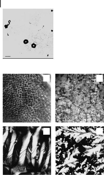
Figure 10.15. Electron micrograph of DNA nanoparticles formed in the presence of spermidine. Poly(dA-dT).poly(dA-dT) solution (3 lM DNA phosphate) in 10 mM NaCl, 1 mM sodium cacodylate, and 200 lM spermidine was placed on a carbon-coated grid and counterstained with 1% uranyl acetate. The bar indicates 100 nm.
A B
C D
Figure 10.16. Effects of spermine and N1-acetylspermine on the liquid crystalline phase transitions of calf thymus DNA. (A) DNA (25 mM in Na cacodylate buffer) was mixed with 1 mM spermine and incubated on a glass slide at 22 C for 15 min (1000). A planar cholesteric phase with a three-dimensional network is observed. (B) The glass slide in
(A) was incubated for 12 h at 37 C and viewed
through the plate under crossed polars (2000). Fingerprint texture with antiparallel grain boundaries is found. (C) Large pitch cholesteric phase was observed when the glass slide was further incubated for 24 h at 37 C (1800).
(D) DNA was mixed with 1 mM N1-acetylsper- mine and incubated for 48 h at 37 C (450).
A crystalline phase is obtained. (Reproduced with permission from Ref. [59].)
10.3 Characterization of DNA Nanoparticles 271
The coverslips are sealed with a solution of polystyrene and plasticizer in xylene to prevent dehydration. Textures are allowed to stabilize for a few minutes to a few hours. Recent studies indicate the formation of two types of liquid crystalline phase: a cholesteric phase and a columnar hexagonal phase in DNA treated with a natural polyamine (Fig. 10.16) [59]. The thermodynamic stability of cholesteric phases formed from DNA can be determined by monitoring different phases under a microscope as a function of temperature, salt concentration, and DNA dilution. It is of interest to examine whether the ability of DNA to form different liquid crystalline phases is related to the efficacy of different agents to facilitate the cellular transport of DNA and oligonucleotides.
10.3.3
Atomic Force Microscopy
Atomic force microscopy (AFM) is the preferred technique for visualizing DNA nanoparticles and making quantitative three-dimensional measurements. AFM, which was invented in 1986, expanded the application of scanning tunneling microscopy to nonconductive, soft, and living biological samples [85–88]. AFM has several capabilities, including imaging topographic details of surfaces from the submolecular level to the cellular level [89], monitoring the dynamic processes of single molecules in physiologically relevant solutions [90], measuring molecular interactions [91], and characterizing the mechanical properties of single molecules or single nanostructures [92]. In general, mixing DNA and condensing agent in solution forms DNA nanoparticles. However, nanoparticles have also been prepared on a solid surface for imaging. When the nanoparticles are formed in solution, they can be immobilized onto a freshly cleaved mica surface and AFM imaging done in air or in solution. In general, imaging in air is much easier than imaging under solution conditions, and can provide valuable structural and morphological information about DNA nanoparticles in three dimensions. For example, the size, height, shape, and volume of the particles can be easily obtained for spherical or rod-like structures, after obtaining the AFM images. For toroidal structures, which are commonly formed during DNA condensation, the toroidal radius and volume can be determined by AFM software as described by Rackstraw et al. [92] (Fig. 10.17). Cross sections of the ring-like particles can be taken at 45 separations, and toroidal radii determined from the cross sections at half-maximum height in order to minimize tip convolution effects. The volume of single nanoparticles can be calculated using the equation:
Volume = 2p2r2R,
(3)
where the values of r and R are as defined in the legend to Fig. 10.17.
In the second case, the condensates can be formed directly on the substrate and imaged by AFM directly in air or under solution. There are many attractive features with regard to imaging under solution. The most obvious one is the ability to follow the dynamic condensation process in physiologically relevant buffers in real time.
272 10 Nanotechnology in Nonviral Gene Delivery
r
R
A B C D
Figure 10.17. Representation of a toroidal structure used to calculate the condensate volume using Eq. (3). In this figure, r = {(A–D)(B–C)}/4, and R = {(B–C)/2} + r.
Compared to electron microscopy, the ability to image in solution under native conditions is the most striking feature of AFM. AFM can be operated in both tapping mode and contact mode to image the DNA nanoparticles. Tapping mode is more commonly utilized, because the destructive effect of the AFM tip on the DNA nanoparticles (which are usually soft) is much less in tapping mode than in contact mode imaging.
10.3.3.1DNA Nanoparticle Studies by AFM
Examples of DNA nanoparticle formation in the presence of PPI dendrimer (generation 4) and spermidine are shown in Fig. 10.18. The DNA structure gradually changes from fully uncondensed plasmid structures to partially condensed flowerlike structures in the presence of 2.5 lM PPI dendrimer. In the presence of 10 lM spermidine, compact, fully condensed nanoparticles are observed. Using AFM, Iwataki et al. [93] have observed competition between compaction of single chains and bundling of multiple chains in giant DNA molecules with spermidine as a condensing agent. When the concentration of DNA was <1 lM in base-pair units, individual DNA molecules condensed into a compact structure, whereas a thick fiber-like assembly of multiple chains occurs when the concentration of DNA is increased to 10 lM. In other studies, different morphologies, including flowers, disks, toroids, and branch-like structures are observed by AFM at different stages of DNA condensation or under different condensation conditions [94–96].
DNA nanoparticle formation in the presence of poly-l-lysine (PLL), modified PLL, spermidine, and PEI has been extensively studied using AFM [18, 96–101]. Hansma and collaborators [18, 96, 101] employed AFM to study the extent of DNA condensation in approximately 100 different complexes of DNA with PLL or PLL attached to the glycoproteins, asialoorosomucoid (AsOR), or orosomucoid. They found that DNA condensation is efficient with 10-kDa PLL covalently attached to AsOR at a lysine:nucleotide (Lys:nt) ratio of 5:1 or higher. Large numbers of toroids and short
10.3 Characterization of DNA Nanoparticles 273
Figure 10.18. AFM images of DNA nanoparticle formation from plasmid pGL3 in the presence of PPI dendrimer and spermidine.
(A) Plasmid DNA on mica surface; (B) partially
condensed plasmid DNA in the presence of 2.5 lM generation-4 PPI dendrimer; (C) plasmid DNA condensed in the presence of 10 lM spermidine.
rods with contour lengths of 300–400 nm are produced under these conditions. Furthermore, it has been found that PLL-AsOR enhanced gene expression in the mouse liver approximately 10to 50-fold as compared with PLL alone. The study by Wolfert and Seymour [99] on the influence of the molecular weight of PLL on the size of DNA nanoparticles shows that the smallest PLL (MW: 3970) produce more homogeneous complexes with diameters ranging from 20 to 30 nm as compared to the large complexes (120–300 nm) formed in the presence of high-molecular-weight PLL (MW: 224,500). DNA nanoparticles formed with low-molecular-weight PLL have significantly lower cytotoxicity than those formed with high-molecular-weight PLL. Interestingly, PEI is more efficient than PLL for nanoparticle formation for gene delivery applications [45, 94, 100, 101]. AFM studies show DNA aggregation and partially condensed nanoparticles in the presence of PLL, whereas PEI can provoke nanoparticles of 30–60 nm. Rings, extended linear structures, toroids, rods, and other intermediates are found in AFM studies of nanoparticles provoked by PLL-polymer conjugates.
Han et al. [102] show condensation of plasmid DNA by a water-soluble lipopolymer into spherical particles of approximately 50 nm diameter. Lim et al. [103] confirm by AFM the formation of self-assembling biodegradable complexes between DNA and poly[a-(4-aminobutyl)-l-glycolic acid], a non-toxic polymeric gene carrier.
In addition to cationic DNA condensing agents, neutral amphiphiles that possess a DNA molecular recognition unit with a hydrophobic region and a PEG unit have also been shown to condense plasmid DNA successfully [78, 79]. AFM studies show a range of collapsed states from partial collapse to the condensed supramolecular toroidal structures, which have an outer and inner diameter of approximately 100 nm (range 106 22 nm) and 20 nm (range 21 11 nm), respectively. These amphiphiles represent the first step toward identifying the design requirements for a selective DNA binding/condensation agent. Isobe et al. [104] have studied the condensation of plasmid DNA with functionalized fullerenes by AFM. Gallyamov et al. [105]
274 10 Nanotechnology in Nonviral Gene Delivery
have investigated the process of compaction of high-molecular-weight T4 DNA directly in an AFM liquid cell. AFM images of globules formed by compaction of DNA in water–alcohol environments at high isopropanol concentration (80%) have also been obtained. By using the technique of deconvolution of the AFM images, it is shown that the globule contains only one closely packed DNA molecule. Our recent studies demonstrate toroid formation of a plasmid DNA in the presence of polyamine analogues (Fig. 10.5) [33].
DNA condensation occurring on surfaces has also been studied [106–111]. Dimitriadis et al. [106] used AFM to study the conformation of DNA adsorbed from aqueous solutions containing monovalent (Na+), divalent (Ca2+), and trivalent (spermidine3+) cations on different solid substrates (hydrophobic, moderately hydrophilic, and hydrophilic surfaces). It is shown that the substrate surface energy mediated DNA/DNA interaction, leading to the formation of a variety of structures. It has also been shown that the condensed DNA structures are formed on hydrophobic surfaces even in the absence of multivalent cations. The study by Allen et al. [108] on DNA–protamine complexes bound to mica by AFM shows that the morphology of the structures varies depending on the sample preparation method. For example, interstrand, side-by-side fasciculation of DNA and toroidal-like structures are observed for complexes formed in solution, following direct mixing of the DNA and protamine. Large aggregates of DNA are also observed occasionally. However, if the DNA is first attached to the mica surface, prior to addition of the protamine, only well-defined toroidal structures are formed, without DNA fasciculation or aggregation. The mean diameter of the toroids is 39.4 nm. The structures indicate that the condensed DNA is stacked vertically by four to five turns with each coil containing as little as 360–370 bp of B-form DNA. Fang et al. [110, 111] showed the two-dimen- sional condensation of DNA molecules on cationic lipid membrane by AFM. It was found that the fluidity of cationic membrane promoted the close packaging of both linear and circular DNA molecules, independent of the length of the DNA. The average interhelical distance for closely packed DNA is over twice the average diameter of DNA.
It is noteworthy that different nanoparticle structures are formed with DNA, depending on whether the condensation process is performed in solution or on a solid surface. For example, Allen et al. [108] show distinct toroidal structures when protamine is added to DNA on a mica surface. However, when DNA and protamine are mixed and incubated in solution before transfer onto mica surface, a network of DNA with a few thin toroids is found. Fang et al. [110] reported similar results when they used silanes as condensing agents. When silanes and DNA are preincubated in solution, looped structures including flower and sausage shapes are observed, whereas on a silicon surface silanes condense DNA into distinct toroids, whose size depends on the length of the DNA.
10.3.3.2Limitation of AFM Technique
Compared to transmission electron microscopy, AFM not only provides direct threedimensional images of DNA nanoparticles, but also has the capability of operating in solution, thereby providing a new avenue to determine the structures of biological
10.3 Characterization of DNA Nanoparticles 275
specimens at high resolution in physiologically relevant ionic and pH conditions. However, like any physical technique, AFM is not perfect, and has limitations based on the sample preparation method. As previously discussed, most DNA nanoparticles for nonviral gene delivery are prepared in solution. In order to image these nanoparticles by AFM, they have to be first transferred and immobilized onto a substrate, which in most cases is mica, and then the substrate fastened to the AFM instrument. A common sample preparation follows a two-step procedure:
1.Deposit an aliquot of the solution of DNA nanoparticles on a freshly cleaved mica surface (modified or unmodified).
2.Rinse and blow dry the sample on the mica surface after a predetermined incubation period.
While following this basic procedure, details of sample preparation differ from laboratory to laboratory. For example, Sun et al. [112] use a filter paper to remove the residual solution after depositing the condensation solution onto the mica surface with an incubation period of 1 min. This procedure may interfere less with the immobilization of the DNA condensates on the surface than rinsing with water, which can either wash away the DNA condensates or induce decondensation. Fang et al. [111] find that rinsing with water prior to drying the mica surface fails due to the rapid decondensation of the condensates. To avoid decondensation, they dry the samples with compressed air after a 5-minute incubation, and then conduct the AFM imaging without rinsing. This procedure can prevent decondensation of the condensates on the mica surface, with the limitation that a high concentration of condensing agents may interfere with the AFM imaging. In most cases, immobilization of DNA condensates is mainly due to the electrostatic interaction between the DNA condensates and the mica surface. Therefore, when the DNA condensates are overall positively charged, the condensates can usually adsorb and adhere to the unmodified mica with overall negative charge. However, when the DNA condensates are overall negatively charged or the interaction between positively charged DNA condensates and unmodified mica surface is too weak to immobilize the DNA condensates, an appropriate procedure must be adopted to obtain successful immobilization. A common procedure employed is to modify the mica surface. For example, Dunlap et al. [94] have modified mica to be positively charged (poly-l-ornithine-coat- ed mica) to immobilize the incomplete condensates with overall negative charge while bare mica with negative charge is used to immobilize the completely saturated condensates, which are positively charged. Furthermore, to enhance the adhesion of some condensates to the surface, Dunlap et al. [94] use another procedure, in which they first submerge the prepared mica substrate in the diluted condensate solution, centrifuge it for 10 min, and then rinse the substrate for imaging. By this procedure, the condensates and substrate surface are forced to be in close contact with each other, resulting in stronger interaction.
It is evident from the above examples that the development of an appropriate procedure to prepare the DNA condensates on the mica surface for imaging is key to the success of the AFM imaging. Without successful immobilization of the DNA condensates on the mica surface, it is misleading to conclude that no condensates
276 10 Nanotechnology in Nonviral Gene Delivery
are formed, based on the result that no condensates are observed by AFM. It may also be wrong to conclude that some condensates are formed in solution only, based on the AFM results. As previously discussed, uncondensed DNA or partially condensed DNA may get fully condensed on the mica surface [104–108].
AFM can be used for the characterization of particles of a wide size range, from 1 to 8000 nm. This technique also allows resolution of single double-stranded DNA molecules on the mica surface.
10.4
Mechanistic Considerations in DNA Nanoparticle Formation
DNA is a highly charged molecule in solution and interacts with solvent and other solutes over a potentially long distance. The negatively charged phosphate groups of DNA molecule in solution repel each other, resulting in a worm-like chain with persistence length of approximately 50 nm [113]. The conformation of DNA in solution depends on solvent–polymer interaction and on interaction between the different segments of the polymer. Condensation occurs as a result of the interplay of different interactive forces in the presence of a condensing agent. In the presence of cationic molecules, DNA condensation is predominantly governed by electrostatic interaction between the cations and negatively charged phosphate groups. DNA condensation in solution can be achieved in vitro by simple addition of multivalent cations, cationic polymers, or neutral crowding molecules [9].
Theories based on electrostatic interaction of positively charged ions with DNA postulate ion competition, and hence the driving force for multivalent ion binding to DNA derives from a net gain in entropy by the release of DNA-bound monovalent ions, such as Na+, into solution:
When the bulk Na+ concentration is increased in the medium, the net gain in entropy on releasing the bound Na+ to the solution decreases, and an increased concentration of multivalent ions is required to compete with Na+ to collapse DNA to compact structures, such as toroids.
The dependence of the EC50 (concentration of condensing agent at 50% DNA condensation) value of multivalent ions on the Na+ concentration is usually repre-
sented by a plot of ln[EC50] against ln[Na+]. The slope of this plot is a measure of the binding affinity of counterions with DNA according to the counterion condensation theory [8, 30, 32, 34, 35, 114]:
where c1 and c2 are the concentrations of counterions of charges z1 and z2 contributing to fractional charge neutralization of H1 and H2 and occupying volumes vp1 and
10.4 Mechanistic Considerations in DNA Nanoparticle Formation 277
vp2, respectively, when they are bound to DNA and k is the Debye screening parameter. n= qp2/ekTb where qp is the charge of the proton, e is the bulk dielectric constant, and b is the average linear charge spacing of the polyelectrolyte in the absence of any associated ions. In other words, the parameter n is given by the ratio between the Bjerrum length and the average axial charge spacing, that is, the contour length divided by the number of charge groups. For double-helical B-DNA, n= 4.2, while for the single-stranded DNA, n= 1.8. Manning [34] and Record et al. [35] calculated these values, while Olson and Manning [115] provided a configurational interpretation of this result. k is given by the equation:
k = 3.29z1/2c11/2 (nm–1)
(6)
Using this value of k in Eq. (4) and introducing the first term of Eq. (4) into Eq. (5), the following equation can be derived after rearrangement:
In Eq. (7), r = z1H1 + z2H2, and the approximation of kb ~ (1–e–kb) has been introduced in Eq. (4). The maximum extent of charge neutralization of DNA by a combination of Na+ and multivalent ion has been calculated to be ~91% [8]. Substituting this value for r, the slope of a plot of lnc2 against lnc1 can be calculated to be 1.5 from Eq. (7). Experimentally determined values of the slope are very close to this value for trivalent and tetravalent polyamines. However, this theory breaks down when counterions of >4 positive charges are considered because of the high affinity of these ions for DNA at low Na+ concentration [33]. A theory for calculating the charge neutralization of DNA by polycations, such as PEI and PLL, is yet to evolve.
Counterion fluctuations and hydration forces play an important role in DNA condensation. Thermal fluctuation in the condensed counterion charge density along the DNA chain can lead to nonuniformity. Interaction with a positive polyelectrolyte can lead to inversion of the charge distribution on DNA [116]. At high ionic strength, increased screening of repulsion between the charges causes the configuration of DNA to be more compact. Fenley et al. [117] have studied the condensation of counterions on different conformations of DNA chains, in linear DNA, DNA circles, and closed circular supercoiled DNA configuration. They found that counterion condensation is correlated to the conformation of DNA. More compact DNA condenses more counterions.
Reorganization of water molecules between DNA duplexes in the presence of condensing agents is also an important aspect of DNA condensation. Kankia et al. [118] studied the hydration effect in the condensation of DNA in the presence of Co(NH3)63+ using ultrasonic and densitometric techniques. The binding was found to be accompanied by dehydration from the hydration shells of DNA and Co(NH3)63+, corresponding to the direct contact between the molecules. Conformational change in DNA structure toward a C-form was observed in the presence of Mn2+ ions by electronic, vibrational, and circular dichroism studies [119]. The ability of Mn2+ to disturb the structure of DNA might be via destabilization of hydration
278 10 Nanotechnology in Nonviral Gene Delivery
layers on the double helix and partial breakage of the hydrogen bonds between the base pairs [120, 121].
Two major theories have been advanced to explain the formation of toroidal structures during DNA condensation (Fig. 10.19). Circumferential winding of DNA to form toroids has been considered for toroid formation in earlier studies [9, 32, 33, 120, 121]. Some electron microscope and AFM studies support this model. Hud et al. [122] proposed a kinetic mechanism in which the DNA molecule in solution is coiled with a constant radius into a series of equally sized loops, which form toroids in the presence of condensing agents. This model is in good agreement with several observations in the literature. According to this model, spontaneous formation of DNA loops in the presence of condensing agents provides a nucleation event for toroid formation. This is kinetically favored because of random polymer fluctuation. The size of the initial loop is suggested to be a factor in determining the size of the condensate [122]. During DNA toroid formation, the growth of the loop proceeds inward and outward equally, keeping the toroid diameter the same as the nucleation loop. The most probable diameter of the loop that spontaneously forms from a DNA molecule is calculated to be 50 nm under conditions at which the ionic strength of the medium is >1 mM. Conwell et al. [83] have compared the condensates formed at low ionic strength and in the presence of additional salt and shown that toroid thickness is dependent on salt concentration. In the presence of added salt, secondary growth of the toroid to the outside occurs, increasing the size of the toroid from the initial loop diameter.
A B
Figure 10.19. Schematic representation of (A) circumferential winding and (B) constant loop models for the formation of toroidal nanoparticles of DNA.
An alternate mechanism of coil–globule transition has also been suggested as the cause of condensation [33, 123]. The theory based on the coil-to-globule transition of flexible polymer in poor solvents is valid only at low DNA concentrations [124, 125]. Since the DNA concentration is very high inside the condensates, this theory cannot fully explain the actual process of condensation. In the condensed state, DNA exists as liquid crystals with lattice spacings close to the bulk DNA liquid crystals at the same osmotic pressure [59, 123]. Condensates can be considered as small liquid crystals with free energy contributed from the bulk and due to the finite size [126]. The contribution from the finite size includes surface energy and elastic energy. The compromise between these energies is suggested to be a factor in determining the shape of the condensate.
10.5 Systemic Gene Therapy Applications 279
Crowding-induced DNA condensation occurs in the presence of neutral polymers such as polyethylene glycol and polyethylene oxide. DNA compaction in bacterial cells is reported to be crowding-induced [126]. This form of condensation is favored at high ionic strength, where electrostatic repulsion among the chain is screened to favor condensation. The ionic strength dependence of crowding-induced DNA condensation shows the existence of two states. At high ionic strength, the concentration of condensing agent required for condensation is independent of the ionic strength. However, at low ionic strength, the critical concentration of condensing agent is sensitive to ionic strength [127].
10.5
Systemic Gene Therapy Applications
While most of the nonviral gene delivery vehicles are being developed using in vitro cell culture models, systemic gene delivery poses numerous challenges. Overcoming serum endonuclease-mediated degradation of plasmid DNA, prevention of the aggregation of nanoparticles in the presence of serum proteins, and the need for tissue-specific targeting are major requirements for administering plasmid constructs for therapeutic purposes [128]. In addition to the use of liposomes, cationic lipids, polymers, and peptides/proteins, attempts have been made to hydrophobize DNA and assemble it into nanometeror micron-sized particles with substantial improvement in transgene expression [129]. Encapsulation of plasmid DNA in lipid envelope and coating with PEG allows extended circulation after intravenous administration and results in accumulation of 10% of the injected dose in distal tumor [130]. Chenosy et al. [131] describe plasmid DNA encapsulation in cationic lipids and stabilization with PEG with significant decrease in plasma clearance. Cui and Mumper [132] have reported entrapping plasmid DNA using emulsifying wax and a cationic surfactant. The stable, uniform nanoparticles (100—160 nm) are produced and plasmid DNA coated on the surface of these preformed cationic nanoparticles or entrapped inside. Tail vein injections in mice show that 40% of the entrapped plasmid DNA remains in circulation, compared to 16% of naked DNA, 30 min after injection.
An example of successful nonviral vehicle used in gene therapy is leuvectin [133]. Leuvectin is a plasmid DNA/lipid complex composed of an expression vector encoding human interleukin-2 (IL-2) in a vehicle containing 5:1 mass ratio with DMRIE/DOPE lipid (1,2-dimyristyloxypropyl-3-dimethylhydroxyethyl ammonium bromide/dioleoylphosphatidyl ethanolamine). In vitro transfection of IL-2 plasmid DNA/DMRIE/DOPE complex results in the expression of sustained levels of biologically active IL-2 [134]. In a murine model of renal cell carcinoma, direct intratumoral administration of an IL-2 plasmid DNA/DMRIE/DOPE complex has resulted in complete tumor regression in the majority of mice. Leuvectin has been tested in phase I and II clinical trials [135]. Results of these studies suggest its potential use in prostate cancer, renal cell carcinoma, and melanoma patients. Other promising vectors include DOTAP:cholesterol cationic liposome, which has been used to deliver p53 and FHIT genes to primary and metastatic lung cancers [136].
280 10 Nanotechnology in Nonviral Gene Delivery
PEI-based aerosol formulations of gene therapy hold promise for pulmonary dysfunctions such as cystic fibrosis, a1-antitrypsin deficiency, pulmonary hypertension, asthma, and lung cancer [137]. PEI-based aerosol is stable during nebulization and results in highly efficient transfection and therapeutic response in several animal lung tumor models in conjunction with p53 and IL-12 genes. However, activation of the immune and inflammatory system is a real problem in the administration of nonviral vectors to the lung [138]. Current research is focused on improvement of gene delivery and expression by modification of delivery vehicles as well as by addition of physical techniques such as electroporation and ultrasound [139, 140].
10.6
Future Directions
Nanotechnology is a multidisciplinary field undergoing rapid expansion with the promise of new developments in medicine, genomics, communications, and robotics. At the nanometer scale, the self-ordering forces and properties of materials seem to be different from those at the macroscale. Theoretical understanding of the properties of DNA at the nanoscale and the techniques to deliver DNA as nanoparticles to specific cell types and disease states need to be perfected. Innovative ideas such as DNA self-assembly on metal carbonate nanoparticles and dissolution of the metal carbonate by acids to form DNA microcapsules are also under development [141]. In current AFM studies of DNA condensation, mainly topographic information is explored, although a variety of information including mechanical properties, magnetic properties, and thermodynamic information can be obtained from AFM. Experiments involving pulling on single DNA molecules and condensing agents and elasticity measurements by AFM could provide valuable insights into the dynamic changes in DNA morphology under different conditions. Two-dimensional mapping of sample elasticity could be achieved by recording force curves while the AFM tip scans across the sample [142]. Surprisingly, reports on the mechanical properties of DNA condensates, such as elasticity and stiffness, are scant in the literature. It has been accepted that the condensed DNA is transported into cells by endocytosis and escapes into the cytoplasm from endosomes. Subsequent transfer from the cytoplasm to the nucleus has been thought to be a critical barrier because the condensates often change their structure after endocytosis. It would be valuable to investigate the morphological and mechanical properties of the DNA condensates in relation to the stability of the condensates after endocytosis, and finally in relation to the gene transfection efficiency.
Acknowledgments
This work was supported by NIH grants CA042439, CA073058, and CA080163 from the National Cancer Institute, and a grant from the Susan G. Komen Breast Cancer Research Foundation.
N-1 (-2,3-dioleoyloxy) propyl)-N, N, N-trimethylammoniumethyl sulphate
DOTMA
N-1- (2,3-dioleoyloxy) propyl)-N, N, N-trimethylammonium chloride
EM
electron microscope
ODN
oligodeoxyribonucleotide
PAMAM
polyamidoamine
PEG
polyethylene glycol
PEI
polyethylenimine
PLL
poly-l-lysine
PPI
polypropylenimine
References
1Vijayanathan, V., Thomas, T., Thomas, T. J. (2002) DNA nanoparticles and development of DNA delivery vehicles for gene therapy.
Biochemistry 41, 14085–14094.
2 Huang, L., Hung, M.-C., Wagner, E. (1999)
Nonviral Vectors for Gene Therapy. Academic
Press, San Diego, CA.
3 Varmus, H. (1988) Retroviruses. Science 240,
1427–1435.
4Bukrinsky, M. I., Haggerty, S., Dempsey, M. P., Sharova, N., Adzhubel, A., Spitz, L., Lewis, P., Goldfarb, D., Emerman, M., Stevenson, M.
(1993) A nuclear localization signal within HIV-1 matrix protein that governs infection of non-dividing cells. Nature 365, 666–669.
5Amalfitano, A. (2003) Use of multiply deleted adenovirus vectors to probe adenovirus vector performance and toxicities. Curr. Opin.
Mol. Ther. 5, 362–366.
6Chuah, M. K., Collen, D., Van den Driessche, T. (2003) Biosafety of adenoviral vectors.
Curr. Gene Ther. 3, 527–543.
7Reid, T., Warren, R., Kirn, D. (2002) Intravascular adenoviral agents in cancer patients: lessons from clinical trials. Cancer Gene Ther. 9, 979–986.
8Wilson, R. W., Bloomfield, V. A. (1979) Coun- terion-induced condensation of deoxyribonucleic acid. A light scattering study. Biochemistry 18, 2192–2196.
9 Bloomfield, V. A. (1996) DNA condensation,
Curr. Opin. Struct. Biol. 6, 334–341.
10Blessing, T., Remy, J. S., Behr, J. P. (1998) Monomolecular collapse of plasmid DNA into stable virus-like particles, Proc. Natl. Acad. Sci. U.S.A. 95,1427–1431.
11Montign, W. J., Houchens, C. R., Illenye, S., Gilbert, J., Coonrod, E., Chang, Y. C., Heintz, N. H. (2001) Condensation by DNA looping facilitates transfer of large DNA molecules into mammalian cells. Nucleic Acids Res. 29, 1982–1988.
12Blagbrough, I. S., Geall, A. J., Neal, A. P. (2003) Polyamines and novel polyamine conjugates interact with DNA in ways that can be exploited in non-viral gene therapy. Biochem. Soc. Trans. 31, 397–406.
13Bloomfield, V.A. (1991) Condensation of DNA by multivalent cations: considerations on mechanism. Biopolymers 31, 1471–1481.
282 10 Nanotechnology in Nonviral Gene Delivery
14 Thomas, T. J., Bloomfield, V. A. (1983) Collapse of DNA caused by trivalent cations: pH and ionic specificity effects. Biopolymers 22, 1097–1106.
15Widom, J., Baldwin, R. L. (1983) Monomolecular condensation of k-DNA induced by cobalt hexamine. Biopolymers 22, 1595–1620.
16Thomas, T. J., Bloomfield, V. A. (1985) Quasielastic laser light scattering and electron microscopy studies of the conformational transitions and condensation of poly(dA-dT).poly (dA-dT). Biopolymers 24, 2185–2194.
17Liu, G., Molas, M., Grossmann, G. A., Pasumarthy, M., Perales, J. C., Cooper, M. J., Hanson, R. W. (2001) Biological properties of poly-l-lysine-DNA complexes generated by cooperative binding of the polycation. J. Biol. Chem. 276, 34379–34387.
18Golan, R., Pietrasanta, L. I., Hsieh, W., Hansma, H. G. (1999) DNA toroids: stages in condensation. Biochemistry 38, 14069–14076.
19Thomas, R. M., Thomas, T., Wada, M., Sigal, L. H., Shirahata, A., Thomas, T. J. (1999) Facilitation of the cellular uptake of a
20Wu, C. H., Wilson, J. M., Wu, G. Y. (1989) Targeting genes: delivery and persistent expression of a foreign gene driven by mammalian regulatory elements in vivo. J. Biol. Chem. 264, 16985–16987.
21Zauner, W., Ogris, M., Wagner, E. (1998) Poly- lysine-based transfection systems utilizing receptor-mediated delivery. Adv. Drug Deliv. Rev. 30, 97–113.
22Lim, Y. B., Choi, Y. H., Park, J. S. (1999) A self-destroying polycationic polymer: biodegradable poly(4-hydroxy-L-proline ester).
J. Am. Chem. Soc. 121, 5633–5639.
23Zuber, G., Dauty, E., Nothisen, M., Belguise, P., Behr, J. P. (2001) Towards synthetic viruses, Adv. Drug Deliv. Rev. 52, 245–253.
24Luo, D., Aaltzman, W.M. (2000) Synthetic DNA delivery systems. Nat. Biotechnol. 18, 33–37.
25Rudolph, C., Muller, R. H., Rosenecker, J. (2002) Jet nebulization of PEI/DNA polyplexes: physical stability and in vitro gene delivery efficiency. J. Gene Med. 4, 66–74.
26Wagner, E., Zenke, M., Cotten, M., Beug, H., Birnstiel, M. L. (1990) Transferrin-polycation conjugates as carriers for DNA uptake into cells. Proc. Natl. Acad. Sci. U.S.A. 78, 3410–3414.
27Felgner, P. L., Gadek, T. R., Holm, M., Roman, R., Chan, H. W., Wenz, M., Northrop, J. P., Ringold, G. M., Danielsen, M. (1987) Lipofection: a highly efficient, lipidmediated DNA-transfection procedure.
Proc. Natl. Acad. Sci. U.S.A. 84, 7413–7417.
28Kumar, V. V., Singh, R. S., Chaudhuri, A. (2003) Cationic transfection lipids in gene therapy: successes, set-backs, challenges and promises. Curr. Med. Chem. 10, 1297–306.
29Matsui, H., Johnson, L. G., Randell, S. H., Boucher, R. C. (1997) Loss of binding and entry of liposome-DNA complexes decreases transfection efficiency in differentiated airway epithelial cells. J. Biol. Chem. 272, 1117–1126.
30Vijayanathan, V., Thomas, T., Shirahata, A., Thomas, T. J. (2001) DNA condensation by polyamines: a laser light scattering study of structural effects. Biochemistry 40, 13644–13651.
31B.ttcher, C., Endisch, C., Fuhrhop, J. H., Catterall, C., Eaton, M. (1998) High yield preparation of oligomeric C-type DNA toroids and their characterization by cryoelectron microscopy. J. Am. Chem. Soc. 120, 12–17.
32Bloomfield, V. A. (1997) DNA condensation by multivalent cations. Biopolymers 44, 269–282.
33Vijayanathan, V., Thomas, T., Antony, T., Shirahata, A., Thomas, T.J. (2004) Formation of DNA nanoparticles in the presence of novel polyamine analogues: a laser light scattering and atomic force microscopic study.
Nucleic AcidsRes. 32, 127–134.
34Manning, G. S. (1978) The molecular theory of polyelectrolyte solutions with applications to the electrostatic properties of polynucleotides. Q. Rev. Biophys. 11, 179–246.
35Record, M. T. Jr., Anderson, C. F.,
Lohman, T. M. (1978) Thermodynamic analysis of ion effects on the binding and conformational equilibria of proteins and nucleic acids: the roles of ion association or release, screening, and ion effects on water activity.
Q. Rev. Biophys. 11, 103–178.
36Pedroso de Lima, M. C., Simoes, S., Pires, P., Faneca, H., Duzgunes, N. (2001) Cationic lipid-DNA complexes in gene delivery: from biophysics to biological applications. Adv. Drug Deliv. Rev. 47, 277–294.
37Sugiyama, M., Matsuura, M., Takeuchi, Y., Kosaka, J., Nango, M., Oku, N. (2004) Possible mechanism of polycation liposome (PCL)-mediated gene transfer. Biochim. Biophys. Acta 1660, 24–30.
38Brown, M. D., Schatzlein, A. G.,
Uchegbu, I. F. (2001) Gene delivery with synthetic (non viral) carriers. Int. J. Pharm. 229, 1–21.
39Templeton, N. S., Lasic, D. D., Frederik, P. M., Stray, H. H., Roberts, D. D., Pavlakis, G. N. (1997) Improved DNA: liposome complexes for increased systemic delivery and gene expression, Nat. Biotechnol. 15, 647–652.
40Juliano R. L., Akhtar, S. (1992) Liposomes as a drug delivery system for antisense oligonucleotides. Antisense Res. Dev. 2, 165–176.
41Zelphati, O., Szoka, Jr., F. C. (1996) Mechanism of oligonucleotide release from cationic liposomes, Proc. Natl. Acad. Sci. U.S.A. 93, 11493–11498.
42Simoes, S., Moreira, J. N., Fonseca, C., Duzgunes, N., De Lima Pedroso, M. C. (2004) On the formulation of pH-sensitive liposomes with long circulation times. Adv. Drug Deliv. Rev. 56, 947–965.
43Filion, M.C., Philip, N. C. (1998) Major limitations in the use of cationic liposomes for DNA delivery. Int. J. Pharm. 162, 159–170.
44Boussif, O., Lezoualch, F., Zanta, M.A., Mergny, M.D., Scherman, D., Demeneix, B., Behr, J.P. (1995) A versatile vector for gene and oligonucleotide transfer into cells in culture and in-vivo-polyethylenimine. Proc. Natl. Acad. Sci. U.S.A. 92, 7297–7301.
45Kichler, A., Leborgne, C., Coeytaux, E., Danos, O. (2001) Polyethylenimine-mediated gene delivery: a mechanistic study. J. Gene Med. 3, 135–144.
46Klemm, A. R., Young, D., Lloyd, J. B. (1998) Effects of polyethyleneimine on endocytosis and lysosome stability. Biochem. Pharmacol. 56, 41–46.
47Godbey, W. T., Wu, K. K., Mikos, A. G. (1999) Tracking the intracellular path of poly(ethylenimine)/DNA complexes for gene delivery.
Proc. Natl. Acad. Sci. U.S.A. 96, 5177–5181.
10.6 References 283
48Kircheis, R., Wightman, L., Wagner, E. (2001) Design and gene delivery activity of modified polyethylenimines. Adv. Drug Deliv. Rev. 53, 341–358.
49Ogris, M., Brunner, S., Schuller, S., Kircheis, R., Wagner, E. (1999) Pegylated DNA/transferrinPEI complexes: reduced interaction with blood components, extended
circulation in blood and potential for systemic gene delivery. Gene Ther. 6, 595–605.
50Petersen, H., Fechner, P. M., Martin, A. L., Kunath, K., Stolnik, S., Roberts, C. J., Fischer, D., Davies, M. C., Kissel, T. (2002) Polyethylenimine-graft-poly(ethylene glycol) copolymers: influence of copolymer block structure on DNA complexation and biological activities as gene delivery system. Bioconjug. Chem. 13, 845–854.
51Oupicky, D., Ogris, M., Howard, K. A, Dash, P. R., Ulbrich, K., Seymour, L. W.
(2002) Importance of lateral and steric stabilization of polyelectrolyte gene delivery vectors for extended systemic circulation. Mol. Ther.
5, 463–472.
52Tomalia, D. A., Baker, H., Dewald, J., Hall, M., Kallos, G., Martin, S., Roeck, J., Ryder, J., Smith, P. (1985) A new class of polymers: starbust-dendritic macromolecules. Polym. J. (Tokyo) 17, 117–132.
53Haensler, J., Sz.ka, F. (1993) Polyamidoamine cascade polymers mediate efficient transfection of cells in culture. Bioconjug. Chem. 4, 372–379.
54Zinselmeyer, B. H., Mackay, S. P., Schatzlein, A. G., Uchegbu, I. F. (2002) The lower generation poly(propylenimine) dendrimers are effective gene transfer agents.
Pharm. Res. 19, 960–967.
55Choi, Y., Mecke, A., Orr, B. G., Holl, M. M. B., Baker, J. R., Jr. (2004) DNA-directed synthesis of generation 7 and 5 PMAM dendrimer nanoclusters. Nano Lett. 4, 391–397.
56Santhakumaran, L. M., Thomas, T.,
Thomas, T. J. (2004) Enhanced cellular uptake of a triplex forming oligonucleotide by nanoparticle formation in the presence of polypropylenimine dendrimers. Nucleic Acids Res. 32, 2102–2112.
284 10 Nanotechnology in Nonviral Gene Delivery
57Bielinska, A., Kukowska-Latallo, J. F., Johnson, J., Tomalia, D. A., Baker, J. R. Jr. (1996) Regulation of in vitro gene expression using antisense oligonucleotides or antisense expression plasmids transfected using starburst PAMAM dendrimers. Nucleic Acids Res. 24, 2176–2182.
58Evans, H. M., Ahmad, A., Ewert, K., Pfohl, T., Martin-Herranz, A., Bruinsma, R. F., Safinya, C. R. (2003) Structural polymorphism of DNA-dendrimer complexes. Phys. Rev. Lett. 91, 075501.
59Saminathan, M., Thomas, T., Shirahata, A., Pillai, C. K., Thomas, T. (2002) Polyamine structural effects on the induction and stabilization of liquid crystalline DNA: potential applications to DNA packaging, gene therapy and polyamine therapeutics. Nucleic Acids Res. 30, 3722–3731.
60Brewer, L., Corzett, M., Balhorn, R. (1999) Protamine-induced condensation and decondensation of the same DNA molecule. Science 286, 120–123.
61Balhorn, R., Brewer, L., Corzett, M. (2000) DNA condensation by protamine and argi- nine-rich peptides: analysis of toroid stability using single DNA molecules. Mol. Reprod. Dev. 56, 230–234.
62Sparrow, J. T., Edwards, V. V., Tung, C., Logan, M. J., Wadhwa, M. S., Duguid, J., Smith, L. C. (1998) Synthetic peptide-based DNA complexes for nonviral gene delivery.
Adv. Drug Deliv. Rev. 30, 115–131.
63Brown, M. D., Gray, I.E., Tetley, L., Santovena, A., Rene, J.,Schatzlein, A. G., Uchegbu, I. F. (2003) In vitro and in vivo gene transfer with poly(amino acid) vesicles.
J. Control. Release 93, 193–211.
64Wagner, E., Cotton, M., Mechtler, K., Kirlappos, H., Birnstiel, M. L. (1991) DNAbinding transferrin conjugates as functional gene-delivery agents: synthesis by linkage of polylysine or ethidium homodimer to the transferrin carbohydrate moiety. Bioconjug. Chem. 2, 226–231.
65Midoux, P., Monsigny, M., (1999) Efficient gene transfer by histidylated polylysine/ pDNA complexes. Bioconjug. Chem. 10, 406–411.
66Hansma, H. G. (2001) Surface biology of DNA by atomic force microscopy. Annu. Rev. Phys. Chem. 52, 71–92.
67Schaffer, D. V., Lauffenburger, D. A. (1998) Optimization of cell surface binding enhances efficiency and specificity of molecular conjugate gene delivery. J. Biol. Chem. 273, 28004–28009.
68Chiou, H. C., Tangco, M. V., Levine, S. M., Robertson, D., Kormis, K., Wu, C. H., Wu, G. Y. ( 1994) Enhanced resistance to nuclease degradation of nucleic acids complexed to asialoglycoprotein-polylysine carriers. Nucl. Acids Res. 22, 5349–5446.
69Adami, R. C., Rice, K. G. (1999) Metabolic stability of glutaraldehyde cross-linked peptide DNA condensate. J. Pharm. Sci. 8, 739–746.
70Ouyang, M., Remy, J., Sz.ka, F. C. (2000) Controlled template-assisted assembly of plasmid DNA into nanometric particles with high DNA concentration. Bioconjug. Chem. 11, 104–112.
71McKenzie, D. L., Kwok, K. Y., Rice, K. G. (2000) A potent new class of reductively activated peptide gene delivery agents. J. Biol. Chem. 275, 9970–9977.
72Kwok, K. Y., Park, Y., Yang, Y., McKenzie, D. L., Liu, Y., Rice, K. G. (2003) In vivo gene transfer using sulfhydryl cross-linked PEG-pep- tide/glycopeptide DNA co-condensates.
J. Pharm. Sci. 92, 1174–1185.
73Borchard, G. (2001) Chitosans for gene delivery. Adv. Drug Deliv. Rev. 52, 145–150.
74Aral, C., Akbuga, J. (2003) Preparation and in vitro transfection efficiency of chitosan microspheres containing plasmid DNA:poly(L- lysine) complexes. J. Pharm. Pharm. Sci. 6, 321–326.
75Mansouri, S., Lavigne, P., Corsi, K., Benderdour, M., Beaumont, E.,
Fernandes, J. C. (2004) Chitosan-DNA nanoparticles as non-viral vectors in gene therapy: strategies to improve transfection efficacy.
Eur. J. Pharm. Biopharm. 57, 1–8.
76Richardson, S. C., Kolbe, H. V., Duncan, R. (1999) Potential of low molecular mass chitosan as a DNA delivery system: biocompatibility, body distribution and ability to complex and protect DNA. Int. J. Pharm. 178, 231–243.
77Erbacher, P., Zou, S., Bettinger, T., Steffan, A. M., Remy, J. S. (1998) Chitosan-
based vector/DNA complexes for gene delivery: biophysical characteristics and transfection ability. Pharm. Res. 15, 1332–1339.
78Vasilevskaya, W., Khokhlov, A. R., Matsuzawa, Y., Yoshikawa, K. (1995) Collapse of single DNA molecule in poly(ethylene glycol) solutions. J. Chem. Phys. 102, 6595–6602.
79Petersen, H., Fechner, P. M., Martin, A. L, Kunath, K., Stolnik, S., Roberts, C. J., Fischer, D., Davies, M. C., Kissel, T. (2002) Polyethylenimine-graft-poly(ethylene glycol) copolymers: influence of copolymer block structure on DNA complexation and biological activities as gene delivery system. Bioconjug. Chem. 13, 845–854.
80Benns, J. M., Maheshwari, A., Furgeson, D. Y., Mahato, R. I., Kim, S. W. (2001) Folate-PEG- folate-graft-poly ethylenimine-based gene delivery. J. Drug Target. 9, 123–139.
81Faraasen, S., Voros, J., Csucs, G., Textor, M., Merkle, H. P., Walter, E. (2003) Ligand-specif- ic targeting of microspheres to phagocytes by surface modification with poly(l-lysine)- grafted poly(ethylene glycol) conjugate.
Pharm. Res. 20, 237–246.
82Bloomfield, V. A. (1981) Quasi-elastic light scattering applications in biochemistry and biology. Annu. Rev. Biophys. Bioeng. 10, 421–450.
83Conwell, C. C., Vilfan, I. D., Hud, N. V. (2003) Controlling the size of nanoscale toroidal DNA condensates with static curvature and ionic strength. Proc. Natl. Acad. Sci. U.S.A. 100, 9296–9301.
84Hud, N. V., Downing, K. H. (2001) Cryoelectron microscopy of lambda phage DNA condensates in vitreous ice: the fine structure of DNA toroids. Proc. Natl. Acad. Sci. U.S.A. 98, 14925–14930.
85Binnig, G., Quate, C. F., Gerber, C. (1986) Atomic force microscope. Phys. Rev. Lett. 56, 930–933.
86Hansma, P. K., Elings, V. B., Marti, O., Bracker, C. E. (1988) Scanning tunneling microscopy and atomic force microscopy: application to biology and technology. Science 242, 209–216.
87Marti, O., Elings, V., Haugan, M., Bracker, C. E., Schneir, J., Drake, B., Gould, S. A., Gurley, J., Hellemans, L.,
Shaw, K. (1988) Scanning probe microscopy of biological samples and other surfaces.
J. Microsc. 152, 803–809.
10.6 References 285
88Radmacher, M., Tillamnn, R. W., Fritz, M., Gaub, H. E. (1992) From molecules to cells: imaging soft samples with the atomic force microscope. Science 257, 1900–1905.
89Engel, A., Muller, D. J. (2000) Observing single biomolecules at work with the atomic force microscope. Nat. Struct. Biol. 7, 715–718.
90Zlatanova, J., Lindsay, S. M., Leuba, S. H. (2000) Single molecule force spectroscopy in biology using the atomic force microscope.
Prog. Biophys. Mol. Biol. 74, 37–61.
91Florin E. L, Moy, V. T., Gaub H. E. (1994) Adhesion forces between individual ligandreceptor pairs. Science 264, 415–417.
92Rackstraw, B. J., Martin, A. L., Stolnik, S., Roberts, C. J., Garnett, M. C., Davies, M. C., Tendler, S. J. B. (2001) Microscopic Investigations into PEG-cationic polymer-induced DNA condensation. Langmuir 17, 3185–3193.
93Iwataki, T., Kidoaki, S., Sakaue, T., Yoshikawa, K., Abramchuk, S. S. (2004) Competition between compaction of single chains and bundling of multiple chains in giant DNA molecule. J. Chem. Phys. 120, 4004–4011.
94Dunlap, D. D., Maggi, A., Soria, M. R., Monaco, L. (1997) Nanoscopic structure of DNA condensed for gene delivery. Nucleic Acids Res. 25, 3095–3101.
95Fang, Y., Hoh, J. H., (1998) Early intermediates in spermidine-induced DNA condensation on the surface of mica. J. Am. Chem. Soc. 120, 8903–8909.
96Hansma, H. G., Golan, R., Hsieh, W., Lollo, C. P., Mullen-Ley, P., Kwoh, D. (1998)
DNA condensation for gene therapy as monitored by atomic force microscopy. Nucleic Acids Res. 26, 2481–2487.
97Lin, Z. Wang, C., Feng, X., Liu, M., Li, J., Bai, C. (1998) The observation of the local ordering characteristics of spermidine-con- densed DNA: atomic force microscopy and polarizing microscopy studies. Nucleic Acids Res. 26, 3228–3234.
98Maurstad, G., Danielsen, S., Stokke, B. T. (2003) Analysis of compacted semiflexible polyanions visualized by atomic force microscopy: influence of chain stiffness on the morphologies of polyelectrolyte complexes.
J. Phys. Chem. 107, 8172–8180.
286 10 Nanotechnology in Nonviral Gene Delivery
99Wolfert, M. A., Seymour, L. W. (1996) Atomic force microscopic analysis of the influence of the molecular weight of poly-l-lysine on the size of polyelectrolyte complexes formed with DNA. Gene Ther. 3, 269–273.
100Marschall, P. Malik, N., Larin, Z. (1999) Transfer of YACs up to 2.3 Mb intact into human cells with polyethylenimine. Gene Ther. 6, 1634–1637.
101Sitko, J. C., Mateescu, E. M., Hansma, H. G. (2003) Sequence-dependent DNA condensation and the electrostatic zipper. Biophys. J.
84, 419–431.
102Han, S. Mahato, R. I., Kim, S. W. (2001) Water-soluble lipopolymer for gene delivery.
Bioconjug. Chem. 12, 337–345.
103Lim, Y.-B. Han, S.-O., Kong, H.-U., Lee, Y., Park, J.-S., Jeong, B. K., Sung, W. (2000) Biodegradable polyester, poly[-(4-aminobutyl)- l-glycolic acid], as a non-toxic gene carrier.
Pharm. Res. 17, 811–816.
104Isobe, H., Sugiyama, S., Fukui, K., Iwasawa, Y., Nakamura, E. (2001) Atomic force microscope studies on condensation of
plasmid DNA with functionalized fullerenes.
Angew. Chem. (Int. Ed.) 40, 3364–3367.
105Gallyamov, M. O., Pyshkina, O. A., Sergeyev, V. G., Yaminsky, I. V. (2000)
T4 DNA condensation in water-alcohol media.
Poverkhnost 7, 88–91.
106Dimitriadis, E. K., Pascual, J., Horkay, F. (2004) Morphology of DNA adsorbed from solutions on hydrophobic surfaces. 227th ACS National Meeting, Anaheim, CA.
107Koltover, I., Wagner K., Safinya, CR. (2001) DNA condensation in two dimensions. Proc. Natl. Acad. Sci. U.S.A. 97, 14046–14051.
108Allen, M. J., Bradbury, E. M., Balhorn, R. (1997) AFM analysis of DNA-protamine complexes bound to mica. Nucleic Acids Res. 25, 2221–2226.
109Martin, A. L., Davies, M.C., Rackstraw, B. J., Roberts, C. J., Stolnik, S., Tendler, S. J. B., Williams, P. M. (2000) Observation of DNApolymer condensate formation in real time at a molecular level. FEBS Lett. 480, 106–112.
110Fang, Y., Yang, J. (1997) Two-dimensional condensation of DNA molecules on cationic lipid membranes. J. Phys. Chem. 101, 441–449.
111Fang, Y., Spisz, T. S., Hoh, J. H. (1999) Ethanol-induced structural transitions of DNA on mica. Nucleic Acid Res. 27, 1943–1949.
112Sun, X.-G., Cao, E.-H, Zhang, X.-Y., Liu, D., Bai, C., (2002) The divalent cation-induced DNA condensation studied by atomic force microscopy and spectra analysis. Inorg. Chem. Commun. 5, 181–186.
113Bloomfield, V. A., Crothers, D. M., Tinoco, I. Jr. (1999) Nucleic Acids Structures,
Properties, and Functions. University Science Books, Sausalito, CA.
114Manning, G. S., Ray, J (1998) Counterion condensation revisited. J. Biomol. Struct. Dyn. 16, 461–476.
115Olson, W. K., Manning, G. S. (1976) A configurational interpretation of the axial phosphate spacing in polynucleotide helices and random coils. Biopolymers 15, 2391–2405.
116Nguyen, T. T., Shklovskii, B. I. (2002) Model of inversion of DNAcharge by a positive polymer: fractionalization of the polymer charge.
Phys. Rev. Lett. 89, 018101.
117Fenley, M. O., Manning, G. S., Marky, N. L., Olson, W. K. (1998) Excess counterion binding and ionic stability of kinked and branched DNA. Biophys. Chem. 74, 135–152.
118Kankia, B. I., Buckin, V., Bloomfield, V. A. (2001) Hexammine cobalt(III)-induced condensation of calf thymus DNA: circular dichroism and hydration measurements.
Nucl eic Acids Res. 29, 2795–2801.
119Polyanichko, A. M., Andrushchenko, V. V., Chikhirzhina, E. V., Vorob’ev, V. I., Wieser, H. (2004) The effect of manganese (II) on DNA structure: electronic and vibrational circular dichroism studies. Nucleic Acids Res. 32, 989–996.
120Marx, K. A., Reynolds, T. C. (1983) Ion competition and micrococcal nuclease digestion studies of spermidine-condensed calf thymus DNA. Evidence for torus organization by circumferential DNA wrapping. Biochim. Biophys. Acta 741, 279–287.
121Kornyshev, A. A., Leikin, S. (1998) Symmetry laws for interaction between helical macromolecules. Biophys. J. 75, 2513–2519.
122Hud, N. V., Downing, K. H., Balhorn, R. (1995) A constant radius of curvature model for the organization of DNA in toroidal condensates. Proc. Natl. Acad. Sci U.S.A. 92, 3581–3585.
123Pelta, J. Jr., Durand, D., Doucet, J., Livolant, F. (1996) DNA mesophases induced by spermidine: structural properties and biological implications. Biophys. J. 71, 48–63.
124Yoshikawa, K., Takahashi, M., Vasilevskaya, V. V., Khokhlov, A. R. (1996) Large discrete transition in a single DNA molecule appears continuous in the ensemble. Phys. Rev. Lett. 76, 3029–3031.
125Ubbink, J., Odijk, T. (1995) Polymerand saltinduced toroids of hexagonal DNA. Biophys. J. 68, 54–61.
126Cunha, S., Woldringh, C. L., Odijk T. (2001) Polymer-mediated compaction and internal dynamics of isolated Escherichia coli nucleoids. J. Struct. Biol. 136, 53–66.
127de Vries R. (2001) Flexible polymer-induced condensation and bundle formation of DNA and F-actin filaments. Biophys. J. 80, 1186–1194.
128Chesnoy, S., Huang, L. (2000) Structure and function of lipid-DNA complexes for gene delivery. Annu. Rev. Biophys. Biomol. Struct.
29, 27–47.
129Hara, T., Tan, Y., Huang, L. (1997) In vivo gene delivery to the liver using reconstituted chylomicron remnants as a novel nonviral vector. Proc. Natl. Acad. Sci. U.S.A. 94, 14547–14552.
130Monck, M. A., Mori, A., Lee, D., Tam, P., Wheeler, J. J., Cullis, P. R., Scherrer, P. (2000) Stabilized plasmid-lipid particles: pharmacokinetics and plasmid delivery to distal tumors following intravenous injection. J. Drug Target. 7, 439–452.
131Chesnoy, S., Durand, D., Doucet, J., Stolz, D. B., Huang, L. (2001) Improved DNA/emulsion complex stabilized by poly (ethylene glycol) conjugated phospholipid. Pharm. Res. 18, 1480–1484.
132Cui, Z., Mumper, R. J. (2002) Plasmid DNAentrapped nanoparticles engineered from microemulsion precursors: in vitro and in vivo evaluation. Bioconjug. Chem. 13, 1319–1327.
133Kaushik, A. (2001) Leuvectin Vical Inc. Curr. Opin. Investig. Drugs. 2, 976–981.
10.6 References 287
134Hoffman, D. M., Figlin, R. A. (2000) Intratumoral interleukin 2 for renal-cell carcinoma by direct gene transfer of a plasmid DNA/ DMRIE/DOPE lipid complex. World J. Urol. 18, 152–156.
135Galanis, E., Hersh, E. M., Stopeck, A. T., Gonzalez, R., Burch, P., Spier, C.,
Akporiaye, E. T., Rinehart, J. J., Edmonson, J., Sobol, R. E., Forscher, C., Sondak, V. K., Lewis, B. D., Unger, E. C., O’Driscoll, M., Selk, L., Rubin, J. (1999) Immunotherapy of advanced malignancy by direct gene transfer of an interleukin-2 DNA/DMRIE/DOPE lipid complex: phase I/II experience. J. Clin. Oncol. 17, 3313–3323.
136Ramesh, R., Saeki, T., Templeton, N. S., Ji, L., Stephens, L. C., Ito, I., Wilson, D. R., Wu, Z., Branch, C. D., Minna, J. D., Roth, J. A. (2001) Successful treatment of primary and disseminated human lung cancers by systemic delivery of tumor suppressor genes using an improved liposome vector. Mol. Ther. 3, 337–50.
137Densmore, C. L. (2003) Polyethyleneiminebased gene therapy by inhalation. Expert Opin. Biol. Ther. 3, 1083–1092.
138Ferrari, S., Griesenbach, U., Geddes, D. M., Alton, E. (2003) Immunological hurdles to lung gene therapy. Clin. Exp. Immunol. 132, 1–8.
139Fratantoni, J. C., Dzekunov, S., Singh, V., and Liu, L. N. (2003) A non-viral gene delivery system designed for clinical use. Cytotherapy 5, 208–210.
140Hosseinkhani, H., Tabata, Y. (2004) PEGylation enhances tumor targeting of plasmid DNA by an artificial cationized protein with repeated RGD sequences, Pronectin(R). J. Control. Release 97, 157–171.
141Shchukin, D. G., Patel, A. A.,
Sukhorukov, G. B., Lvov, Y. M. (2004) Nanoassembly of biodegradable microcapsules for DNA encasing. J. Am. Chem. Soc. 126, 3374–3375.
142Thompson, J. B., Hansma, H. G.,
Hansma, P. K., Plaxco, K. W. (2002) The backbone conformational entropy of protein folding: experimental measures from atomic force microscopy. J. Mol. Biol. 322, 645–652.


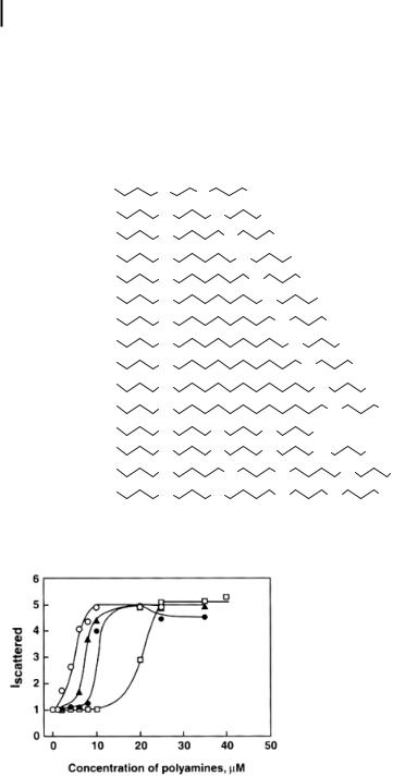
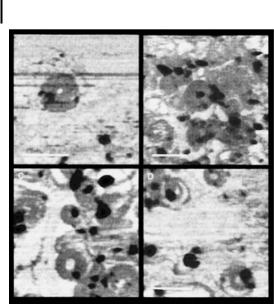

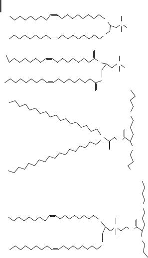
 Control
 ODN + 0.1 M G-4
 ODN + 0.25 M G-4
 ODN + 0.5 M G-4
 ODN + 1 M G-4
 ODN + 2.5 M G-4