344 13 Decorporation of Biohazards Utilizing Nanoscale Magnetic Carrier Systems
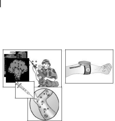
tors (see Fig. 13.1). After an appropriate time interval (perhaps <1 h), the toxin-bear- ing nanospheres are removed from the human blood stream using an extracorporeal, closed-loop tubing system attached to a compact magnetic separator device. This hand-held device passes blood through channels designed to avoid clotting and to enhance suitable laminar flow conditions. Permanent magnets contained within the device magnetically separate the spheres from the blood flow. The detoxified blood is returned into the body and the toxin-bound particles are stored inside the device for bioassay, bioforensics, or disposal.
Figure 13.1. Conceptual rendition of proposed biotoxin detoxification technology. An exposed soldier self-administers nanoparticles and a strap-on magnetic filtration unit is attached by vascular access to his arm to remove the toxin.
If developed successfully such a system would provide a combination of strategic advantages.
1.Uniqueness and therapeutic improvement: The system is highly innovative and would provide, for the first time, a method of selective and efficient removal of a variety of biotoxins from humans with internal contaminations. Toxin removal, not merely binding of blood-toxin alone, is most important mainly because (i) many biohazards, e.g., chemicals and radioactive substances, are not effectively neutralized by simple in vivo antitoxin–toxin binding and (ii) antitoxin–toxin complexes remaining within the body may induce secondary illnesses, e.g., kidney failure from immune complex deposition, and rebound toxicemia from release dissociation and tissue deposition.
2.Biocompatibility: Nontoxic, biodegradable nanospheres are composed of magnetite (or other magnetic material) encapsulated within a biocompatible polymer. Therefore, injection of the magnetic nanospheres with subsequent incomplete or no removal will pose no harm to the subject.

13.2 Technological Need 345
3.Diversity and repeatability: The therapeutic diversity lies in the vast number of already existing and newly designed antitoxins, antibodies, and ligands that can be attached to the biocompatible magnetic nanoparticles. Chronic exposures or exposures with low blood but high tissue toxin concentrations can be treated with repeated injection and removal of antitoxin-bearing particles.
4.System compactness and portability: As the total nanospheres injected for toxin binding can be expected to be less than 2 mg per kilogram body weight, various antitoxin injectants and the actual magnetic removal device can be engineered as a hand-held, single-use, presterilized, selfor helper-applicable unit.
The future biomedical realization of this technology would be an expandable system also applicable to (i) the diagnosis of diseases, e.g., rapid bioassay examination of milligram quantities of concentrated toxins bound to the nanospheres, and (ii) the treatment of diseases in biohazard-exposed humans, e.g., sequestering a wide variety of blood-borne toxins and biohazards in vivo with subsequent removal from the body. Since the nanoparticles will be stabilized against opsonization and macrophage engulfment, they can be circulated within the blood stream for extended periods facilitating in-field use, e.g., by military and first-response personnel, or its application as a mass-screening tool in a triage setting. Importantly, if successful, this detoxification system will provide a unique platform nanotechnology for the treatment of other medical illnesses, such as autoimmune diseases and medication overdoses, in which disease-specific receptors (chelators, ligands) already have been developed but subsequent removal of toxic compound from the blood stream is not yet possible.
This chapter will provide a brief review to familiarize the reader with the pertinent chemical and biomedical engineering background and outline our currently ongoing parallel and sequential efforts to successfully establish this decorporation system. In our discussion, we will focus on the synthesis and in vivo testing of biostabilized and biodegradable magnetic nanospheres, the incorporation of toxin-bind- ing ligands to the magnetic nanospheres, and the development of a prototype magnetic sequestration system.
13.2
Technological Need
Several blood detoxification methods are currently available. The clinically more important ones can be summarized as follows.
Hemodialysis and hemofiltration applies an osmotic gradient across a semipermeable membrane to dialyze/filtrate hydrophilic substances out of the blood. The major limitations are long procedure duration, extracorporeal circulation of large blood volumes requiring large-bore arterial access, nonselective substance removal, and effectiveness limited to hydrophilic substances with lower molecular weight. Its use is mostly restricted to patients with kidney failure and in some medicationrelated intoxications.

346 13 Decorporation of Biohazards Utilizing Nanoscale Magnetic Carrier Systems
Plasmapheresis utilizes extracorporeal, nonspecific exchange of plasma (which is cell-free blood) with albumin or saline solutions. This method removes most of the blood fluid phase and therefore can only be used for a limited period of time and in specific clinical situations where the toxic substance is present in abundant concentration. Its utility is generally restricted to autoimmune diseases.
Extracorporeal immunoabsorption is a variation of hemodialysis in which extracorporeal circulated blood is exposed to a larger exchange surface saturated with immune-absorbent materials (e.g., antibodies). It is a more specific removal method but less effective, requires the circulation of large blood volumes, and is restricted to specific antibody–antigen interactions.
We can perform direct injection of chelators and antibodies, in which, for example, injected antibodies neutralize some actions of circulating antigen (e.g., medication or bacterial toxin interactions). However, complete antigen binding can often not be achieved, and also relatively high antibody dosing is required, increasing the risk of allergic (anaphylactic) and systemic (kidney failure, etc.) side effects. Furthermore, the antibody–toxin complex is not removed from the blood and remaining toxin can dissociate, leading to rebound intoxication.
Obviously, there is currently no adequate detoxification system and, for the majority of biohazard exposures, no therapies other than supportive measures. In agreement with the clinically most successful acute and chronic detoxification system, hemodialysis, we are proposing that a novel versatile detoxification system must remove the offending agent(s) from the blood stream. Simple blood biohazard sequestration within the blood stream as achieved by toxin–ligand or antibody–anti- gen binding will insufficiently protect humans from harmful toxin exposure. This is evidently exemplified in (i) treatment approaches solely based on in vivo antibody– antigen binding, in which safe antibody treatment is problematic due to reduced antibody affinity, systemic side effects (i.e., antibody–antigen complex-mediated diseases, renal failure, etc.), anti-antibody production – all inherent limitations if higher or repeated antibody injections are required; as well as in (ii) cases of radioactive and chemical toxin exposures, where ligand–toxin binding does not alter the toxic activity and natural disease course induced by the offending agents.
However, in contrast to conventional hemodialysis and other, less commonly used detoxification systems, the nanospheres-based system will have important advantages such as:
. Specificity. Only substances binding specifically to the nanoparticle-ligand surface will be removed.
. Removal. Several important advantages exist:
–Dissociation of already formed toxin–antitoxin complexes becomes less important as the compounds are continuously removed from the body.
–Active toxin blood stores are permanently removed; therefore, additional protein-, tissue-, and membrane-bound toxins are continuously released as free toxin into the blood stream and eventually bound to the nanospheres (decreasing toxin body stores).

13.3 Technical Basis 347
–Quantification of removed toxins permits (i) direct estimation of toxin removal efficiency, (ii) a clear estimate of required therapy duration, (iii)
further laboratory identification of biohazard origin (bioforensics), (iv) improved antitoxin design, etc.
. Quantitative binding. Quantitative toxin binding is facilitated by large nano- particle-antitoxin binding capacity; this becomes especially useful when onlylow affinity antitoxins are available.
. Nontoxicity. Nanoparticles remaining within the body are metabolized physiologically without adverse effects.
. Efficiency of removal. Strong magnetic field gradients will allow high-yield first-pass removal of antitoxin–toxin compounds.
. Convenience of usage. Two-step detoxification, simplified for mass usage by a nonverbal visual guide: injection of nanospheres followed by simple needle insertion of filtration unit; miniaturized portable or large-scale hospital-based designs.;
. Safety of usage. No risk of disease transmission (as in antibody treatment, blood transfusion, etc.); no blood loss; closed-loop, preheparinized and presterilized, single-use system avoids blood contamination, allowing selfor helper-applied usage by nonmedical personnel, and preservation of the sample toxin.
. Repeatability. Re-exposures to biohazards or reaccumulation of toxin from body tissue stores can conveniently be treated with repeated detoxification sessions.
If successful, the most apparent and dramatic advancement is the introduction of a robust, hand-held biodetection and treatment system that can provide a concentrated multianalyte for high-sensitivity bioassay. As such, it is possible to determine the in vivo, presymptomatic presence of pathogens through highly selective sequestration and magnetically assisted separation without harmful side effects to surrounding healthy tissues and cells.
The technology is radically novel, but the components have some precedent as was described above. We seek to integrate known technology with advancements and novel design strategies in our laboratories to engineer a powerful detoxification system. Even in its simplest form, a system of in vivo detoxification using biostabilized nanospheres will have a scale of diverse applications making it attractive to many military or civilian applications.
13.3
Technical Basis
Upon first inspection of the technology concept several questions arise as to its feasibility and applicability. Such concerns are addressed directly below.
34813 Decorporation of Biohazards Utilizing Nanoscale Magnetic Carrier Systems
13.3.1
Difference Between Drug Sequestration and Drug Delivery Using Nanospheres and Microspheres
There are many examples of R&D in nanospheres and microspheres systems for the goal of drug delivery; the reader is pointed to Davis [1] and Douglas et al. [2] and the work of H feli [3] and L4bbe et al. [4]. At first inspection, drug delivery and drug removal using nanoparticles might be considered parallel research. However, there are important differences in the development of these two systems (Tab. 13.1). First, drug delivery systems must optimize the encapsulation of drug material within the nanoparticle. As such, the best configuration is to use higher volume-to-surface-area particles. Drug removal requires the antithesis – high surface-area-to-volume ratios
–in order to maximize the surface functionality (capacity) of the nanospheres for toxin removal. Also, drug delivery systems often do not require long circulation times in the body and thus may not require robust surface properties to avoid opsonization. In detoxification, drug removal will require circulation of the nanospheres in the blood stream for several cycles in order to maximize capture of toxins and permit removal. Next, because the drug is encapsulated within the microspheres matrix for drug delivery systems, the surface can be freely conjugated. In contrast, nanospheres for toxin removal will require the surface to be conjugated with stabilizing ligands and ligands to sequester toxins. The reason for this is to maximize the kinetics for toxin removal – a surface effect. Finally, drug delivery systems are often not magnetic, instead relying on surface ligands to bind to tissue sites. Systems that are magnetic must maximize the magnetic moment of particles in order to minimize the externally applied magnetic gradient and field. For drug removal, the magnetic component is important for filtration but may be adjusted depending on the
design of the magnetic filtration system. That is, a high-aspect-ratio filtration chamber can be fabricated to produce blood flow rates that range from < 1 cm s–1 to >100 cm s–1 depending on the actual capture efficiencies. Because of the versatility in designing the filtration system, we envision the magnetic moment of the particles to be much less than required by magnetic drug delivery systems (<10 emu g–1 as opposed to >30 emu g–1 in drug delivery systems).
Table 13.1 Contrast between drug delivery and the proposed toxin removal system based on magnetic nanoparticles.
Property |
Drug delivery |
Toxin removal |
|
|
|
Size |
High volume-to-surface-area |
High surface-area-to-volume |
Magnetic moment |
>30 emu/g |
<10 emu/g |
Surface functionality |
Not necessary |
Stabilized against opsonization, |
|
|
anti-toxins anchored to surface |
Circulation time |
Minutes |
>1 h |
emu: electromagnetic unit.

13.3 Technical Basis 349
13.3.2
Vascular Survival of Nanospheres
There has been initial work investigating particle pharmacokinetics within our group and by others. Particles without appropriate surface characteristics are immediately removed (within minutes) by the reticuloendothelial system (liver, spleen, etc.) [5], and particles that are too large will cause capillary occlusion and cause serious adverse effects and death after systemic injection into the animal ([6]; unpublished data obtained in our monkey experiments using commercially available cellu- lose-based microspheres). However, success in liposome and nanoparticle systems identifies the importance of hydrophilic polyethylene glycol (PEG)-derivatives in prolonging intravascular survival [7, 8]. Particles coated with PEG chains offer steric and charge stability (near neutral) and prevent antibody formation, opsonization, and phagocytosis [8, 9]. Investigations found that the blood circulation times of particles increase as the molecular weight of covalently linked PEG increases. Five hours after systemic injection, only one-third of 20-kDa PEG-conjugated poly(lactic- co-glycolic acid) nanospheres (140 nm) had been captured by the liver in comparison to uncoated particles [8]. Similar prolongation was summarized by Allen et al. [10, 11] and described by Li et al. [7] and Dunn et al. [12]. However, exact mechanisms for macrophage avoidance are not known and discrepancies exist as to the best PEG length or derivative. Most recent evidence has demonstrated a 20-h halflife for circulation, exemplifying the great progress scientists have made in this area [13].
13.3.3
Toxicity of Components
Nontoxic polymeric nanoand microspheres have been described in the literature and comprise a category of natural and synthetic biopolymers. Biopolymers include but are not limited to poly(lactic acid), poly(lactic-co-glycolic acid), poly(ethylene glycol), poly(caprolactone), albumin, and dextran. Biopolymers are chemically degraded at rates dependent on the particle size, surface properties, cross-linking density, and the molecular weight of the polymer [14]. The acute toxicity of several biopolymers has been evaluated [15] and suggests no ill effects due to the polymers. Instead, the polymers are degraded and metabolized into harmless fragments [16]. Poly(lactic acid), poly(lactic-co-glycolic acid) and poly(caprolactone) are FDA-approved for injection in several forms. From our own work, histopathological examinations of lung, liver, brain, and spleen of a series of monkeys and rats exposed to systemic injections of magnetic particles identified no early toxicological changes (tissue changes, coagulation, and extravasation) and no capillary obstructions were observed in examined animal organs after adjustment of particle size and injection modus.
The presence of magnetic particles incorporated within the polymeric matrix introduces a second source of potential toxicity. However, studies have shown [17, 18] that, in the long term, the magnetite crystals are in part metabolized, increasing hepatic and splenic ferritin stores, and in part incorporated into red blood cells.

350 13 Decorporation of Biohazards Utilizing Nanoscale Magnetic Carrier Systems
Thus, provided that the injected dose of magnetic iron is below the toxic dose threshold, they are safe. (The toxic dose threshold is 10 mg kg–1 body mass or 750 mg in standard man [16]. Also, normal serum levels in blood are 80–180 mg dL–1 and action levels are >500 mg dL–1.) We estimate that about 100 mg of nanoparticles will be injected to treat a typical biohazard-exposed subject, a fraction of which will be composed of iron (if 50% loading of nanoparticle with elemental iron, then the patient may be exposed to 50 mg of unbound iron). Not only is this amount of injected magnetite much smaller than the dose leading to toxic iron effects but, more importantly, our detoxification system will extract most of the injected magnetite as part of the toxin removal process. Therefore, body storage will only be a minor deposition method. Superparamagnetic iron oxide is approved by the FDA for injection.
13.3.4
Magnetic Filtration of Nanospheres from Circulation
Magnetic separator units for industrial use are available commercially and have been used for various research related immunoseparations [19–23]. However, a magnetic separator suitable for our application of clearing nanospheres from blood flow has not been designed. Based on the performance of commercial separators and our own preliminary investigations, we will implement technically straightforward engineering for the design of the first prototype biomedical separators. Our proposed “filtration” system utilizes small permanent magnets attached to the body of a specialized closed-loop catheter system. The actual design of the device is part of the proposed research as several options are plausible given predefined conditions. Common to all design options, the blood will be diverted from the body to an array of tiny (few hundred micrometer diameter) flow tubes. The tubes will be immersed in a magnetic field gradient, causing the magnetic nanospheres to deflect towards and collect at the tube wall. The precise geometry of the tubing system (size, material, coating, length, shape, etc.) will be defined in our research to minimize interactions with blood coagulation (i.e., thrombosis) and blood cells (i.e., destruction). In addition, our analyses will identify different design strategies for different modes of operation (e.g., in-field vs. unit-based) and user level of training (e.g., selfvs. helperapplied).
Magnetic separation can be illustrated with our following simple experiment. A suspension of monodisperse nanospheres, 400 nm in diameter (measured moment 50 emu g–1, =1.4 g cm–1) was contained in 0.9% saline in a 0.6-cm diameter vial. A hand-held commercially available magnet (0.4 T at surface) was placed against the outside wall of the vial and all particles rapidly deflected to the inner wall close to the magnet surface within 3 s. Assuming constant particle velocity (here 0.6 cm per 3 s) within the contained fluid, we plot the trajectories of particles under flow conditions (Fig. 13.2). The plot estimates the length of tubing that would be needed to deflect particles flowing at 50, 10, and 1 cm s–1 in 1-mm diameter tubes. For rapid flow velocities, a tube >20 cm immersed in the magnetic field would be required to separate the particles from the flow. However, for flow rates of 10 cm s–1, easily achieved with microflow tubing designs, only approximately 5 cm of tubing is neces-

352 13 Decorporation of Biohazards Utilizing Nanoscale Magnetic Carrier Systems
plications of magnetic carriers [http://www.magneticmicrosphere.com] and attendance has grown rapidly.
For the detoxification technology, we need to synthesize nanospheres with (i) optimal size to avoid obstructing capillary blood flow and immediate vascular clearance, (ii) adequate size uniformity to facilitate modeling and quality control and assurance, and (iii) optimal surface properties to prolong vascular circulation. Specifically, the nanospheres must be single populations between 100 and 5000 nm. If bimodal or multimodal populations are synthesized then the diameters of the individual populations must be sufficiently different to allow for some physical separation method (e.g., filtration). The surface charge must be neutral or slightly negative (<10 mV, measured by zeta-potential) due to surface PEGylation in order to increase blood circulation time and minimize bioclearance by opsonization and phagocytosis.
13.4.1.1Nanosphere Size
The literature presents many articles describing the synthesis of nanospheres and microspheres composed of natural and synthetic polymers. Many types of polymers have been investigated for potential in vivo applications. By far the most discussed polymers are poly(lactic acid) and poly(lactic-co-glycolic acid) macromonomers. There is a plethora of literature too numerous to cite that describes synthesis methods, drug encapsulation, release kinetics, biostability, and in vivo properties such as macrophage engulfment, biodegradation, and organ disposition. The Journal of Controlled Release contains many articles every year on the subject matter.
The method of nanospheres formation is straightforward. The polymer is dissolved in a volatile organic solvent and added to an aqueous solution and stirred vigorously. An emulsion of oily droplets forms. Surfactants in the solution promote the stabilization of the emulsion until the volatile organic solvent dissolves in the solution and evaporates, leaving a solidified sphere of polymer. Magnetic spheres contain additional magnetic powders in the organic solution. The size of the spheres depends on process parameters. Some process parameters are outlined in Tab. 13.2.
A popular type of polymer composition is polystyrene. Polystyrene spheres are formed by a completely different method called emulsion polymerization [30] and will not be mentioned further except to say that they are uniform in size with a wellknown surface chemistry and functionalization. They are not appropriate for in vivo use due to the toxicity of polystyrene, but their uniform properties make them attractive model systems during feasibility studies.
The limits set by physical removal of particles flowing in the blood are rather broad. To maximize surface receptor density we seek particles with sufficiently high surface-area-to-volume ratios. To be discussed in the next section, high surface-area- to-volume ratio nanospheres would imply using the smallest particle possible or those <100 nm. However, as one reduces the nanosphere size, it becomes more difficult to sustain the magnetite content or magnetic moment. In other words, the magnetic moment drops at a rate faster than would be expected based on the reduction in particle volume as the diameter is reduced. We have had success in separating 400-nm polystyrene magnetic nanoparticles from blood (specific magnetization=50 emu g–1). Thus, we hypothesize that a nanoparticle 100–400 nm in size will

13.4 Technology Specifications 353
Table 13.2 Some process parameters in the synthesis of magnetic nano-/microspheres.
Polymers |
Poly(d-lactide), poly(l-lactide), poly(d,l-lactide), |
|
poly(lactic–co-glycolic acid), polymers and copolymers linked to |
|
polyethylene glycol, poly(caprolactone) |
Surfactants |
Poly(vinyl alcohol), poloxamer, polyethylene glycol, tocopherol |
|
polyethylene glycol succinate, sodium dodecyl sulfate |
Volatile organics |
Dichloromethane, chloroform, acetone, ethyl acetate |
Mixing speed |
1,000–20,000 rpm, 15–95 W insonation |
Mixing modality |
High-speed homogenization, ultrasonic insonation, paddle mix- |
|
ing, magnetic stir bar |
Polymer/organic solvent ratio |
1–0% |
Oil/water ratio |
1–10% |
Mixing time |
Several seconds to minutes depending on modality to form ini- |
|
tial emulsion, several hours to harden |
Polymer MW |
2–300 kDa |
Surfactant MW |
PVA 10–150 kDa |
Surfactant concentration |
PVA 0.1–10% |
Magnetic phase |
Magnetite Fe3O4, maghemite c-Fe2O3, passivated Fe, |
|
passivated Co |
Temperature |
Unknown, may reduce coalescence |
|
|
be optimal in this program. There is a parallel requirement that the size of the particles not be broadly distributed (high polydispersity). The reason for this is to facilitate modeling of the system and ensure predictability in particle properties such as magnetic moment, surface area, receptor density per batch size, surface charge, etc.
13.4.1.2Surface Properties
Covalent attachment of biologically active compounds to polymers and polymeric spheres became one of the methods for alteration and control of biodistribution, pharmacokinetics, and, often, toxicity of these compounds [31]. One of the most popular polymeric materials used for this purpose is polyethylene glycol (PEG). It possesses an ideal array of properties: excellent solubility in aqueous solutions [32], extremely low immunogenicity and antigenicity [33], and has the advantage of being nontoxic and was approved by the FDA for internal use in humans [34]. Gref [27] provides an excellent discussion on PEG biochemistry and protein interaction.
Jeon and Andrade [35] proposed a mathematical model taking into account the four types of interactions between a protein and hydrophobic substrate. They stated that the best conditions for protein repulsions were found to be long PEG chain length and high surface density. If D is the distance from the anchorage to the substrate of the two terminally attached PEG chains, in the case of small proteins (approximately 4 nm in diameter), D should be around 1 nm, whereas for larger proteins (6–8 nm), D should be around 1.5 nm [35].
The challenge to our program is to determine the proper PEG chain length and surface coverage to increase circulation half-lives to permit in vivo binding of the
354 13 Decorporation of Biohazards Utilizing Nanoscale Magnetic Carrier Systems
toxin and removal of the toxin-loaded nanospheres. PEGs come in various forms. They can be made linear or branched, and with different molecular weight (chain length), and partial substitution (e.g., polyethylene glycol–polypropylene glycol coblock polymers). The literature contains discrepancies as to the best choice of chain length, and computer models [35–38] suggest potentially subtle but important differences to explain PEG behavior towards protein adsorption. One study [8] concludes that polystyrene nanospheres with longer PEG chains, those greater than 10,000 Da, survive longest in the rat, but does not show direct evidence of surface coverage. Another study [12] concentrates on showing the importance of surface coverage density but does not compare these results with those as a function of PEG length. Importantly, long-chain PEGs may sterically interfere with each other during the surface bonding procedure or during copolymerization. Thus, the surface may not be able to accommodate the long-chain PEGs to the 100% surface coverage needed to avoid opsonization. Another study [39] suggests that vascular survival is not enhanced by conjugating PEG chains longer than 5000 Da, while Yamoaka et al. [13] refute this suggested lack of dependency. These discrepancies necessitate confirmatory and supplemental study.
13.4.1.3Biodegradability
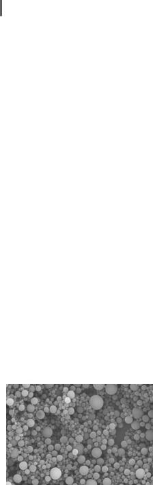
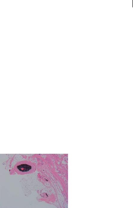
We are adapting the parameters found to prolong the vascular survival of the model polystyrene-based nanoparticles (PEG chain length, PEG surface density, receptor site density, biokinetics) to biodegradable magnetic nanospheres. There are several biopolymers to choose from in synthesizing the nanoparticles. There is a plethora of data on poly(lactic acid) (PLA) and poly(lactic-co-glycolic acid) (PLGA) polymers. Gref et al. [40] describe how to synthesize PLGA–PEG nanoparticles <150 nm using an oil-in-water solvent evaporation technique. Li et al. [7] describe a similar method. H feli et al. [3] describe an oil-in-water method for synthesizing PLA microspheres >3 lm, and our work has shown that this method can yield less polydisperse nanospheres from 900 nm to 3000 nm (Fig. 13.3). Mosqueira et al. [41] describe the preparation of PLA–PEG nanoparticles with variable PEG content and chain length. Only H feli et al. [3] describe incorporating magnetite into the PLA or PLGA particles so we must understand how magnetite affects the size and chemistry of the biodegradable spheres, especially the submicron spheres.
PLA
Figure 13.3. Poly(lactic acid) microspheres. Note the variance in sphere diameter.

13.4 Technology Specifications 355
13.4.1.4Surface Receptors
The nanospheres must have (i) large surface area and surface functional sites for attachment of surface receptors (e.g., antibodies, chelating agents for radionuclides, or ligands for chemical toxins) in sufficient number to reduce or eliminate completely the concentration of toxins in the blood, and (ii) the placement of such surface receptors where they can interact readily with the blood but not facilitate protein adsorption onto the particle surface. The first criteria are interrelated with the design of the nanosphere size since particle diameter is intrinsically related to surface area. To estimate the target receptor site densities we provide an example. Simulating a class A agent Bacillus anthracis exposure, we inject 0.6 mg of lethal factor (LF) into a rat (300 g weight, 25 mL blood volume) which leads to a LF blood concentration of 24 ng mL–1 or 7.5 pmol per rat (MW 80–90 kDa). Nanospheres we used in our laboratory have a surface receptor capacity of at least 1 mequiv mg–1 or 1 lmol mg–1 for univalent ligands. Thus, an anticipated injection of 10 mg of nanoparticles into the rat would have a capacity of 10 lmol barring steric hindrance. This value is on the order of 106 times greater than the necessary theoretical capacity needed to bind quantitatively LF toxin in the blood. Assuming a steric hindrance offered by an 80to 90-kDa LF protein (15 nm diameter projected image) on a 400-nm magnetic nanoparticle (10 mg injection), we can expect a binding capacity of 0.7 nmol LF protein – still a factor of 100 greater than necessary for quantitative LF removal from the blood. Therefore, a goal is to maintain this order of receptor site density (1 mequiv mg–1) during the synthesis of biodegradable nanospheres from PEG based copolymers.
There are two options to attaching the antibodies or chelating ligands to the surface: (i) attach them directly to the particle surface using short chain functional groups such as carboxyl and epoxy bridges, or (ii) attach them to the PEG or PEGderivative chains extending from the particle surface. The shortcomings of the first procedure are that the receptor groups may be sterically hindered from encountering the toxins present in solution due to the long-PEG-chain neighbors. Also, since the receptors would be attached directly to the surface they would be competing with the PEGs for surface coverage. This condition inherently limits the surface density of PEGs and facilitates opsonization and macrophage removal of the nanospheres in vivo. Therefore, option (ii) appears to be the most appropriate choice. It has been demonstrated by our research partners and others that the terminal groups of PEG chains can be activated and covalently bonded to other functional groups. Our own research has shown that we can attach streptavidin onto the terminal groups of PEG (300 and 2000 Da). However, we believe it may be difficult to cap the activated terminus of the PEG completely during the receptor attachment step. This may result in a charged surface and increased opsonization. Instead, we are pursuing a method by which the coblock polymer used to make the nanospheres (e.g., PLA–PEG) contains a suitable end group for direct attachment of receptor. For instance, we have synthesized PLA–PEG–biotin copolymer (Fig. 13.4) based on the technique of Salem et al. [42]. Streptavidin was attached after incubation in buffer solution [43]. Similarly, biotinylated antibodies could be directly attached to the streptavidin [44] or attached during the coblock polymer synthesis by substituting

356 13 Decorporation of Biohazards Utilizing Nanoscale Magnetic Carrier Systems
the biotinylated antibody for biotin. Although preliminary studies are promising, there are still limitations that need to be addressed, including efficiency of antibody conjugation and effect of conjugation of the PEGs on circulation of the nanospheres in vivo.
Figure 13.4. Biodegradable coblock polymer with biotinylated end group.
The model system that we are demonstrating in initial experiments is the strepta- vidin–biotin pair. However, this system provides a lower limit in determining the necessary residence times for nanosphere flow (i.e., the amount of time or blood volumes to which the nanospheres must be exposed to ensure removal of toxins). The follow-up system in subsequent research will concentrate on more realistic systems that display weaker binding constants. These antigen–antibody systems will present a realistic expectation of this system based on the accomplished receptor site densities and vascular survival. Therefore, a goal is to determine if it is possible to functionalize identified candidate magnetic nanoparticles with their respective candidate antibodies and that the antigen–antibody capture efficiency remains stable when applied to the living animal conditions. This would ensure that it is possible to detoxify the antigen from the animal blood.
Inherent with the research challenge of receptor site density is the determination of the suitability of current antibodies to maximize surface density. Antibodies are quite large molecules (tens to hundreds of kilodaltons), but their active components can be quite small. Thus, it is important to quantitatively evaluate whether antibody fragments can enhance the receptor site density achievable (due to steric effects) while maintaining its specificity and stability. In other words, the development of a reliable technology for developing receptors for future implementation of the nanoparticle technology is critical – and a obligatory prerequisite for optimization of receptor density (i.e., maximizing receptor capacity while minimizing opsonization/ engulfment risk). Importantly, it is possible that trying to optimize receptor capacity with conventional antibodies is not useful, since the system cannot be manufactured with conventional antibodies. It is plausible that a reduction in the size of the antibody or fragment needed to achieve selective bonding to the toxin would enhance the manufacturing output of the antibody.
An important aspect of the proposed technology is the possibility for generic attachment of a variety of antitoxins to the nanoparticle substrate. For instance, by
13.4 Technology Specifications 357
coupling streptavidin to the terminal groups of the PEGs one has a generic method of attaching biotinylated antibodies to the nanoparticle’s surface. We seek to describe methods of generic attachment for chemical toxin ligands and radiological toxins. We initially focus on the streptavidin receptor for biotinylated targets. Successful designs will be moved from the streptavidin–biotinylated protein system of sequestration to the biotinylated antibody–antigen system to more realistically determine binding kinetics in vitro and in vivo. If encouraging results are obtained, we will test the ability of the magnetic nanospheres to deplete the antigens in complex mixtures, such as rat blood. Positive results would serve as the prelude to the development of animal models as part of subsequent testing. We note the success of Sakhalkar et al. [44] in selective binding of in vivo targets with similar nanosphere composition.
13.3.2
Magnetic Filtration of Toxin-Bound Magnetic Nanospheres
We are designing an extracorporeal magnetic filtration unit, a small external catheter system, that (i) allows dialysis-like blood circulation through a well-defined tubing or microchannel system while generating no blood clots or coagulation events, (ii) will permit quantitative removal of the freely circulating, toxin-bound nanospheres via application of hand-held permanent magnets, (iii) is easily portable and in-field applicable (i.e., the entire device is hand-held), and (iv) is designed for administration by nonmedical staff or trained medics (both designs will be developed).
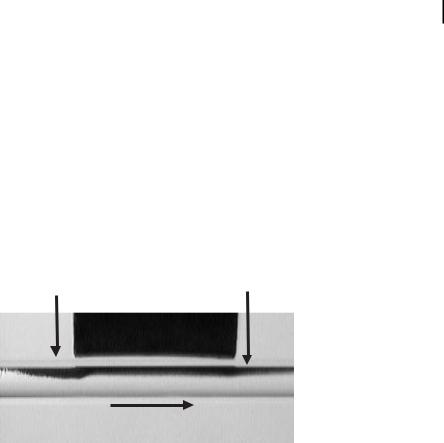
The current knowledge of perfusion devices and blood flow is sufficient for the development of such a filtration device without additional scientific discovery. Our own research provides evidence for particle filtration in high-velocity blood flow using small, permanent magnets (Fig. 13.5). Thus, a carefully orchestrated series of experiments utilizing existing technology should be successful for production of the device. The device is predicated on:
Figure 13.5. Pathology of magnetic particles (black, peak vel.=105 cm s–1) using a 20-mm permanent magnet placed 8–10 mm from artery.
35813 Decorporation of Biohazards Utilizing Nanoscale Magnetic Carrier Systems
1 The ability to successfully separate the particles from the blood. This will require a sufficient magnetic field to draw the particles to the wall and hold
them there under shear flow.
2A flow rate of at least 100–200 ml min–1 will be required for total body cleansing. Lower flow rates can easily be obtained from a large-bore venous puncture at mid-arm level. A simple siphon hand-pump will ensure proper flow
rate. Higher flow rates are achieved from a commonly performed femoral arterial puncture with a double-bore needle [double-lumen (inflow and outflow) catheterization avoids a second vascular puncture]. A trained technical person should be capable of performing such a procedure.
3Anticoagulation locally in the perfusion chamber by dissolution of heparin from the walls of the tubes into the flowing fluid, which should permit adequate anticoagulation locally without inducing it systemically. Shear rates and stresses will be kept at levels compatible with minimizing both clot formation and thrombosis in the design of the magnetic field so that particle removal or “filtration” is quantitative and highly efficient, minimizing the length of the device and its size and weight. The design will include a clotfiltering device at the blood return port.
Further design aspects depend on future investigations using the prototype magnetic field chamber. Certainly, the inherent physical properties will vary for each magnetic filtration device variation necessary to accommodate different biomedical and user applications and these design characteristics cannot be predicted a priori purely on theoretical grounds. However, realistic modeling of various future design strategies will become possible if the first prototype separation device is characterized and its performance tested in living animals and healthy volunteers. Important design strategy questions can then be adequately answered; examples are:
1What are the most feasible vascular access modes (venous, bivenous, arter-
ial?) and site (antecubital, brachial, femoral?) for easy and fast (selfvs. helper?) use?
2What are the optimal catheter (single vs. dual lumen, cannulation size?) and tubing system (coating, flexibility, filter system, branching?) for unobstructed blood circulation and to avoid blood clotting?
3What is the most favorable magnetic device design (one magnet/catheter unit or a “snap-on” tubing design?), allowing easy fixation and portability?
Our first prototype detoxification system will be designed to utilize safe and practical venous access at the inner mid-arm regions (antecubital) of an exposed human via 14-gauge, dual-lumen needle insertion (selfor helper-applied) which can easily be performed by trained nonmedical personnel. Using this approach, the careful estimation of practical low blood flow rates at about 40 ml min–1 will allow total body blood (estimated 6 L) turnover with removal of toxin in about 150 min. Variations of this approach will permit faster detoxification; for example, helper-applied femoral artery needle access at the groin site can reduce the clearance time by a factor of 8–10, establishing blood clearance within approximately 15 min. More com-
13.4 Technical Progress 359
plex vascular access and device design strategies can be instituted when exposed humans are treated in medical units or hospital settings.
13.4
Technical Progress
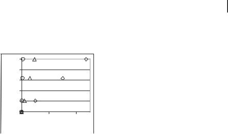
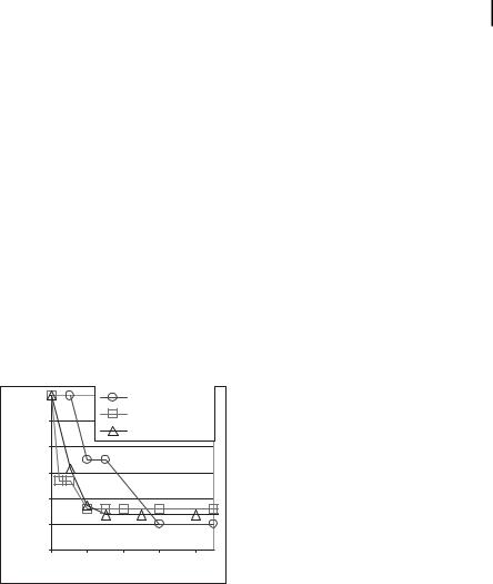
This program is in its infancy and we are pursuing proof-of-principle experiments. In vitro sequestration of a biotinylated enzyme from simple fluids and whole rat blood was performed under static and dynamic flow conditions. Particles were composed of nanocrystalline maghemite (c-Fe2O3) encapsulated in polystyrene nanospheres. Several variations were tested including various PEG lengths (MW 330–6000) and particle sizes (250–3000 nm). Streptavidin, the model receptor, was either bonded to the carboxylated terminal group of the PEG or attached directly to the nanoparticle surface. Biotinylated horseradish peroxidase (HRP) was used as the model “toxin.” The results (Fig. 13.6) indicate a reduction of the free enzyme to about 50% maximum levels in the blood in all tests. Equilibrium was reached within 20–30 min. We have more recently achieved 72% separation of biotinylated HRP from saline in heparinized whole rat blood [43].
|
1.00 |
|
|
in vitro, water |
|
|
0.90 |
|
|
in vitro, blood |
|
|
|
|
flow cell, water |
||
free HRP |
|
|
|
||
0.80 |
|
|
|
|
|
0.70 |
|
|
|
|
|
of |
|
|
|
|
|
|
|
|
|
|
|
fraction |
0.60 |
|
|
|
|
|
|
|
|
|
|
|
0.50 |
|
|
|
|
|
0.40 |
|
|
|
|
|
0 |
10 |
20 |
30 |
40 |
|
|
|
Time (min) |
|
|
Figure 13.6. Horseradish peroxidase (HRP) “toxin” levels in water and blood after HRP and nanosphere injection.
We synthesized biodegradable PLA–PEG–biotin–streptavidin nanospheres and microspheres and achieved up to 42% separation of the biotinylated HRP from normal saline (0.25 mg nanospheres per milliliter of heparinized rat blood, 0.375 mg HRP mL–1 blood [43]). The surface charge is neutral from pH 4 to pH 9 making them, to a first order, suitable for in vivo trials. We have encapsulated rhodamine-B and measured detection limits in whole rat blood. The signal-to-noise levels are suitable for quantification.
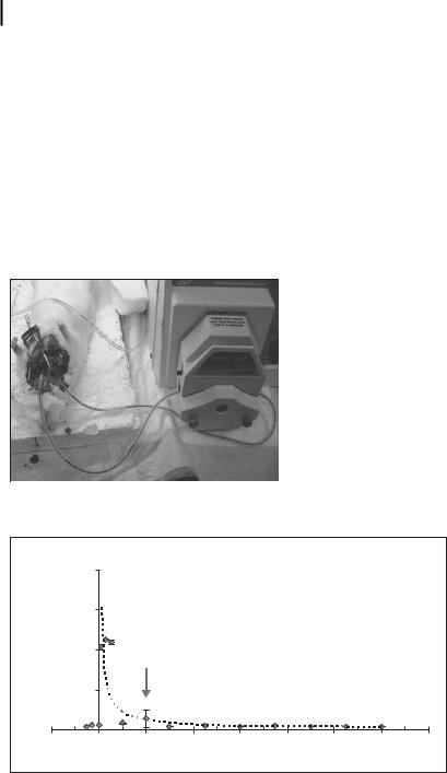
In vivo experiments, performed on retired breeder rats, included (i) the design of a closed-loop, adjustable-flow blood recirculation unit permitting blood turnover and sampling over several hours in the live animal; (ii) kinetic studies of several candi-
360 13 Decorporation of Biohazards Utilizing Nanoscale Magnetic Carrier Systems
date magnetic nanospheres and toxins; (iii) first magnetic filtration experiments. In the latter investigations, continuous extracorporeal blood circulation was achieved via carotid–jugular cannulation and external pump support with filtration of magnetic nanospheres using 1-mm-diameter closed-loop tubing and a single NdFeB magnet (0.4 T at surface, 18 mm diameter) (Fig. 13.7). Procedural blank experiments monitored the circulation half-life (T1/2) of biotinylated HRP in the rat model (Fig. 13.8), showing that the HRP levels decrease immediately (T1/2=15–20 min). A longer circulating marker would be useful. Experimental results on toxin sequestration and rat pathology are pending.
Toward the development of the magnetic filter, we employed computational fluid dynamics and computational magnetic field models developed by The Department of Energy to predict the capture efficiency of magnetic microparticles passing through a simple magnetic field profile. This design provides the simplest case of a
Figure 13.7. Continuous extracorporeal blood circulation in the rat.
|
|
|
Rat: 12/22/03 2B |
|
|
|
|
|
|
4.0 |
|
|
|
|
|
|
|
Absorbance |
3.0 |
|
|
|
|
|
|
|
2.0 |
|
|
|
|
|
|
|
|
1.0 |
|
|
|
|
y = 3.0828x-0.8627 |
|
||
|
|
|
|
|
|
|||
|
|
|
|
|
|
R2 = 0.8343 |
|
|
|
0.0 |
|
|
|
|
|
|
|
-20 |
0 |
20 |
40 |
60 |
80 |
100 |
120 |
140 |
|
|
|
|
Time (min) |
|
|
|
|
Figure 13.8. Removal of biotinylated HRP in the rat model during normal circulation [34].