


95
4
Nanostructured Collagen Mimics in Tissue Engineering
Sergey E. Paramonov and Jeffrey D. Hartgerink
4.1
Introduction
A human body consists of approximately 1014 cells. This huge number of cells is organized and maintained at several levels of scale. First, cells must maintain their proper phenotype and not dedifferentiate into more generic cell types or undergo programmed cell death. Next, groups of cells are organized into tissues, which make up organs, and these organs are brought together to make a complete individual. This organization is maintained in large part by the extracellular matrix (ECM). ECM along with the cells it surrounds are the two necessary components of any multicellular tissue. ECM is a very complex biological environment that provides cells not only with a surface on which to attach to move, but also with chemical and biological information stored in the form of bioactive molecules, enzymes, and growth factors. Proper tissue functioning depends strongly on the correct ECM structure and composition, and several severe diseases (for example, osteogenesis imperfecta) may arise when the ECM is corrupted. ECM is a diverse material but its primary components are a collagen scaffold that provides the support for cells and a highly hydrated polysaccharide gel that allows the rapid diffusion of nutrients and resists compressive forces.
Recent advances in medicine have dramatically advanced reconstructive surgery and given rise to a whole new area of research: tissue engineering. Tissue engineering attempts to replace or rebuild damaged, diseased, or aged human tissues with those grown outside the body. The tissue growth requires cells, and the recipient can often provide his or her own cells, thus eliminating problems of finding a suitable donor and immune rejection of those donated organs. One key aspect of successful tissue engineering is the proper reconstruction of ECM of a particular tissue. This is a very difficult task, since the ECM is an exceptionally complex biomaterial. In order to tackle this problem it needs to be broken in several more simple tasks. Much research in tissue engineering currently concentrates on scaffold design. The choice of a scaffold is crucial, since it provides a surface on which cells can grow and a shape to which to conform. The variety of currently used scaffolds can be divided into two categories, those with a synthetic origin and those with a biological origin. Synthetic scaffolds include simple polymers such as poly-lactic acid (PLA), poly-gly-
Nanofabrication Towards Biomedical Applications. C. S. S. R. Kumar, J. Hormes, C. Leuschner (Eds.) Copyright 2005 WILEY-VCH Verlag GmbH & Co. KGaA, Weinheim
ISBN 3-527-31115-7

96 4 Nanostructured Collagen Mimics in Tissue Engineering
colic acid (PGA), PLA–PGA blends, and also inorganic ceramics such as hydroxyapatite (HA).The main design principle in this case is the biodegradability and nontoxicity of the polymer and its degradation products. Traditional advantages of synthetic polymers include precise control over structure and function, good mechanical qualities, and relatively low production costs. The large class of biocompatible synthetic polymers includes poly(acrylic acid) and its derivatives, poly(ethylene oxide) and its copolymers, poly(lactic acid), poly(vinyl alcohol), polyphosphazene, and polypeptides [1].
Although these scaffolds show very exciting properties they lack chemical functionality that can be used for final tuning of these materials upon the needs of a particular tissue; typically they act only to provide macroscopic structure on which cells may grow. Biologic scaffolds typically contain collagen that has been harvested and chemically purified. Various types of naturally occurring and chemically synthesized polymers have been studied and tested as scaffolds for tissue engineering. Among the naturally occurring polymers are the scaffolds based on collagen, hyaluronate, fibrin, alginate, and several others. Harvested collagen is a widely used tissue engineering scaffold. Although it is potentially immunogenic, collagen meets many of the requirements for an artificial matrix, such as biodegradability and cell recognition. Collagen has been successfully utilized for reconstruction of skin [2] and as a tissue scaffold for many different cell types [3]. We believe that ECM reconstruction should begin with a collagen mimic which can be prepared in a synthetic way. These synthetic collagen fibers should be able to be modified to display cell signaling peptides (such as RGD, IKVAV) or entire proteins such as growth factors. The artificially produced collagen can include different proteins, responsible for cell differentiation and proliferation. If this can be done it will be an important step toward replicating the complete ECM and the preparation of designer tissues for medical applications.
This article will discuss the structure and properties of natural collagen and then focus on how this knowledge has been used in attempts at creating synthetic collagen. Next we will discuss tissue engineering approaches which have attempted to mimic one or more properties of the extracellular matrix, and we will conclude with a discussion of our approach to the preparation of collagen mimics.
4.2
Collagen Structural Hierarchy
Collagen is the most abundant protein in the human body and constitutes about 25% of the total protein mass. Collagen has a complicated structure with many levels of hierarchy. At the lowest level is the amino acid sequence, which is found to have a highly conserved three amino acid repeat of Gly-X-Y where positions X and Y (in this case Y refers to a general amino acid, not to tyrosine) contain a high percentage of the imino acids proline and hydroxyproline respectively (Fig. 4.1). Proteins with this sequence are found to preferentially adopt a proline type II helical conformation that allows three of these peptides to assemble into the next level of hierar-
4.3 Amino Acid Sequence and Secondary Structure 97
chy, the triple helix. These helices are bundled into fibrils and are often covalently cross-linked through modified lysine residues. Finally, these fibrils are bundled into macroscopic fibers (Fig. 4.2).
O |
|
O |
H |
(S) |
N |
N |
|
|
N |
|
(S) |
O
(R)
HO
Figure 4.1. The chemical structure of the most common collagen triplet: glycine-proline-hydroxyproline.
When produced in vivo collagen is first synthesized as a propeptide that consists of a long triple-helical central region flanked by N and C terminal regions which assist in folding and prevent high-order assembly. This procollagen is excreted into the extracellular space, where it is enzymatically cleaved, leaving only the central triple helix, which then undergoes further assembly into larger structures called fibrils [4]. The dimensions of fibrils vary greatly from 10 to 300 nm in diameter and can be many hundreds of micrometers in length. These fibrils gain additional strength by covalent cross-linking of modified lysine residues. The fibril structure has a characteristic banding pattern revealed in electron microscopy images that results from the packing of triple helices that are offset from one another. The final stage of assembly involves packing of fibrils into macroscopic fibers that are large enough to be observed in a light microscope.
4.3
Amino Acid Sequence and Secondary Structure
Collagen has a unique secondary structure which consists of three poly(proline-II)- like chains. These three chains form a triple helix in which the peptide chains are wound around one another forming a right-handed superhelix [5, 6]. The geometrical parameters of poly(proline-II)-like conformation and the triple-helical structure are slightly different. The triple helix is characterized by 2.9 & rise per residue and 3.33 residues per turn with dihedral angles u = –75 and j= 145 , whereas poly(pro- line-II)-like conformation is described by the same angles but a looser helix with 3.1 & per residue and 3 residues per turn [5, 7, 8]. The collagen structure varies from tissue to tissue and about 15 types of collagen molecules have been found. A single collagen chain consists of a sequence of a trimeric amino acid repeat Gly-X-Y. Typically X positions are occupied by Pro and Y positions are occupied by 4-hydroxypro- line; together these amino acids comprise about 20–25% of a collagen single chain.
98 4 Nanostructured Collagen Mimics in Tissue Engineering
Figure 4.2. From left to right the collagen triple helix, collagen fibril, and collagen fiber.
The presence of Gly at every third position is required to allow tight packing of three single chains to form a triple helix. Proline and hydroxyproline prevent the formation of other secondary structures and provide necessary backbone conformation for triplex formation. The three single polypeptide chains are held together by hydrogen bonds that reside inside the triple helix. Hydrogen bonds are formed between the carbonyl oxygen of the amino acid in the X position and the amido hydrogen of glycine from an adjacent chain, while the carbonyl oxygen of glycine and amino acids in the Y position are oriented toward the exterior of the helix (Fig. 4.3). Supercoiled structure formation requires the three chains to be shifted by one residue relative to each other. This allows one of the three chains to have a Gly residue facing the interior of the triple helix at all times. Glycine, the smallest of the amino
Figure 4.3. View of the collagen triple helix with the interchain hydrogen bonds identified.

4.4 Experimental Observation of the Collagen Triple Helix 99
acids, is required in this position due to the very tight packing of the helices against one another. The structural environment of Gly and residues in the X and Y positions is very different: side chains of amino acids in the X and Y positions point away from triple helix while Gly is completely buried inside the supercoiled structure.
4.4
Experimental Observation of the Collagen Triple Helix
Studying collagen and collagen model peptides is a relatively complex task. Direct observation of collagen triple helical structure is difficult and restricted to a few experimental methods. Among the most used techniques are circular dichroism spectroscopy (CD) and nuclear magnetic resonance (NMR). X-ray structure analysis, although very powerful for direct structure determination, is limited due to the difficulty of preparing quality crystals, and only a few structures have been reported.
CD is a very robust tool that has been used to observe protein secondary structure for decades. A review of this technique was published by Sreerama and Woody in 2000 [9]. It features a large negative peak around 200 nm and a small positive peak between 215 and 227 nm [Fig. 4.4(a)] [10]. Unfortunately, it is hard to distinguish between true triple helix, present in a solution, and a polyproline-II-like single chain. The latter exhibits a very similar CD spectrum with a small positive CD band at about 228 nm and a large negative band at about 205 nm [11]. Nevertheless, both the collagen spectrum and the spectrum of the polyproline-II-like conformation differ significantly from the CD spectrum of unordered polypeptides, which have a dynamic structure that accepts a wide range of conformations. Their characteristic spectra have a small negative band near 225 nm and a somewhat more intense negative band near 200 nm [11, 12].
The ambiguity of collagen CD data can be addressed by exploring temperature dependencies of the optical rotation and the CD molar ellipticity. During the temperature transition the triple-helical structure falls apart, releasing the three polypeptide chains that were held together by hydrogen bonds. This transition can be easily seen by monitoring the nonlinear, cooperative change in magnitude of either the positive or the negative band in the CD spectra with increasing temperature [Fig. 4.4(b)]. In contrast, polypeptide chains that are in a polyproline-II-like or “random coil” conformation undergo a linear thermal transition and their temperature-dependent optical rotation curves appear as straight lines.
The presence of a triple helical structure in solution can also be observed by NMR spectroscopy. Folding of three polypeptide chains results in a unique spin system with interchain nuclear Overhauser effects (NOEs) that cannot be observed for unfolded chains [13]. Studies by Brodsky et al. [14] indicate that the spin system of a model (Pro-Hyp-Gly)10 peptide can be identified by means of double quantum-fil- tered correlation spectroscopy (DQF-COSY) and total correlation spectroscopy (TOCSY) spectra. Glycines can be distinguished from Pro and Hyp by the amide proton and by a pair of CaH resonances. Although it has the same connectivity

100 4 Nanostructured Collagen Mimics in Tissue Engineering
a)
b)
Figure 4.4. (a) CD spectrum of typical triple helix. (b) Temperature dependence of natural collagen unfolding as observed by CD at 220 nm.

4.5 Folding Kinetics 101
pattern as Pro, Hyp is easily distinguished by the CcH which is shifted downfield by the OH group. Triple helix formation can be proven by analyzing NOE spectra of peptides. NOEs occur between two atoms at distances smaller than 5 &. The spin– spin coupling constants depend on dihedral angle and along with distances derived from NOEs can be used to determine peptide structure. Unfortunately it is very difficult to distinguish between interchain and intrachain NOEs in triple helix since both can lie within 5 & distance. One way to solve this problem has been to compare observed NOEs of backbone with those calculated from peptide models based on X-ray structural data [14]. The other approach includes insertion of labeled Gly (15N-Gly, 13C-Gly) in the middle range of a sequence and observation of heteronuclear 1H-15N correlation spectra as well as 15N relaxation and heteronuclear NOEs [13, 15].
4.5
Folding Kinetics
Collagen folding kinetics attract much attention since they provide an approach for understanding the complex process of triple helix formation. Collagen contains a large number of imino acids, which participate in the folding process. It is known that an imide bond of Pro can adopt cis conformation more easily than an amide bond and a great number of Pro may exist in cis conformation in an unfolded polyPro chain [16]. Thus cis-trans isomerization of peptide bonds, involving proline and hydroxyproline, can limit the rate of triple helix propagation in collagen. During the formation of a triple helix all cis imino bonds must be converted to the trans conformation. This type of folding is referred to as a “zipper-like propagation” [17]. The folding of natural collagen has been characterized for type I and III collagen [18]. Collagen folding starts with the self-association of three collagen propeptides. The C-terminus of these propeptides is rich in Gly-Pro-Hyp repeats and these repeat units initiate triple helix formation. The propagation of the triple-helix conformation, limited by cis-trans isomerization, from the C to the N terminus of the protein finishes the formation of a collagen triple-helical structure. Studies on collagen model peptides reveal that the rate-limiting step depends on concentration. At high concentration the rate-limiting step is cis-trans isomerization that follows after the nucleation reaction where three monomeric peptide chains combine to initiate triple helix formation. At low concentration the rate-limiting step is the nucleation reaction while the propagation step comparatively is fast. Xu et al. [19] studied the early steps of folding of a collagen-like peptide with a C-terminal (Gly-Pro-Hyp)4 and N- terminal 18-residue sequence from a1(I) chain of type I collagen. They found that the peptide adopts a rigid triple helical conformation, with a melting temperature near 26 C [20]. The kinetic scheme they derived suggests that 40–50% of the starting monomer population needs to have the required trans bonds in order to begin helix formation. They argue that little accumulation of intermediates, such as partially folded dimers, is possible since third-order reaction is sufficient to describe the folding from the isomerized monomer to a triple helix. A negative activation energy was observed for this third-order folding reaction, which is similar to the folding of

102 4 Nanostructured Collagen Mimics in Tissue Engineering
globular proteins or a-helical coiled coils. The negative activation energy suggests that an entropic barrier plays greater role than the enthalpic barrier and suggests the existence of a rapid pre-equilibration between two or three collagen chains before the rate-limiting nucleation step. Boudko et al. [21] studied the kinetics of collagen formation in a broad range of concentrations in order to elucidate the nucleation mechanism. They noticed a shift from third to first order with increasing concentration and attributed this to a transition of the rate-limiting step from nucleation to propagation. They argued that the observed third-order rate constant for (Pro-Pro-Gly)10 at low concentration would include the simultaneous collision of three single peptide chains, which is an extremely infrequent event. That led them to assume that the formation of a very unstable dimer, which is in fast pre-equilibri- um with monomeric chain, exists prior to the nucleation of a triple helix. The authors stress that the dimers are very unstable and they appear as two-stranded short nuclei of a suitable conformation for addition of the third chain. By fitting the concentration dependence of initial rates of single chains the rate of triple helix propagation was found to be k= 0.0007 s-1 at 7 C. They found this propagation rate to be in good agreement with the rate of cis-trans isomerization obtained by B chinger et al. The extreme slowness of the collagen folding process, compared to other biological molecules such as a-helical coiled coils or DNA [22], may be explained by taking into account the great instability of collagen dimers. These dimers were never detected, whereas DNA or a-helical coiled coils have well-known doublestranded structures. The instability of collagen dimers may also explain the necessity of nucleation domains in collagen.
4.6
Stabilization Through Sequence Selection
Recently many research efforts have been focused on stabilizing collagen and on how the different variations in primary collagen structure affect the overall stability of a triple helix. It is well known that Gly has to be present as every third residue to allow three collagen chains to pack into triplex. However, the X and Y positions may contain various amino acids, which are not restricted to imino acids. Nevertheless, stable triple-helical structure requires about 20% of imino acids to be present in the X and Y positions. There are more than 400 possible Gly-X-Y combinations, but only about 25 possible triplets occur with more than 1% frequency. Some residues appear more frequently in the X (e.g., Phe, Leu, Glu) or Y positions (e.g., Arg, Lys), and some are rarely or never found in collagen (e.g. ,Trp, Tyr). Among imino acids, Pro is usually found in the X position while Hyp, which results from posttranslational modifications of Pro, is usually in the Y position. Pro-Hyp-Gly is the most stabilizing triplet and frequently occurs in the collagen sequence. Any substitution of Pro or Hyp drops the stability of the triple helix, yet many other residues are found therein. The reason for their occurrence may therefore be sought in their ability to induce the self-assembly of other levels of the collagen hierarchy, such as fibril and fiber formation. For example, the electrostatic charges or hydrophobicity of amino acids
4.6 Stabilization Through Sequence Selection 103
can help to mediate the staggered association of collagen triple helices [23]. Non Gly-Pro-Hyp triplets also bear biological functions, such as providing recognizable regions for collagenases, providing binding sites for integrins, proteoglycans, and fibronectin [23], and allowing covalent cross-linking through lysine side chains.
Substitution of Gly in the Gly-X-Y triplet with any other residue provokes a dramatic decrease of collagen stability. It was found that the genetic disease osteogenesis imperfecta results from such substitution. In this disease the defective mineralization of type I collagen either in a1 or a2 chain leads to the formation of brittle, fragile bones. The severity of osteogenesis imperfecta depends on the actual mutations: for some substituting amino acids (Ser, Cys) it can be nonlethal, while for others (Asp and Val) it is lethal in all cases, unless the substituting amino acids are located near the N-terminus. Studies by Long et al. [24] indicate that a single Gly to Ala substitution in the model peptide (Pro-Hyp-Gly)10 still allows the formation of a triple helix but lowers its thermal stability by 30 C. X-ray data obtained by Bella et al. [8] for this peptide show that the peptide is slightly distorted at the site of substitution and water bridges replace the usual interchain hydrogen bonds. Persikov et al. [25] determined the order of relative thermal stability of different peptide sequences X-Pro- Hyp. In the case of X = Gly the melting temperature was 44.5 C, and it decreased dramatically for any other residue. In some cases the authors were not able to detect any formation of a triple-helical structure and the corresponding melting temperatures were estimated by linear extrapolation of the data obtained in trimethylamine N-oxide. Based on their data the following order of relative thermal stability can be derived: Gly > Ser > Ala > Cys > Arg > Val > Glu > Asp, with Asp possessing an estimated melting temperature of –17 C.
A propensity scale for different amino acids in the X and Y positions were studied using host peptides with a stabilizing peptide sequence [26]. Variable guest Gly-X-Y units were inserted in the middle of a (Gly-Pro-Hyp)3-Gly-X-Hyp-(Gly-Pro-Hyp)4 or a (Gly-Pro-Hyp)3-Gly-Pro-Y-(Gly-Pro-Hyp)4 sequence and their thermal stabilities were studied by CD spectroscopy. Without doubt Pro in the X position gives the most stable triple-helical structure, with a melting temperature (Tm) of 47.3 C. The presence of any other residue in the X position drops the stability and the lowest Tm= 31.9 C is observed in the case of X = Trp. Next to Pro the most stabilizing set of amino acids includes all charged amino acids (Arg, Glu, Asp and Lys) as well as Gln and Ala. The stabilization effect may arise from side-chain interactions with accessible backbone carbonyl groups. Nonpolar residues Met, Val, Ile, and Leu together with Ser and Asn form less stable triple helices, and the presence of Thr, His, and Cys in the X position lowers the stability even more. Gly and the aromatic residues are the most destabilizing residues, which is most likely due to unfavorable conformations and steric interactions.
The variations of amino acid residues in the Y position show that the most stabilizing residues are Hyp (Tm= 47.3 C) and Arg (Tm= 47.2 C), while aromatic residues are again the most destabilizing (Trp, Tm= 26.1 C). The remainder of the amino acids falls in a continuous range of transition temperatures with no special preferences for charged or nonpolar residues.

104 4 Nanostructured Collagen Mimics in Tissue Engineering
Comparison of the effects of different amino acids in the X and Y positions reveals that the greatest stabilization occurs when X = Pro and Y = Hyp, while the presence of Gly or aromatic residues significantly destabilizes the triple helix. The lack of correlation between charged and nonpolar residues suggests that the interchain interactions and solvent exposure are different for the X and Y sites of a triplehelical structure. The relationship between frequency of amino acid occurrence in fibrillar collagen sequences and transition temperature of corresponding host–guest peptides allows one to assume that this frequency is related to the triple helical stability. The presence of destabilizing residues may be necessary for fibril formation and binding of various extracellular matrix components, and at the same time it provides the appropriate denaturation temperature of the triple helical structure close to the upper limit temperature of the body.
4.7
Stabilization via Hydroxyproline: The Pyrrolidine Ring Pucker
As was noted earlier, the posttranslational hydroxylation of Pro occurs in the Y position, and studies of model peptides reveal that the presence of Hyp in the X position actually destabilizes triple helical structure [27]. To understand this observation one must understand the mechanism by which Hyp in the Y position leads to stabilization. Recent findings suggest two alternative explanations. The first explanation states that the stabilization arises from the hydrogen bonds formed between the hydroxyl group of Hyp and carbonyl oxygen of Gly of an adjacent chain [8, 28]. The second explanation suggests that the electronegativity of hydroxyl group plays major role by favoring the appropriate pyrrolidine ring conformation of Hyp, while hydrogen bonds do not contribute as much.
The pyrrolidine ring of Pro and Hyp can exist in two distinct conformations or puckerings (Fig. 4.5). These two different puckered forms are usually referred to as Cc-exo (up) and Cc-endo (down). The differences between the two arise from the displacement of the Cb and the Cc atoms from the mean plane of the ring. A downpuckering of prolines occurs in the X position while those in the Y position adopt up-puckering. Such alternation of puckering X(endo)-Y(exo) is observed in X-ray crystal structures of collagen model peptides [29]. The puckering pattern strongly correlates with the backbone u dihedral angles and together they suggest that the proper ring puckering with Pro down in the X position and Pro or Hyp up in the Y position is very important for the triple helix formation.
The strongest evidence for this mechanism was obtained by Raines and coworkers [30]. They synthesized a polypeptide containing 4(R)-fluoroproline (fProR), (Pro- fProR-Gly)10, and found that the substitution of hydroxyl group with fluorine greatly increases the melting temperature to about 90 C, which is the highest among known collagen-like polypeptides. The fact that fluorine is more electronegative and cannot act as a hydrogen bond donor supports the idea that stereoelectronic factors are the primary reason for helix stabilization, as opposed to hydrogen bonding. Furthermore, an analogous study by the same group on a sequence containing 4(S)-fluoro-
HO
(R)
N
Figure 4.5.
|
4.7 Stabilization via Hydroxyproline: The Pyrrolidine Ring Pucker 105 |
||||
|
|
O |
|
|
|
|
|
H |
|
|
|
R |
|
N |
OH |
"Down" |
|
|
(R) |
||||
H (S) |
|||||
|
|
|
|||
(S)
O
O
(S) |
|
(R) H |
"Up" |
|
N |
||
|
|
||
H |
|
|
|
|
OH |
|
|
|
R |
|
|
|
|
|
“Up” and “down” conformation of hydroxyproline.
proline (fProS), (Pro-fProS-Gly)7, showed that the transition, if any, occurs at a temperature below 2 C.
Barone and coworkers [31] performed quantum mechanical and molecular mechanical computations on model polypeptides, (Pro-Pro-Gly)n, in order to investigate the importance of vicinal and long-range interresidue effects on triple helical stability. They found that the inclusion of interresidue interactions does not stabilize the X-down/Y-up conformation. Moreover, the X-down/Y-down isomer was always more stable than the X-down/Y-up isomer, which is found in triple helix. Thus, the preference for X-down/Y-up in collagen cannot be an intrinsic property of the Pro-Pro-Gly sequence. This adds weight to the stabilizing role of Hyp or fProR in the Y position. It was shown by the same authors [32] that the presence of electronegative 4(R) substituent favors the up-puckering and moreover the relative stability of the up isomer increases with an increase of substituent electronegativity. Proper preorganization of the peptide backbone, which has Hyp in the Y position, decreases the entropic penalty for triple helix formation. This finding agrees with the experimentally observed enhancement in triple helix stability in the case of fProR-containing sequences.
The effects of fProR and fProS in the X position have been studied by several groups [33]. As one would expect these two residues preferentially adopt two different puckering with fProR-up and fProS-down and their effect on the triple-helix stability should be opposite. Indeed, it was shown that (fProS-Pro-Gly)7 has a transition temperature around 33 C, whereas (fProR-Pro-Gly)7 does not form a stable triple-helix. With the increase of the peptide length the transition temperature also increases and (fProS-Pro-Gly)10 undergoes such a transition at 58 C while the triple-helix formation for (fProR-Pro-Gly)10 is still undetectable. Overall it seems more likely that the stabilization impact of Hyp originates in the stereoelectronic preference of hydroxylated pyrrolidine ring to adopt the proper up ring puckering.

1064 Nanostructured Collagen Mimics in Tissue Engineering
4.8
Triple Helix Stabilization Through Forced Aggregation
Several attempts have been made to force or enhance triple helix formation using preorganized molecular scaffolds. In this approach the stabilization effect is achieved by covalently fixing three polypeptide chains together in close proximity, thus reducing the entropic costs for nucleation and increasing local peptide concentration.
For example 1,2,3-propanetricarboxylic acid was prepared by Germann and Heidmann [34] to bridge three polypeptide chains, which were found to form a triple helix. A more conformationally constrained organic molecule was used as a template by Goodman et al. [35]. They synthesized a derivative of the Kemp triacid (KTA) [36] (Fig. 4.6). This acid contained three axial carboxylic acid functionalities parallel to each other, which were further modified to include Gly spacers in order to make the resulting structure more flexible and reduce steric hindrance. The authors showed that the KTA-based template stabilized the triple helical structure. It
was found that the KTA-[Gly-(Gly-Pro-Hyp)5-NH2]3 transition temperature was 70 C, which was 52 C higher than that of a simple Ac-(Gly-Pro-Hyp)5-NH2. Moreover, this scaffold was found to induce triple-helical folding of the very short peptide, containing only three Gly-Pro-Hyp repeats. Another chiral triacid scaffold, based upon a cone-shaped cyclotriveratrylene (CTV), was used by Liskamp et al. [37] to enforce triple helical folding of both model and native collagen sequences. As was stated in the preceding paragraphs, the substitution of imino acids in a Gly-Pro-Hyp polypeptide by non-imino acids greatly destabilizes triple helix. Therefore the authors were interested in studying the influence of a scaffold on the stability of native collagen sequences containing a large percentage of non-imino residues. The data obtained showed that (+)-CTV was more potent than (–)-CTV in enhancing the correct folding. In the case of the native collagen sequence, no folding was measurable for (–)-CTV, while the (+) stereoisomer was shown to be suitable for inducing triple helix formation.
Recently, tris(2-aminoethyl)amine (TREN) coupled with the succinic acid spacers (suc) was incorporated to assemble triple helices containing Gly-Nleu-Pro sequences (Nleu: N-isobutylglycine) [38]. CD and NMR studies along with thermal denaturation and molecular modeling revealed that the TREN-[suc-(Gly-Nleu-Pro)n-NH2]3 where n= 5 and 6 formed a stable triple helix in water. Comparison between the TREN and KTA scaffolds indicates that the TREN induces triple helix formation more effectively than the KTA scaffold. Most likely the observed enhance in stability is due to the greater flexibility of the TREN molecule.
Another approach to stabilizing the triple helix used by Tirrell et al. [39] is to attach long-chain fatty acids to the N or C terminal end of a collagen model peptide, creating a peptide amphiphile. The fatty acid tails have a strong propensity to aggregate in water, which brings together the attached peptide chains, resulting in an effective increase in local peptide concentration and thus helps to stabilize the triple helix. Their peptide amphiphiles consisted of two parts: a “head group” composed of a peptide with a collagen-like sequence, and a “tail group” made of a saturated
4.8 Triple Helix Stabilization Through Forced Aggregation 107
O
|
OH HO |
O |
|
N |
|
COOH |
O |
OH |
|
H |
|
|
|
|
|
H |
|
COOH |
||
|
O |
|
|
N |
|
|
|
|
|
N |
|
|
|
|
|
|
|
|
|
|
|
|
|
|
H |
O |
COOH |
|
|
|
|
N |
||
|
|
|
|
|
||
|
|
|
|
|
O |
|
|
KTA |
|
|
TREN-(suc-OH)3 |
||
|
|
|
-O |
|
|
|
|
MeO |
|
O |
O |
|
|
|
O |
|
|
OMe |
|
|
O
-O
O
O-
MeO O
(+/-)CTV[CH2CO2-Bu4N+]3
Figure 4.6. Chemical structures of KTA, TREN, and CTV, which have been used to tether peptide strands together and enhance the triple helix character.
carbon chain. The “head group” has a propensity to adopt triple-helical conformation, thus providing a structural element that can be recognized by cells and other biomolecules. The hydrophobic “tail group” provides a proper register of peptide strands and can induce triple-helical folding due to the self-association in polar solvents. Furthermore, tail hydrophobicity can be beneficial for interaction with other biological surfaces (e.g., cell membrane).
According to their monolayer observations, CD and NMR data peptide amphiphiles were indeed able to fold into highly ordered polyproline-II-like triple-helical structures. CD melting curves confirmed that the peptide amphiphiles experience

108 4 Nanostructured Collagen Mimics in Tissue Engineering
temperature transition corresponding to the unfolding of three peptide chains. It was also shown that the presence of hydrophobic tails greatly stabilizes the triplehelical structure. The denaturation temperatures of simple peptides were 15–20 C below the transition temperatures of alkylated analogs.
Different tail lengths result in different nanostructures of peptide amphiphiles [40]. All amphiphiles with single tails form spheroidal micelles. Increasing the tail length to 16 carbons in the case of double-tail amphiphiles results in a significant change in the aggregate structure from spheroidal micelles to strands, presumably formed by aggregation of spheroidal micelles into disk-like micelles that stack upon each other. Amphiphiles with alkyl tails 18 and 20 carbons in length show a chain melting transition, and the triple-helical structure of the peptide head group is disrupted by a crystalline organization of aliphatic tails.
4.9
Extracellular Matrix and Collagen Mimics in Tissue Engineering
Mimics of the extracellular matrix can be thought of in two categories: those that mimic the function of the ECM and those that mimic the structure of ECM, and to some extent these categories overlap. The efforts to mimic ECM function have largely been focused on the development of polymer matrices that can be degraded over time and those that provide biomolecular recognition abilities. Without biomolecular recognition, ECM and cell surface proteins are absorbed nonspecifically on the biomaterial surface, thus allowing uncontrolled cell growth. Therefore biomaterials have been modified to include peptide or protein chains in order to direct specific cellular responses and mediate cell–scaffold interactions [41]. Before the discovery of binding domains in the ECM proteins, long chains of fibronectin, vitronectin, and laminin were attached to the scaffold surface and were shown to promote cell adhesion and proliferation [42]. Later, the attachment of the short peptide sequences proved to be more efficient than attachment of the whole proteins, since the proteins tend to adopt distorted or randomly folded conformations upon adhesion to the scaffold and their receptor-binding domains were not sterically accessible.
There are several peptide sequences derived from the signaling domain of fibronectin and laminin which, surprisingly, have been found to function largely independently of the complete protein. Among them are RGD, REDV, IKVAV, and YIGSR. These peptide sequences have been attached to a number of substrates. For example, Saltzman et al. [43] attached the RGD sequence to the backbone of the PEG polymer. They found this hybrid polymer to promote aggregation of mechanically dissociated fetal brain cells, pheochromocytoma cells (PC12), and neuroblastoma cells. An endothelial cell adhesion receptor REDV was immobilized on the polymer surface, which is not cell-adhesive otherwise [44]. It was shown that such a derivatization of the polymer allows only endothelial cells to attach and spread on the surface, while fibroblasts and vascular smooth muscle cells stay intact. The lami- nin-derived oligopeptides YIGSR and a 19-mer IKVAV-containing sequence were attached to fluorinated ethylene propylene films [45]. This resulted in a higher per-

4.9 Extracellular Matrix and Collagen Mimics in Tissue Engineering 109
centage of receptor-mediated cell attachment on the functionalized film surfaces than on nonfunctionalized ones.
Another method for enhancing the biomimetic nature of these scaffolds is to include enzymatically degradable peptide sequences in the polymer backbone to enable the degradation of the polymer by proteases [46]. The fibroblast collagenase recognition site APGL was incorporated into poly(ethylene glycol). The resulting material was demonstrated to be readily degradable by the targeted protease.
The effects of peptide attachment to the biomaterials have been studied in many tissues including bone, neural, and cardiovascular tissue. It seems that such a modification of scaffolds is very important for successful tissue regeneration since the attached peptides modulate cell adhesion, proliferation, and migration. However, several problems such as the synthesis of materials with appropriate mechanical properties and the design of adhesion molecules suitable for particular cell types still remain unsolved [41].
Attempts to mimic the structure of ECM, with the idea that function follows form, are less common, perhaps because of the complexity of the structures found within the ECM. Peptide amphiphiles have been used to make a nanostructured fibrous scaffold reminiscent of extracellular matrix [47, 48]. In this approach the fibrous nature of the ECM is mimicked, but the structure and organization of the fibers themselves is not. The design of the peptide amphiphile included a hydrophobic tail region and a peptide head group. The peptide head group consisted of three regions. Four consecutive cysteine residues were introduced in the sequence in order to be able to covalently capture self-assembled structure through cross-linking disulfide bonds. The second region included a phosphoserine residue, which after self-assembly is exposed to the aqueous environment and can induce nucleation of calcium phosphate on the exterior of the self-assembled fiber, thus giving rise to the formation of composite organic/inorganic bone-like structures. The authors also included the RGD sequence as a third part, which has been found to be very important in cell adhesion processes [49]. Transmission electron microscopy (TEM) examination of self-assembled peptide amphiphile at acidic pH revealed a network of fibers with a diameter of 6 nm. These fibers are thought to be made of cylindrical micelles in which the alkyl tails were situated inside the fiber and the peptide region was exposed on the exterior of the structure. The orientation of the peptide on the surface of the fiber allows this system to be easily tailored to present various chemical functionalities on the surface of the fiber. Mineralization experiments showed that peptide amphiphiles were able to induce nucleation of hydroxyapatite on their surface. It was found that the hydroxyapatite crystal growth was preferentially oriented along the long axes of nanofibers, a phenomenon widely observed in biomineral growth in vivo [50]. In another study by the same group oppositely charged peptide amphiphiles were shown to self-assemble into fibers by forming salt-bridged pairs at neutral pH [48]. The fact that self-assembly takes place at neutral pH strongly suggests that the positively and negatively charged amphiphiles are mixed in one fiber as opposed to forming homomeric fibers. This mixing reduces the highly unfavorable opposite charge concentration and lowers electrostatic repulsion.

1104 Nanostructured Collagen Mimics in Tissue Engineering
4.10
“Sticky Ends” and Supramolecular Polymerization
Our strategy for the preparation of collagen mimics falls into the category of ECM mimics which attempt to copy the structure of the ECM. Our approach is to design short peptides with a general sequence of (XYG)7 which can self-assemble into triple helices and, depending on the selected sequence, carry out further assembly into much longer triple-helical strands. If successful, this will address one of the significant problems in chemical collagen synthesis, which is that natural collagen peptides are typically on the order of 1000 amino acids in length while conventional solid-phase peptide synthesis is limited to approximately 100 amino acids.
In general a peptide with sequence (XYG)n will assemble into a triple helix, assuming that n is large enough and X and Y are selected to have a high proportion of proline and hydroxyproline respectively. This provides a desirable conformation and also satisfies the hydrogen bonding between peptides. Because of the requirement that a glycine residue must be in the interior of the triple helix at all times and that this glycine residue is contributed from alternating peptide strands, a triple helix composed of three identical peptides is naturally offset by one amino acid (Scheme 4.1).
XYGXYGXYGXYGXYGXYGXYG
XYGXYGXYGXYGXYGXYGXYG
XYGXYGXYGXYGXYGXYGXYG
Scheme 4.1. A 21-amino-acid triple helix. The requirement for glycine to pack in the interior of the triple helix causes a single amino acid offset between peptides. X and Y refer to any amino acid.
Table 4.1. Five peptides examined. N-termini and C-termini were prepared as free amine and carboxylic acid respectively. O, hydroxyproline; P, proline; G, glycine; E, glutamic acid; K, lysine.
Peptide |
Sequence |
Net charge |
|
|
|
1 |
PPGPPGPPGPPGPPGPPGPPG |
0 |
2 |
PPGPOGPPGPOGPPGPOGPPG |
0 |
3 |
POGPPGEPGPOGPKGPOGPPG |
0 |
4 |
POGPPGEPGPOGPEGPOGPPG |
–2 |
5 |
POGPPGKPGPOGPKGPOGPPG |
+2 |
This means that any given section perpendicular to the long axis of the triple helix must have one glycine, one amino acid from position X, and one amino acid from position Y. It has been demonstrated that proper selection of charged amino acids in a-helical-coiled coils can force the two helices out of register with one another and create “sticky ends” [51]. These sticky ends can then be used for further assembly of peptides which results in a non-covalent or supramolecular polymerization. The same idea may be applied with respect to a collagen triple helix. Proper selection of

4.10 “Sticky Ends” and Supramolecular Polymerization 111
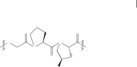
charged amino acids such as lysine and glutamic acid may lead to preferred pairings that create a larger amino acid offset than a single amino acid. To test this idea we have prepared five peptides with an XYG repeat as shown in Tab. 4.1.
Peptides 1 and 2 were designed as control peptides to examine the baseline level of triple helix formation when no charged residues were employed. Peptide 1 contains no hydroxyproline while peptide 2 contains three hydroxyproline residues in the Y position. Peptides 3–5 all contain three hydroxyproline residues in Y positions and also have two charged residues, one in an X position and one in a Y position. Peptide 3 has both glutamic acid and lysine while peptide 4 has only glutamic acid and peptide 5 has only lysine. If peptide 4 is dissolved at a pH of 7, the charges on its glutamate side chains would be expected to discourage assembly through electrostatic repulsion, as shown in Scheme 4.2.
POGPPGEPGPOGPEGPOGPPG
POGPPGEPGPOGPEGPOGPPG
POGPPGEPGPOGPEGPOGPPG
POGPPGEPGPOGPEGPOGPPG
POGPPGEPGPOGPEGPOGPPG
POGPPGEPGPOGPEGPOGPPG
Scheme 4.2. Alternate assembly modes for peptide 4 in which multiple glutamate residues are unfavorably aligned with one another while glycine alignment is enforced.
However, if peptide 5 is added to the solution in a 1:1 ratio it may be expected that this electrostatic repulsion could be converted to a favorable attraction between oppositely charged glutamate and lysine, as shown in Scheme 4.3. This leads to large overhanging, “sticky” ends which can lead to further assembly of the peptides. This self-assembly can continue indefinitely and is the basis for the preparation of very long triple-helical strands.
POGPPGEPGPOGPEGPOGPPG
POGPPGKPGPOGPKGPOGPPG
POGPPGEPGPOGPEGPOGPPG
POGPPGEPGPOGPEGPOGPPGPOGPPGKPGPOGPKGPOGPPG POGPPGKPGPOGPKGPOGPPGPOGPPGEPGPOGPEGPOGPPG
POGPPGEPGPOGPEGPOGPPGPOGPPGKPGPOGPKGPOGPPG
Scheme 4.3. Proposed mechanism of assembly of peptides 4 and 5. Assembly of staggered triple helices is promoted by charge pairing between the oppositely charged glutamic acid
and lysine residues. The first three peptides assemble, forming “sticky ends”. Additional peptides can then assemble, propagating the sticky ends.

112 4 Nanostructured Collagen Mimics in Tissue Engineering
Peptide 3 functions in a fashion similar to the mixture of peptides 4 and 5 but the complementary charges are both found within the same peptide.
Peptides 1–5 were prepared on an Advanced ChemTech APEX396 multipeptide synthesizer using standard 9-fluorenylmethoxycarbonyl (FMOC) solid-phase peptide synthesis protocols. The N-termini and C-termini were prepared as free amines and carboxylic acids respectively. The peptides were purified by reversed-phase high-per- formance liquid chromatography and their masses confirmed by MALDI-TOF MS (matrix-assisted laser desorption/ionization time of flight mass spectrometry). Solutions of each peptide were prepared at a concentration of 6 mM and the pH adjusted to 7 using NaOH. The CD spectrum was collected on each sample in a 0.01-mm cuvette. The spectrum of a mixed solution of peptides 4 and 5 was also examined, in which the concentration of each peptide was 3 mM for a total peptide concentration of 6 mM (Fig. 4.7). Unsurprisingly, the peptide with the weakest signal at 225 nm was peptide 1, which lacks any hydroxyproline residues. In contrast, peptide 2 with three hydroxyproline residues showed a significant positive peak at 225 nm and negative peak below 200 nm. Amongst the three peptides containing charged amino acids, peptide 4 had the weakest triple-helical signal. This is expected because of the unfavorable charge pairing between ionized glutamate residues that would result from the formation of a triple helix. Peptide 3, which has the ability to align itself in such a way as to form cooperative charge pairs between lysine and glutamate, had a significantly enhanced signal at 225 nm as compared to peptide 4. The CD of peptide 5, however, was surprising. We had expected this peptide to behave in a fashion similar to peptide 4, with only weak triple helix signature due to unfavorable charge pairing between positively charged lysine amino acids, whereas in fact peptide 5 had one of the strongest triple-helical spectra. The sixth experiment involved mixing the negatively charged peptide 4 with the positively charged peptide 5. If there was no interaction between these peptides one would expect a CD spectrum that was an average of the individual spectra of 4 and 5 by themselves. Instead, we saw a very strong triple-helical spectrum that was somewhat more intense than the spectrum of peptide 5 by itself. Initial inspection of these data may lead one to believe that this spectrum is simply the result of peptide 5 dominating the signal; however, it should be kept in mind that in the mixed sample peptide 5 is at half the concentration as compared to that of the isolated experiment. Therefore, the strong signal that we see must be the result of an interaction between peptides 4 and 5 in which peptide 4 gains an enhanced triple-helical character when in association with peptide 5, as suggested in Scheme 4.3.
To examine the nanostructure of this mixed peptide system we utilized negativestain TEM. If our supposition regarding the assembly of these peptides as outlined in Scheme 4.3 is accurate, we would expect to find fibrous material which is the result of a supramolecular polymerization. Images taken at a magnification of 50K do reveal a fibrous structure with lengths in excess of 100 nm (see Fig. 4.8). Assuming a triple-helical assembly reminiscent of collagen, this indicates a length in excess of 300 amino acids. However, the diameter of these fibers is greater than the 1.5 nm expected for a triple helix. It may be that as the triple helix is extended a second level of self-assembly takes place, resulting in the bundling of triple helices into
4.10 “Sticky Ends” and Supramolecular Polymerization 113
a
b
Figure 4.7. CD data from peptides 1–5 and a 1:1 mixture of peptides 4 and 5. Peptides are at a concentration of 6 mM, in 10 mM phosphate buffer, pH = 7 at 2 C. Peptides 4 and 5 were mixed in a 1:1 ratio for a total peptide concentration of 6 mM.
(a) Entire CD spectrum to 180 nm. (b) Expanded view of the critical region near 225 nm.
114 4 Nanostructured Collagen Mimics in Tissue Engineering
fibril-like super structures. However, without more detailed analysis this theory cannot yet be confirmed.
Figure 4.8. Negatively stained TEM image of a mixture of peptides 4 and 5. This image shows what appears to be an assortment of nanofibers approximately 100 nm in length and 10 nm in diameter.
4.11
Conclusion
The extracellular matrix is a rich and complicated environment. Mimicking its nanostructure and function remains a significant challenge despite a great deal of knowledge regarding its chief constituent, collagen. Much of the challenge in recreating collagen will be in unraveling how the higher levels of association – the fibril and fiber – are controlled. This understanding is likely to be obtained not from small peptide studies but with larger systems. We hope that our work will help to provide access to the mechanism of fibril and fiber assembly and ultimately to synthetic materials that can be used in sophisticated tissue engineering approaches.
Abbreviations
The usual single and three letter codes for the amino acid abbreviations were used. CD – circular dichroism spectroscopy
CTV – cyclotriveratrylene DNA – deoxyribonucleic acid
DQF-COSY – double quantum-filtered correlation spectroscopy ECM – extracellular matrix
FMOC – 9-fluorenylmethoxycarbonyl fProR – 4(R)-fluoroproline
fProS – 4(S)-fluoroproline HA – hydroxyapatite KTA – Kemp triacid

References 115
Nleu – isobutylglycine
NMR – nuclear magnetic resonance NOE – nuclear Overhauser effect PC12 – pheochromocytoma cells PGA – poly-glycolic acid
PLA – poly-lactic acid suc – succinic acid
TE – tissue engineering
TEM – transmission electron microscopy TOCSY – total correlation spectroscopy TREN – tris(2-aminoethyl)amine
References
1 K. Y. Lee, D. J. Mooney, Hydrogels for tissue engineering, Chem. Rev. 2001, 101, 1869.
2F. A. Auger, M. Rouabhia, F. Goulet,
F. Berthod, V. Moulin, L. Germain, Tissueengineering human skin substitutes developed from collagen-populated hydrated gels: clinical and fundamental applications, Med. Biol. Engin. Comput. 1998, 36, 801.
3P. M. Kaufmann, S. Heimrath, B. S. Kim, D. J. Mooney, Highly porous polymer matrices as a three-dimensional culture system for hepatocytes, Cell Transplant. 1997, 6, 463; D. Seliktar, R. A. Black, R. P. Vito,
R. M. Nerem, Dynamic mechanical conditioning of collagen-gel blood vessel constructs induces remodeling in vitro, Ann. Biomed.
Eng. 2000, 28, 351.
4K. E. Kadler, D. F. Holmes, J. A. Trotter, J. A. Chapman, Collagen fibril formation,
Biochem. J. 1996, 316, 1.
5A. Rich, F. H. C. Crick, The molecular structure of collagen, J. Mol. Biol. 1961, 3, 483.
6G. N. Ramachandran, Treatise on Collagen, Academic, New York, 1964.
7P. M. Cowan, S. McGavin, The structure of poly-L-proline, Nature 1955, 176, 501.
8J. Bella, M. Eaton, B. Brodsky, H. M. Berman, Crystal and molecular structure of a collagenlike peptide at 1.9 A resolution, Science 1994, 266, 75.
9N. Sreerama, R. W. Woody, in Circular dichroism: principles and applications, 2nd ed. (Eds.: N. Berova, K. Nakanishi, R. W. Woody), John Wiley & Sons, Inc., New York, 2000, pp. 601.
10M. G. Venugopal, J. A. M. Ramshaw, E. Braswell, D. Zhu, B. Brodsky, Electrostatic interactions in collagen-like triple-helical peptides, Biochemistry 1994, 33, 7948; F. R. r. Brown,
J.P. Carver, E. R. Blout, Low temperature circular dichroism of poly (glycyl-L-prolyl-L-ala- nine), J. Mol. Biol. 1969, 39, 307.
11D. D. Jenness, C. Sprecher, W. C. J. Johnson, Circular dichroism of collagen, gelatin, and poly(proline) II in the vacuum ultraviolet,
Biopolymers 1976, 15, 513.
12A. A. Adzhubei, M. J. E. Sternberg, Lefthanded polyproline II helices commonly occur in globular proteins, J. Mol. Biol. 1993, 229, 472; D. J. Russell, G. Pearce, C. A. Ryan,
J.D. Satterlee, Proton NMR assignments of systemin, J. Protein Chem. 1992, 11, 265.
13B. Brodsky, M. Li, C. G. Long, J. Apigo,
J.Baum, NMR and CD studies of triple-heli- cal peptides, Biopolymers 1992, 32, 447.
14M. Li, P. Fan, B. Brodsky, J. Baum, Twodimensional NMR assignments and conformation of (Pro-Hyp-Gly)10 and a designed collagen triple-helical peptide, Biochemistry 1993, 32, 7377.
15J. Baum, B. Brodsky, Real-time NMR investigations of triple-helix folding and collagen folding diseases, Folding Des. 1997, 2, R53;
K.H. Mayo, NMR and x-ray studies of collagen model peptides, Biopolymers (Pept. Sci.)
1996, 40, 359; C. G. Long, M. H. Li, J. Baum,
B.Brodsky, Nuclear magnetic resonance and circular dichroism studies of a triple-helical peptide with a glycine substitution, J. Mol. Biol. 1992, 225, 1.

116 4 Nanostructured Collagen Mimics in Tissue Engineering
16J. F. Brandts, H. R. Halvorson, M. Brennan, Consideration of the possibility that the slow step in protein denaturation reactions is due to cis-trans isomerism of proline residues, Biochemistry 1975, 14, 4953; T. E. Creighton, Possible implications of many proline residues for the kinetics of protein unfolding and refolding, J. Mol. Biol. 1978, 125, 401.
17J. Engel, D. J. Prockop, The zipper-like folding of collagen triple helices and the effects of mutations that disrupt the zipper, Annu. Rev. Biophys. Biophys. Chem. 1991, 20, 137.
18P. Bruckner, E. Eikenberry, Procollagen is more stable in cellulo than in vitro, Eur. J. Biochem. 1984, 140, 397; H. P. Bachinger,
P.Bruckner, R. Timpl, D. J. Prockop, J. Engel, Folding mechanism of the triple helix in type-
IIIcollagen and type-III pN-collagen. Role of disulfide bridges and peptide bond isomerization, Eur. J. Biochem. 1980, 106, 619.
19Y. Xu, M. Bhate, B. Brodsky, Characterization of the nucleation step and folding of a collagen triple-helix peptide, Biochemistry 2002, 41, 8143.
20W. Yang, M. L. Battineni, B. Brodsky, Amino acid sequence environment modulates the disruption by osteogenesis imperfecta glycine substitutions in collagen-like peptides, Biochemistry 1997, 36, 6930; X. Liu, S. Kim, Q.-H. Dai, B. Brodsky, J. Baum, Nuclear magnetic resonance shows asymmetric loss of triple helix in peptides modeling a collagen mutation in brittle bone disease, Biochemistry 1998, 37, 15528.
21S. Boudko, S. Frank, R. A. Kammerer,
J.Stetefeld, T. Schulthess, R. Landwehr,
A. Lustig, H. P. Bachinger, J. Engel, Nucleation and propagation of the collagen triple helix in single-chain and trimerized peptides: translation from third to first order kinetics,
J. Mol. Biol. 2002, 317, 459.
22E. Durr, H. R. Bosshard, Folding of a threestranded coiled coil, Protein Sci. 2000, 9, 1410;
D.Porschke, M. Eigen, Co-operative nonenzymic base recognition. 3. Kinetics of the helix-coil transition of the oligoribouridylic– oligoriboadenylic acid system and of aligoriboadenylic acid alone at acidic pH, J. Mol. Biol. 1971, 62, 361.
23D. J. S. Hulmes, A. Miller, D. A. D. Parry,
K.A. Piez, Analysis of the primary structure of collagen for the origins of molecular packing, J. Mol. Biol. 1973, 79, 137.
24C. G. Long, E. Braswell, D. Zhu, J. Apigo,
J.Baum, B. Brodsky, Characterization of col- lagen-like peptides containing interruptions in the repeating Gly-X-Y sequence, Biochemistry 1993, 32, 11688.
25A. V. Persikov, J. A. M. Ramshaw, B. Brodsky, Collagen model peptides: sequence dependence of triple-helix stability, Biopolymers 2000, 55, 436.
26A. V. Persikov, J. A. M. Ramshaw,
A.Kirkpatrick, B. Brodsky, Amino acid propensities for the collagen triple-helix,
Biochemistry 2000, 39, 14960.
27J. Engel, D. J. Prockop, Does bound water contribute to the stability of collagen? Matrix Biol. 1998, 17, 679.
28J. Bella, B. Brodsky, H. M. Berman, Hydration structure of a collagen peptide, Structure 1995, 3, 893.
29R. Berisio, L. Vitagliano, L. Mazzarella,
A.Zagari, Crystal structure of a collagen-like polypeptide with repeating sequence Pro- Hyp-Gly at 1.4 A resolution: implications for collagen hydration, Biopolymers 2000, 56, 8.
30S. K. Holmgren, K. M. Taylor, L. E. Bretscher,
R.T. Raines, Code for collagen’s stability deciphered, Nature 1998, 392, 666.
31R. Importa, F. Mele, O. Crescenzi, C. Benzi, V. Barone, Understanding the role of stereoelectronic effects in determining collagen stability. 2. A quantum mechanical/molecular mechanical study of (proline-proline- glycine)n polypeptides, J. Am. Chem. Soc. 2002, 124, 7857.
32R. Importa, C. Benzi, V. Barone, Understanding the role of stereoelectronic effects in determining collagen stability. 1. A quantum mechanical study of proline, hydroxyproline, and fluoroproline dipeptide analogues in aqueous solution, J. Am. Chem. Soc. 2001, 123, 12568.
33M. Doi, Y. Nishi, S. Uchiyama, Y. Nishiuchi, T. Nakazawa, T. Ohkubo, Y. Kobayashi, Characterization of collagen model peptides containing 4-fluoroproline; (4(S)-fluoro- proline-Pro-Gly)10 forms a triple helix, but (4(R)-fluoroproline-Pro-Gly)10 does not,
J. Am. Chem. Soc. 2003, 125, 9922;
J. A. Hodges, R. T. Raines, Stereoelectronic effects on collagen stability: the dichotomy of 4-fluoroproline diastereomers, J. Am. Chem. Soc. 2003, 125, 9262.

34H. P. Germann, E. Heidemann, A synthetic model of collagen: an experimental investigation of the triple-helix stability, Biopolymers 1988, 27, 157.
35Y. Feng, G. Melacini, J. P. Taulane,
M.Goodman, Acetyl-terminated and tem- plate-assembled collagen-based polypeptides composed of Gly-Pro-Hyp sequences. 2. Synthesis and conformational analysis by circular dichroism, ultraviolet absorbance, and optical rotation, J. Am. Chem. Soc. 1996, 118, 10351.
36D. S. Kemp, K. S. Petrakis, Synthesis and conformational analysis of cis,cis-1,3,5-trimethyl- cyclohexane-1,3,5-tricarboxylic acid, J. Org.
Chem. 1981, 46, 5140.
37E. T. Rump, T. S. Dirk, D. T. S. Rijkers,
H.W. Hilbers, P. G. De Groot,
R. M. J. Liskamp, Cyclotriveratrylene (CTV) as a new chiral triacid scaffold capable of inducing triple helix formation of collagen peptides containing either a native sequence or Pro-Hyp-Gly repeats, Chem. Eur. J. 2002, 8, 4613.
38J. Kwak, A. De Capua, E. Locardi,
M.Goodman, TREN (tris(2-aminoethyl) amine): an effective scaffold for the assembly of triple-helical collagen mimetic structures,
J.Am. Chem. Soc. 2002, 124, 14085.
39Y.-C. Yu, P. Berndt, M. Tirrell, G. B. Fields, Self-assembling amphiphiles for construction of protein molecular architecture, J. Am. Chem. Soc. 1996, 118, 12515.
40T. Gore, Y. Dori, Y. Talmon, M. Tirrell,
H.Bianco-Peled, Self-assembly of model collagen peptide amphiphiles, Langmuir 2001, 17, 5352.
41H. Shin, S. Jo, A. G. Mikos, Biomimetic materials for tissue engineering, Biomaterials 2003, 24, 4353.
42M. J. Humphries, S. K. Akiyama,
A.Komoriya, K. Olden, K. M. Yamada, Identification of an alternatively spliced site in human plasma fibronectin that mediates cell type-specific adhesion, J. Cell Biol. 1986, 103, 2637.
References 117
43W. Dai, J. Belt, W. M. Saltzman, Cell-binding peptides conjugated to poly(ethylene glycol) promote neural cell aggregation, Bio/Technology 1994, 12, 797.
44J. A. Hubbell, S. P. Massia, N. P. Desai,
P.D. Drumheller, Endothelial cell-selective materials for tissue engineering in the vascular graft via a new receptor, Biotechnology 1991, 9, 568.
45J. P. Ranieri, R. Bellamkonda, E. J. Bekos,
J.T. G. Vargo, J. A. G. Aebischer, Neuronal cell attachment to fluorinated ethylene propylene films with covalently immobilized laminin oligopeptides YIGSR and IKVAV. II.
J.Biomed. Mater. Res. 1995, 29, 779.
46J. L. West, J. A. Hubbell, Polymeric biomaterials with degradation sites for proteases involved in cell migration, Macromolecules 1999, 32, 241.
47J. D. Hartgerink, E. Beniash, S. I. Stupp, Selfassembly and mineralization of peptideamphiphiles nanofibers, Science 2001, 294, 1684; G. A. Silva, C. Czeisler, K. L. Niece,
E.Beniash, D. A. Harrington, J. A. Kessler,
S. I. Stupp, Selective differentiation of neural progenitor cells by high-epitope density nanofibers, Science 2004, 303(5662), 1352.
48K. L. Niece, J. D. Hartgerink, J. J. J. M. Donners,
S.I. Stupp, Self-assembly combining two bioactive peptide-amphiphile molecules into nanofibers by electrostatic attraction, J. Am. Chem. Soc. 2003, 125, 7146.
49M. D. Pierschbacher, E. Ruoslahti, Cell attachment activity of fibronectin can be duplicated by small synthetic fragments of the molecule,
Nature 1984, 309, 30.
50S. Weiner, H. D. Wagner, The material bone: structure-mechanical function relations,
Annu. Rev. Mater. Sci. 1998, 28, 271; W. Traub, T. Arad, S. Weiner, Three-dimensional ordered distribution of crystals in turkey tendon collagen fibers, Proc. Natl. Acad. Sci. U.S.A. 1989, 86, 9822.
51M. J. Pandya, G. M. Spooner, M. Sunde,
J.R. Thorpe, A. Rodger, D. N. Woolfson, Sticky-end assembly of a designed peptide fiber provides insight into protein fibrillogenesis, Biochemistry 2000, 39, 8728.

119
5
Molecular Biomimetics: Building Materials Nature’s Way, One Molecule at a Time1
Candan Tamerler and Mehmet Sarikaya
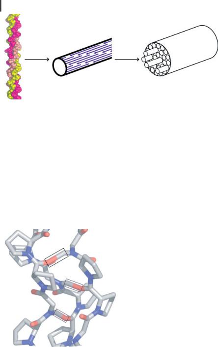
Physical and chemical functions of organisms are carried out by a very large number (billions) of proteins, of differing variety (~105 in humans), through predictable and self-sustaining interactions, developed through evolution. Using biology as a guide, in the molecular biomimetics approach we select, design, genetically tailor, synthesize, and utilize short polypeptides as molecular erectors in self-assembly, ordered organization, and biofabrication of nanoinorganic materials and molecularly hybrid systems in nanotechnology (molecular electronics, magnetics, and photonics) and nanobiotechnology (biosensors, bioassays, and biomaterials). These polypeptides are usually 7–15 amino acids long, and are obtained via combinatorial biology using cell surface or phage display libraries. Once selected, the inorganic binding polypeptides can be further engineered using genetic engineering to tailor their properties for specific material surfaces, morphologies, and crystal chemistries, and for designed applications. The potential of engineered polypeptides in nanotechnology is enormous due to molecular recognition, selfand co-assembly, and manipulation via DNA technologies.
5.1
Introduction
Future functional materials systems, developed either for nanobiotechnology or nanotechnology, could include protein(s) in the assembly, formation, and, perhaps, in the final structure as an integral component leading to specific and controllable functions similar to those seen in biological soft and hard tissues (Fig. 5.1). In the new field of molecular biomimetics – a true marriage of traditional physical and biological fields – hybrid materials could potentially be assembled from the molecular level using the recognition properties of proteins that specifically bind to inorganics [1, 2].
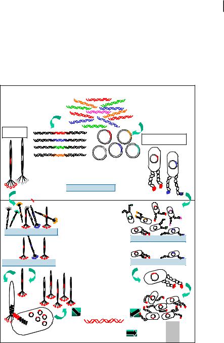
Molecular biomimetics offer three simultaneous solutions to the problem of the control and fabrication of large-scale nanostructures and ordered assemblies of materials in two and three dimensions (Fig. 5.2) [3]. The first is that inorganic-bind-
1)This article is adapted from a recent review paper prepared for Annual Review of Materials Research (Ref. [3]).
Nanofabrication Towards Biomedical Applications. C. S. S. R. Kumar, J. Hormes, C. Leuschner (Eds.) Copyright 2005 WILEY-VCH Verlag GmbH & Co. KGaA, Weinheim
ISBN 3-527-31115-7
120 5 Molecular Biomimetics: Building Materials Nature’s Way, One Molecule at a Time
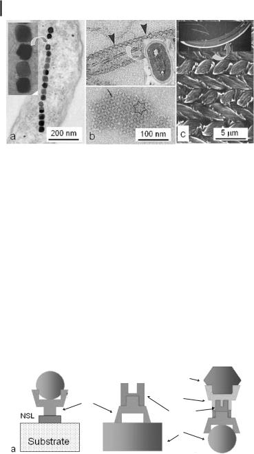
Figure 5.1. Examples of biologically synthesized hybrid materials with a variety of physical properties. (a) Magnetic nanoparticles formed by a magnetotactic bacterium (Aquaspirillum magnetotacticum) are single-crystalline, singledomained magnetite (Fe3O4) particles (inset: higher magnification image revealing cubooctahedral particle shape) (M. Sarikaya, unpublished). (b) Nanostructurally ordered thin-film calcite on the outer layer of an S-layer
bacterium, Synechococcus strain GL24, that serves as a protective coating. (c) Mouse enamel is a hard, wear-resistant material with a highly ordered micro/nano architecture consisting of hydroxyapatite crystallites that assemble into a woven rod structure (SEM image). Each rod is composed of thousands of hydroxyapatite particles (inset: cross-sectional image of a mouse incisor tooth; white region is enamel, backed by grayish dentine) [3].
ing peptides and proteins are selected and designed at the molecular level and through genetics. This allows control at the lowest dimensional scale possible. The second is that such proteins can be used as linkers or molecular erector sets to join synthetic entities, including nanoparticles, functional polymers, or other nanostructures on molecular templates. The third solution is that the biological molecules selfand co-assemble into ordered nanostructures. This ensures a robust assembly process for the construction of complex nanostructures, and possibly hierarchical structures, similar to those found in nature (self-assembly).
|
|
|
|
|
|
|
Material-1 |
|
|
|
|
|
|
Binder |
Binder |
|
|
|
|
|
|
Polypeptide-2 |
|
|
|
|
|
|
Polypeptide-1 |
||
|
|
|
|
|
|
||
|
|
|
|
|
|
|
Linker |
|
|
|
NSL |
|
|
||
|
|
|
|
|
peptide |
||
|
|
|
|
|
|
|
|
|
|
|
|
|
|
|
Material-1 |
|
|
|
|
|
|
|
|
|
|
|
|
Substrate |
b |
c |
|
|
a |
|
|
|
|
||
|
|
|
|
|
|||
|
|
|
|
|
|
|
|
Figure 5.2. Potential utility of inorganic-binding proteins as: (a) linkers for nanoparticle immobilization, (b) functional molecules that assemble on specific substrates, and (c) heterofunctional linkers involving two (or more) binding proteins adjoining several nanoinorganic units. NSL, nonspecific linker [1, 3].
5.1 Introduction 121
Only a few polypeptides have been identified that specifically bind to inorganics. These are mostly biomineralizing proteins extracted from hard tissues followed by isolation, purification, and cloning. Although this approach is difficult, time-con- suming, and has major limitations, several proteins isolated in this fashion have been used as nucleators, growth modifiers, or enzymes in the synthesis of certain inorganics [4–9]. Some examples include amelogenins in mammalian enamel synthesis [5], silicatein, effective in sponge spicular formation [9], and calciteand arago- nite-forming polypeptides in mollusk shells [4, 6, 7]. The preferred approach to
Pre-selection
Generation of libraries:
Insertion of RO into
Randomized genes encoded
Oligonucleotides (RO)
on phage genomes or bacterial plasmids
Phage
Bacterial Cell
Display of random amino acid sequences within a protein residing on the surface of a coat protein (phage) or flagella protein (cell)
Cleaning & Characterization of substrates
Selection
Biopanning library against desired substrate
Washing cycles to distrup weak interactions with the substrate
Elution of bound phages or cells from surfaces
Repeat biopanning several rounds for


 enrichment of tight binders
Replication of phage or growing of cells
Extraction of DNA
Obtaining inorganic-binding aminoacid sequences
HWTR DPGI KT LIGK DPGI GGT
HCRTYW DPGI
Figure 5.3. Principles of phage display (left) and cell surface display (right) protocols adapted for selecting polypeptide sequences with binding affinity to a given inorganic substrate [3].

122 5 Molecular Biomimetics: Building Materials Nature’s Way, One Molecule at a Time
obtaining inorganic-binding polypeptides is to use combinatorial biological techniques [1, 10–13]. In this approach, a large, random library of peptides with the same number of amino acids, but varying compositions, are screened to identify specific sequences that strongly bind to an inorganic material of practical use. In molecular biomimetics, the ultimate goal is to generate a “molecular erector set” in which different proteins, each engineered to bind to a specific surface, size, or morphology of an inorganic compound, promote the assembly of intricate, hybrid structures composed of inorganics, proteins, and even functional polymers [1–3]. Achieving this would be a giant leap towards realizing nanoscale building blocks in which the protein and its binding characteristics are tailored using DNA technologies [14] while the inorganic component is synthesized for its specific functions (e.g., electronic, optical, or magnetic) [15]. These short polypeptides (or small proteins) are called genetically engineered proteins for inorganics (GEPIs) [1, 3]. In the following section, we provide an overview of the display technologies that can be used to select polypeptides that recognize inorganic compounds and highlight unique aspects of using these systems. We next give examples of achievements involving the use of inorganic-binding polypeptides. Finally, we present future prospects in bioand nanotechnologies.
5.2
Inorganic Binding Peptides via Combinatorial Biology
Since the invention of phage display nearly two decades ago [16], display technologies have proven an extraordinarily powerful tool for a myriad of biotechnological and biological applications. These include the characterization of receptor and antibody binding sites, the study of protein–ligand interactions, and the isolation and evolution of proteins or enzymes exhibiting improved or otherwise altered binding characteristics for their ligands. The three most common approaches, phage display (PD), cell surface display (CSD), and ribosome display (RD), have recently been reviewed [17–21]. All technologies are based on the common theme of linking phenotype and genotype. Both PD and CSD rely on the use of chimeric proteins that consist of a target sequence fused within (or to) a protein that naturally localizes on the surface of a bacteriophage (a bacterial virus) or a cell to achieve display (Fig. 5.3). Using standard molecular biology techniques [17], the DNA sequence of the target region (for instance, the active site of an enzyme or the complete sequence of a small polypeptide) can be randomized to create a library of phages or cells, each of which will synthesize a different version of the chimera on its surface. By contacting the library with an immobilized ligand, washing out weak or non-binders and repeating the process to enrich for tight binders, a subset can be selected from the original library exhibiting the ability to interact tightly with the desired ligand, a process is known as biopanning. Because the chimera is encoded within the phage genome or on a plasmid carried by the cell, the identity of the selected sequences (e.g., their amino acid compositions) can be deduced by DNA sequencing (Fig. 5.3).

5.2 Inorganic Binding Peptides via Combinatorial Biology 123
The growing interest in hybrid materials incorporating both inorganic components and peptides or proteins for nanotechnology or nanobiotechnology applications has made PD and CSD very appealing for the isolation of polypeptides capable of binding inorganic materials with high affinity. Others and we adapted both technologies in our laboratory for the selection of inorganic binders for metals, semiconductors, and dielectrics. To date, CSD has been used to identify peptides recognizing iron oxide [22], gold [10], zinc oxide [23], and zeolites [24], whereas PD has been employed to isolate sequences binding to gallium arsenide [11], silica [13], silver [25], zinc sulfide [26], calcite [27], cadmium sulfide [28], and noble metals such as platinum and palladium [S. Dincer et al. 2003, unpublished]. Some of these peptides have been used to assemble inorganic particles [11, 26, 28] and some others for formation (control the nucleation and growth) of the compounds they were selected for [13, 25, 27, 29].
Inorganic materials are very different substrates from proteinaceous ligands and surprisingly little attention has been paid to adapting display technologies developed with biology in mind to the realm of materials science. For instance, many materials rapidly develop an oxide layer on their surface, expose different crystallographic faces to the solvent, and may become chemically or physically modified when incubated in the biological media used during the panning process. To avoid becoming a victim of the first law of directed evolution (“you get what you screen for”) [30], it is therefore imperative to characterize inorganic surfaces before and after panning using spectroscopic and imaging techniques [31]. It may also be useful to monitor wash or elution buffers (e.g., using atomic adsorption spectroscopy to detect metals and metalloids). If evidence of surface modification or deterioration is obtained, buffer conditions should be optimized to guarantee compatibility with the target inorganic material.
Inorganic compounds come in a variety of forms, from polydisperse and morphologically uncharacterized powders to single crystals. The nature of the inorganic substrate may disqualify a particular display technology. For instance, PD is suitable for work with powders even if a gradient centrifugation step is used to harvest complexes between binding phages and particles. On the other hand the CSD system would not be amenable to such an enrichment process since centrifugal forces would shear off the flagella from the cell. Similarly, while both PD and CSD are theoretically suitable for panning on single crystals, tightly bound cells or phages may be very difficult to elute from the material, thereby leading to the loss of high-affinity clones. In such cases, the use of the bacterial system may be advantageous since all binders have an equal likelihood to be recovered following flagellar breakage.
In traditional biological applications of peptide libraries (e.g., antibody epitope characterization, mapping of protein–protein contacts, and the identification of peptide mimics of nonpeptide ligands), three to four biopanning cycles are usually performed in PD while four to five are carried out in CSD. After these cycles of enrichment, the selected sequences typically converge towards a consensus consisting of identical or conservatively replaced residues (e.g., an isoleucine for a leucine). Such consensus sequences reflect precise interactions between the side chains of the protein under study and those of the selected polypeptides. However, all available evi-
124 5 Molecular Biomimetics: Building Materials Nature’s Way, One Molecule at a Time
dence indicates that this rule does not hold true in the case of inorganic binding sequences where similarities rather than a strict consensus are generally observed. This presumably reflects the heterogeneity of the inorganic substrate at the atomic and crystallographic levels and the fact that there are multiple solutions to the problem of inorganic binding. One could, for example, envision binding strategies relying on shape complementarities, electrostatic interactions, van der Waal’s interactions, or various combinations of these mechanisms. Clearly, a better understanding of the rules that govern the binding of polypeptides to inorganic compounds is needed to understand the nature of specificity, predict cross-specificity and affinity, and, ultimately, for the design of hybrid materials exhibiting controlled topology and composition.
5.3
Physical Specificity and Molecular Modeling
The specificity of a polypeptide for an inorganic surface is likely to be rooted in its molecular structure and the atomic (nanoscale) surface structure of the inorganic. Structural information is, therefore, essential not only to elucidate the fundamentals of the recognition process, but also for practical applications. Such knowledge would allow genetic or chemical modifications to create additional functionalities (e.g., attachment of conducting or light-emitting polymers to create hybrid, heterofunctional molecules, ability to bind DNA or proteins), thereby yielding a molecular “tinker-toy”. Ideally, using molecular dynamics/simulated annealing protocols and solution and/or solid-state NMR constraints, one could obtain an averaged lowestenergy structure for as many GEPIs as is feasible, and utilize these structures along with a simulation program in modeling the orientation and binding energetics at
a
b
Au(111)
Au(112)
|
4 |
|
3 |
(Å) |
|
RMSR |
2 |
|
1 |
|
0 |
200 |
400 |
600 |
800 |
c |
Time (ns) |
|
|
|
|
|
Figure 5.4. (a) and (b) are molecular dynamic rendering of gold binding protein (3-repeat GBP-1) on Au{111} and {112} surfaces viewed edge-on, respectively. Coloring corresponds to residue type: polar residues are highlighted in green, charged in blue, and hydrophobic in white. (c) Root-mean-square displacements
(RMSD) of Ca atoms on Au{111} were a result of an equilibration relative to the predicted starting structure (black). The protein is stable after 500 ps. Polar residues (green) exhibit a small RMSD compared with the fluctuations observed in the hydrophobic residues (red) [32]
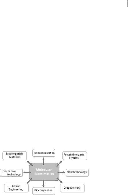
5.4 Applications of Engineered Polypeptides as Molecular Erectors 125
specific interfaces. These data could then be used to rank peptides by interfacial interaction energies, allowing the identification of important side chains and preferred chain alignments for each polypeptide with specific interfaces. Experimental findings together with structural information from simulations should add up to give a coherent understanding of GEPI–inorganic surface interactions.
In a collaborative work, we have recently performed computer structure modeling studies to predict the shape of a GEPI (viz. gold-binding protein, GBP-1) in solution, and present a summary here as an illustration [Fig. 5.4(a)] [32]. This preliminary work was carried out in the hope that any coincidence between the amino acid residues of the peptide and the spacing of the inorganic atomic lattice would shed light on how a GEPI binds to an inorganic surface. Raw amino acid sequence data for three repeats of the 14 amino acid GBP-1 was compared to all known protein structures using FASTA searches and various first-order secondary-structure prediction algorithms (Chou-Fasman and Holley/Karplus) [32]. Figure 5.4(a) shows the threerepeat GBP-1 above a {111} Au atomic lattice that highlights the correspondence of OH– groups to gold atom positions. These initial results suggest that binding repeats of GBP-1 form an anti-parallel b-pleated sheet conformation, which places OH– groups from serine and threonine residues into a regular lattice based on energy minimized in vacuo using X-PLOR [Fig. 54(b)]. We also showed in these preliminary studies that GBP-1 does not bind to Au{112} surface as tightly [Fig. 4(a)], because of the migration of water molecules through the atomic grooves on this crystallographic surface, which decouples the protein from the surface.
5.4
Applications of Engineered Polypeptides as Molecular Erectors
Controlled binding and assembly of proteins onto inorganic substrates is at the core of biological materials science and engineering with wide ranging applications [33, 34] (Fig. 5.5). Protein adsorption and macromolecular interactions at solid surfaces
Figure 5.5 Potential applications of molecular biomimetics in nanotechnology and nanobiotechnology using combinatorially selected and genetically engineered inorganic-binding polypeptides [3].
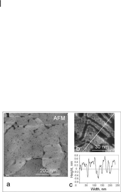
126 5 Molecular Biomimetics: Building Materials Nature’s Way, One Molecule at a Time
play key roles in the performance of implants and hard-tissue engineering. DNA and proteins adsorbed specifically onto probe substrates are used to build microarrays suitable for modern genomics [35], pharmogenetics [36], and proteomics [37, 38]. Engineered polypeptides hybridized with functional synthetic molecules could be used as heterofunctional building blocks in molecular electronics and photonics [39, 40]. The unique advantages of engineered polypeptides, created through molecular biomimetics discussed here, include highly specific molecular surface recognition of inorganics, self-assembly into ordered structures, and tailoring of their molecular structures and functions through molecular biology and genetics protocols. Using inorganic binding polypeptides, one can create molecular erectors sets for potential nanotechnological and nanobiotechnological applications.
5.4.1
Self-Assembly of Inorganic-Binding Polypeptides as Monolayers
One of the central questions related to the genetically engineered proteins is whether they can assemble with a long-range order on a given crystallographic surface of a material in addition to chemically recognizing it. While this aspect of molecular biomimetics is in its nascency, the atomic-force microscopy (AFM) image of Fig. 5.6 shows that it is possible to assemble a one-monolayer-thick gold-binding protein layer on the {111} surface of gold (H.M. Zareie, M. Sarikaya, 2003, unpub-
Figure 5.6. (a) Self-assembled three-repeat GBP-1 on Au{111} (AFM image) showing ordered-domained structure. The angle among the domain boundaries is either 120 or 60 , implying recognition of the symmetry of
the top surface layer on Au{111}. The line across several domains in the AFM image (b) produces a profile with 0.5-nm-high platforms, revealing monolayer thickness of the GEPI film (H.M. Zareie, M. Sarikaya, 2003, unpublished].
5.4 Applications of Engineered Polypeptides as Molecular Erectors 127
lished results). In fact, close inspection of this and other images reveal that GBP-1 assembles into domains with clear and straight boundaries that actually make a 60 / 120 angle, suggesting that the polypeptide actually recognizes the six-fold lattice symmetry on the Au{111} surface. What is also significant in these results is that the assembly process progresses until the whole surface is completely covered. Instead of using the traditional thiol or silane linkers in self-assembled monolayers [41, 42], this result demonstrates self-assembly of GEPI monolayer that may be used as functional linkages – a central premise in this research.
5.4.2
Morphogenesis of Inorganic Nanoparticles via GEPIs
In biomineralization, a significant aspect of biological control over materials formation is via protein/inorganic interactions that control the growth morphology in tissue, such as bone, dentin, mollusk shells, and bacterial and algal particle formation [43–46]. Studies aiming at finding how such proteins affect biomineralization have traditionally focused mostly on templating [4–9], nucleation [47, 48], and enzymatic reactions [8, 9]. With the emergence of combinatorially selected peptides that strongly bind inorganics, a natural step is to examine how these polypeptides affect inorganic formation, and investigate various effects in mineralization (including nucleation, growth, morphogenesis, and enzymatic effects). These studies have been carried out under aqueous environments necessary for biological functions of selected GEPIs, most notably those binding to Au [29] and Ag [25]. The morphology
Figure 5.7. Effect of GEPI on nanocrystal morphology. Two mutants out of more than 50 from a library of gold-binding GEPIs were tested to result in flat gold particle formation as shown in (a) and (d) similar to those formed under acidic (b) or boiling conditions (c). Particles
formed in the presence of vector-encoded alkaline phosphatase and neutral conditions did not result in morphological change of gold particles, as shown in (e) and (f), respectively [29].
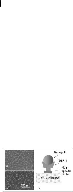
128 5 Molecular Biomimetics: Building Materials Nature’s Way, One Molecule at a Time
of solution-grown gold particles was controlled by the presence of gold-binding GEPIs selected by CSD [29]. More than 50 mutants were tested for their influence on the rate of crystallization of nanogold particles formed by the reduction of AuCl3 with Na citrate. Two mutants accelerated the rate of crystal growth and changed the particle morphology from cubo-octahedral (the usual shape of gold particles grown under equilibrium conditions) to flat, triangular, or hexagonal (Fig. 5.7). Similar crystals usually form under acidic conditions in the absence of gold-binding polypeptides [49]. It may be concluded, therefore, that slightly basic polypeptides may have acted as an acid in flat gold crystallization.
5.4.3
Assembly of Inorganic Nanoparticles via GEPIs
Organization and immobilization of inorganic nanoparticles in twoor three-dimen- sional geometries are fundamental in the utilization of the nanoscale effects [50]. For example, quantum dots could be produced using vacuum techniques (e.g., molecular beam epitaxy, MBE), and such an organization is shown in Fig. 5.8(a) in the GaInAs/GaAs system (T. Pearsall, 1996, unpublished). However, this can only be accomplished under stringent conditions of high temperature, very low pressures, and a toxic environment. A desirable approach would not only synthesize the inorganic particles under less stringent conditions but also assemble/immobilize them via self-assembly using functional molecules as coupling agents. Inorganic particles have been coupled and functionalized using synthetic molecules (e.g., thiols, lipids, and biological molecules, including amino acids, polypeptides, and ligand-function- alized DNA) and assembled to generate novel materials using the recognition properties of these molecules [1, 2, 40, 51]. Nanoparticles synthesized in wet-chemical conditions in the presence of these molecules (e.g., citrate, thiol, silane, lipid, and amino acids) not only cap the particle, resulting in controlled growth, but also prevent their uncontrolled aggregation [40, 52, 53].
Figure 5.8 The AFM images show quantum (GaInAs) dots assembled on GaAs substrate
(a) via high-vacuum (molecular beam epitaxy, MBE) strain-induced self-assembly and (b) via
seven-repeat GBP-1. (c) Schematic illustration of (b), where PS is polystyrene substrate, and the nonspecific binder is glutaraldehyde.

5.5 Future Prospects and Potential Applications in Nanotechnology 129
Synthetic and biological molecules used as coupling agents, however, are nonspecific to a given material. For example, thiols couple gold as well as silver nanoparticles in similar ways [40]. Likewise, citrate ions cap noble metals indiscriminately [40, 52, 53]. A desirable next step in molecular recognition and assembly via molecules would be to use polypeptide sequences that recognize inorganics specifically. In this context, GEPIs could potentially be used for assembly of nanoparticles. In addition to inorganic surface recognition, a further advantage of the GEPI is that it can be genetically fused to other functional biomolecular units or ligands to produce heterobifunctional (or multifunctional) molecular entities [54, 55]. In Fig. 5.8(b), we show the assembly of nanogold particles on GBP-1-coated flat mica surfaces [56]. When seven-repeat GBP-1 was used as the linker, high-density gold particles formed on the surface, resembling a distribution not unlike in that in Fig. 5.8(a). Clearly, in this case, unlike the conditions of the MBE, the assembly was accomplished under ambient conditions of temperature or pressure and in an aqueous solution. Therefore, the homogeneous decoration of the surface with nanogold suggests that proteins may be useful in the production of tailored nanostructures as quantum dots. The recognition activity of the protein would provide the ability to control the particle distribution, while particle preparation conditions would allow size control.
5.5
Future Prospects and Potential Applications in Nanotechnology
Proteins hold great promise for the creation of architectures at the molecular or nanoscale levels [1–3, 40]; the ultimate aim is to use proteins for controlled binding and assembly onto inorganics. Genetically engineered proteins for inorganics (GEPIs) represent a new class of biological molecules that are combinatorially selected to bind to specific inorganic surfaces [1, 3]. The ordered assembly of GEPI on inorganic surfaces could have a significant impact on molecular technology applications, offering several novel practical advantages. Our results described above are the first demonstrations that combinatorially selected polypeptides can selfassemble specifically on a selected inorganic single crystal surface, and that a GEPI may be molecularly recognizing an inorganic surface. Realizing the fact that thiol and silane linkages are the other two major molecular linkers for noble metal and oxide (silica) surfaces that have constituted the field of self-assembled molecules up until now, it is naturally expected that self-assembled GEPI monolayers – SA(GEPI)M – as “molecular erector sets” will open up new avenues for designing and engineering new and novel functional surfaces. We have already demonstrated that inorganic materials can be assembled at the nanoscale by proteins that have been genetically engineered to bind to selected materials surfaces. It is also the first time that engineered proteins were shown to affect crystal morphology [29]. The combinatorial genetic approach is a general one, which should be applicable to numerous surfaces [10–13, 22–27]. The modularity of binding motifs should allow genetic fusion of peptide segments recognizing two different materials. The resulting heterobifunctional molecules could be used to attach different materials to each other and
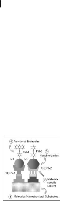
130 5 Molecular Biomimetics: Building Materials Nature’s Way, One Molecule at a Time
may permit assembly of complex nanocomposite and hybrid materials. These results could lead to new avenues in nanotechnology, biomimetics, biotechnology, and crystal and tissue engineering such as in the formation, shape modification, and assembly of materials, and development of surface-specific protein coatings.
One particular potential utility of GEPI as a molecular linker is in nanotechnology. Both nanostructured inorganics and functional molecules are becoming fundamental building blocks for future functional materials such as nanoelectronics, nanophotonics, and nanomagnetics [1–3, 39, 40, 57]. Before nanoscience could be implemented in practical and working systems, however, there are numerous challenges that must be addressed. Some of these challenges include molecular and nanoscale ordering and scale up into larger architectures. A nanotechnological system, for example, could require several components made up of materials of different physical and chemical characteristics. These different materials have to be connected and assembled without an external manipulation. As schematically shown in Fig. 5.9, the components may include two or more inorganic nanoparticles, several functional molecules, and nanopatterned multimaterial substrates, all assembled through the specific interaction with the appropriate GEPI molecules.
Although significant advances have been made in developing protocols for sur- face-binding polypeptides, many questions need to be answered before their robust design and practical applications are effectively realized. These questions include: (i) What are the physical and chemical bases for recognition of inorganic surfaces by the genetically engineered polypeptides? (ii) What are the long-range assembly characteristics, kinetics, and stability of the binding? (iii) What are the molecular mechanisms of engineered polypeptide binding onto (noble) metals compared to those on
Figure 5.9 An illustration of the potential of utility of GEPIs as molecular erectors in functional nanotechnology. Two different GEPIs (GEPI-1 and GEPI-2) assemble on a patterned substrate. One could use either a designer protein, followed by genetic fusion of the respective GEPIs, or directed-assemble GEPIs on a
nanopatterned substrate. Two different inorganic particles I-1 and I-2 are immobilized selectively on GEPI-1 and GEPI-2, respectively. Synthetic molecules (i.e., conducting or photonic) via functionalized side groups or hybridized GEPIs are assembled on the nanoparticles [3].

5.5 Future Prospects and Potential Applications in Nanotechnology 131
nonmetals? (iv) How do surface characteristics affect binding? (v) Based on the insights achieved, can we develop a “road map” in using GEPIs as molecular linkers and open new avenues in self-assembled molecular systems in nanotechnology based on biology? Some of these questions can be answered using the existing nanotechnological and molecular biological tools, and some require adaptation of biology methodologies into molecular biomimetics. For example, new algorithms need to be developed to adapt current protein-folding models into molecular recognition, reconformation, and binding of engineered proteins on inorganic compounds with various crystallographical, surface-topological, and chemical characteristics. Modeling studies coupled with rapid development of the experimental tools – such as high-resolution scanned probe microscopies and spectroscopies, local surface spectroscopies (such as surface-enhanced RAMAN and surface plasmon resonance), x- ray photoelectron spectroscopy, smalland grazing-angle x-ray diffraction, and liquid- and solid-state nuclear magnetic resonance spectroscopy – is expected to lead into new predictive protocols in designing novel polypeptide sequences that specifically bind to desired inorganics and self-assemble on them and can be used as molecular erectors. Considering the present rapid developments in the inorganic-bind- ing polypeptide selection protocols, the increased variety of materials utilized as substrates, and novel modeling adaptations, it is expected that many of the above questions will find answers in the near future with subsequent significant impact in broad multidisciplinary fields with potentially wide-ranging nanotechnology and nanomedicine applications.
Acknowledgments
We thank our colleagues A. Jen, F. Ohuchi, D. Schwartz, F. Baneyx, and B. Traxler (University of Washington, Seattle), J. Evans (New York University), K. Schulten (University of Illinois, Urbana), and S. Brown (University of Copenhagen) for invaluable discussions, and our students and researchers S. Din er, D. Heidel, R. Braun, M. H. Zareie, M. Duman, E. Ventkatasubramanian, V. Bulmus, E. ren, D. Sahin, and H. Fong for technical help. This research was supported by USA-ARO through the DURINT program (PM Dr. R. Campbell) under grant no. DAAD19–01–1-04999 (Defense University Research Initiative on Nanotechnology).
Abbreviations
AFM – atomic force microscopy
CSD – cell surface display
GBP – gold-binding protein
GEPI – genetically engineered polypeptides for inorganics
MBE – molecular beam epitaxy
PD – phage display
PS – polystyrene
RD – ribosomal display

1325 Molecular Biomimetics: Building Materials Nature’s Way, One Molecule at a Time
References
1M. Sarikaya, C. Tamerler, A. Y. Jen, K. Schulten, F. Baneyx, Molecular bio-
mimetics: nanotechnology through biology.
Nature Mater. 2003, 2, 577–585.
2 P. Ball, Life’s lessons in design. Nature 2001, 409, 413–416.
3M. Sarikaya, C. Tamerler, D. T. Schwartz,
F. Baneyx, Materials assembly and formation using engineering polypeptides, Ann. Rev.
Mater. Res. 2004, 34, 373–408.
4M. A. Cariolou, D. E. Morse, Purification and characterization of calcium-binding conchiolin shell peptides from the mollusk, Haliotisrufescens, as a function of development.
J. Comp. Physiol. B. 1988, 157, 717–729.
5M. L. Paine, M. L. Snead, Protein interactions during assembly of the enamel organic extracellular matrix. J. Bone Miner. Res. 1996, 12, 221–226.
6A. Berman, L. Addadi, S. Weiner, Interactions of sea-urchin skeleton macromolecules with growing calcite crystals: A study of intracrystalline proteins. Nature 1988, 331, 546–548.
7A. Weizbicki, C.S. Sikes, J. D. Madura,
B. Darke, Atomic force microscopy and molecular modeling of proteins and peptide binding to calcite. Calcif. Tissue Intl. 1994, 54,
133–141.
8N. Kr.ger, R. Deutzman, M. Sumper, Polycatyonic peptides from diatom biosilica that direct silica nanosphere formation. Science
1999 286, 1129–1132.
9J. N. Cha, K. Shimizu, Y. Zhou,
S. C. Christiansen, B. F. Chmelka, et al., Silicatein filaments and subunits from a marine sponge direct the polymerization of silica and silicones in vitro. Proc. Natl. Acad. Sci. USA
1999, 96, 361–365.
10S. Brown, Metal recognition by repeating polypeptides. Nature Biotechnol. 1997, 15, 269–272.
11S. R. Whaley, D. S. English, E. L. Hu,
P. F. Barbara, M. A. Belcher, Selection of peptides with semiconducting binding specificity for directed nanocrystal assembly. Nature 2000, 405, 665–668.
12D. J. H. Gaskin, K. Strack, E. N. Vulfson, Identification of inorganic crystal-specific sequences using phage display combinatorial library of short peptides: A feasibility study.
Biotech. Lett. 2000, 22, 1211–1216.
13R. R. Naik, L. Brott, S. J. Carlson, M. O. Stone, Silica precipitating peptides isolated from a combinatorial phage display libraries.
J.Nanosci. Nanotechnol. 2002, 2, 95–100.
14D. Voet, J. G. Voet, Biochemistry. 2003, Wiley, New York
15B. Muller, Natural formation of nanostructures: from fundamentals in metal heteroepitaxy to applications in optics and biomaterials science. Surf. Rev. Lett. 2001, 8, 169–228.
16G. P. Smith, Filamentous fusion phage: Novel expression vectors that display cloned antigens on the virion surface. Science 1985, 228, 1315–1317.
17M. Dani, Peptide display libraries: design and construction. J Recept. Signal Transduct. Res.
2001, 21, 469–488.
18K. D. Wittrup, Protein engineering by cell surface display. Curr. Opin. Biotechnol. 2001,
12, 395–399.
19H. M. E. Azzazy, W. E. Jr. Highsmith, Phage display technology: clinical applications and recent innovations. Clin. Biochem. 2002, 35, 425–445.
20W. J. Dower, L. C. Mattheakis, In vitro selection as a powerful tool for the applied evolution of proteins and peptides. Curr. Opin.
Chem. Biol. 2002, 6, 390–398.
21P. Samuelson, E. Gunneriusson, P. &. Nygren,
S.St hl, Display of proteins on bacteria.
J. Biotechnol. 2002, 96, 129–154.
22S. Brown, Engineering iron oxide-adhesion mutants of Escherichia coli phage k-receptor.
Proc. Natl. Acad. Sci.USA 1992, 89, 8651–8655.
23K. Kiargaard, J. K. Sorensen, M. A. Schembri,
P.Klemm, Sequestration of zinc oxide by fimbrial designer chelators. Appl. & Env. Microbiol. 2000, 66, 10–14.
24Nygaard S, Wandelbo R, Brown S. Surfacespecific zeolite-binding proteins. Adv. Mater. 2002, 14, 1853–1856.
25R. R. Naik, S. J. Stringer, G. Agarwal,
S.E. Jones, M. O. Stone, Biomimetic synthesis and patterning of silver nanoparticles. Nature Mater. 2002, 1, 169–172.
26W. S. Lee, C. Mao, C. E. Flynn, A. M. Belcher, Ordering quantum dots using genetically engineered viruses. Science 2002, 296, 892–895.

27C. M. Li, G. D. Botsaris, D. L. Kaplan, Selective in vitro effects of peptides on calcium carbonate crystallization. Crystal Growth & Design 2002, 2, 387–393.
28C. Mao, C. E. Flynn, A. Hayhurst, R. Sweeney,
J.Qi, G. Georgiou, B. Iverson, A. M. Belcher, Viral assembly of oriented quantum dot nanowires. Proc. Natl. Acad. Sci. USA 2003, 100, 6946–6951.
29S. Brown, M. Sarikaya, E. Johnson, Genetic analysis of crystal growth. J. Mol. Biol. 2000, 299, 725–732.
30C. Schmidt-Dannert, F. H. Arnold, Directed evolution of industrial enzymes. Trends. Biotechnol. 1999, 17, 135–136.
31H. Dai, C. K. Thai, M. Sarikaya, F. Baneyx,
D.T. Schwartz. Through-mask anodic patterning of copper surfaces and film stability in biological media. Langmuir, 2004, 20, 3483–3486.
32R. Braun, M. Sarikaya, K. S. Schulten, Genetically engineered gold-binding polypeptides: Structure prediction and molecular dynamics. J. Biomater. Sci. 2002, 13, 747–758.
33S. E. Sakiyama-Elbert, J. A. Hubbell, Design of novel biomaterials. Ann. Rev. Mater. Res. 2001, 31, 183–201.
34B. Ratner, F. Schoen, A. Hoffman, J. Lemons (eds.), Biomaterials Science: Introduction to Materials in Medicine. Academic Press, San Diego, 1996.
35J. R. Epstein, I. Biran, D. R. Walt, Fluorescence based nucleic acid detection and microarrays Anal. Chim. Acta 2002, 469, 3–36.
36M. L. Yarmush, A. Yarayam, Advances in proteomics technologies. Ann. Rev. Biomed. Eng. 2002, 4, 349–373.
37P. Cutler, Protein Arrays: The current state- of-the-art. Proteomics 2003, 3, 3–18.
38M. E. Chicurel, D. D. Dalma-Weiszhausz, Microarrays in pharmagenomics – advances and future promise. Pharmacogenomics 2002, 3, 589–601.
39D. I. Gittins, D. Bethell, D. J. Schiffrin,
R.J. Michols, A nanometer-scale electronic switch consisting of a metal cluster and redox-addressable groups. Nature 2000, 408, 67–69.
40C. M. Niemeyer, Nanoparticles, proteins, and nucleic acids: Biotechnology meets materials science. Angew. Chem. Int. Ed. 2001, 40, 4128–4158.
References 133
41G. M. Whitesides, J. P. Mathias, C. T. Seto, Molecular Self-assembly and nanochemistry- A chemical strategy for the synthesis of nanostructures. Science 1991, 254, 1312–1319.
42A. Ulman (ed.), Self-assembled Monolayers of Thiols. Academic Press, San Diego, 1998.
43R. B. Frankel, R. P. Blakemore (ed.), Iron Biominerals. Plenum, New York, 1991.
44M. Glimcher, M. Nimni, Collagen cross-link- ing and biomineralization. Connect. Tissue Res. 1992, 27, 73–83.
45S. Schultze, G. Harauz, T. I. Beveridge, Participation of a cyanobacterial S layer in finegrain mineral formation. J. Bacteriol. 1992, 174, 7971–7981.
46M. Sarikaya, Biomimetics. Biomimetics: Materials fabrication through biology Proc. Natl. Acad. Sci. USA, 1999, 96, 14183–14185.
47A. M. Belcher et al., Control of crystal phase switching and orientation by soluble molluskshell proteins. Nature 1996, 381, 56–58.
48P. Mukharjee, S. Senapati, D. Mandal,
A.Ahmad, M. I. Khan, et al., Extracellular synthesis of gold nanoparticles by the fungus
Fusarium oxysporum. Chem. Biol. Chem. 2002,
3, 461–463.
49J. T4rkevich, P. C. Stevenson, J. Hillier, A study of the nucleation and growth of processes in the synthesis of colloidal gold.
Trans. Faraday Soc. Disc. 1951, 11, 55–75.
50B. Muller, Natural formation of nanostructures: from fundamentals in metal heteroepitaxy to applications in optics and biomaterials science. Surf. Rev. Lett. 2001, 8, 169–228.
51O. Yamauchi, A. Odani, S. Hirota, Metal ion assisted weak interactions involving biological molecules. From small complexes to metalloproteins. Bull. Chem. Soc. Jpn. 2001, 74, 1525–1545.
52R. Djulali, Y. Chen, H. Matsui, Au nanowires from sequenced histidine-rich peptide. J. Am. Chem. Soc. 2002, 124, 13660–13661.
53Y. Xia, Y. Sun, Shape-Controlled Synthesis of Gold and Silver Nanoparticles. Science 2002, 298, 2176–2179.
54A. R. McMillan, C. D. Pauola, J. Howard,
S.L. Chen, N. J. Zaluzec, et al., Ordered nanoparticle arrays formed on engineered chaperonin protein templates. Nature Mater. 2002, 1, 247–52.
134 |
|
5 Molecular Biomimetics: Building Materials Nature’s Way, One Molecule at a Time |
||
|
|
D. Moll et al. S-layer-streptavidin fusion pro- |
|
M. Sarikaya, H. Fong, D. Heidel, R. Humbert, |
55 |
56 |
|||
|
|
teins as template for nanopatterned molecu- |
|
Biomimetic assembly of nanostructured |
|
|
lar arrays. Proc. Natl. Acad. Sci. USA 2002, 99, |
|
materials. Mater. Sci. Forum 1999, 293, 83–87. |
|
|
14646–14651. |
57 |
National Nanobiotechnology Initiative. 2003, |
http://www.nano.gov.
II
Characterization Tools for Nanomaterials and Nanosystems
Nanofabrication Towards Biomedical Applications. C. S. S. R. Kumar, J. Hormes, C. Leuschner (Eds.) Copyright 2005 WILEY-VCH Verlag GmbH & Co. KGaA, Weinheim
ISBN 3-527-31115-7