

3
Cell Populations
I will treat the behavior of E. coli from the top down, or outside in, beginning with the behavior of cell populations, and then working toward the molecular biology. Imagine an ensemble of self-propelled microscopic particles, moving about in a dilute aqueous medium, robots programmed to respond to external stimuli. How are the robots distributed in space and time?
Chemotactic Rings
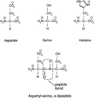
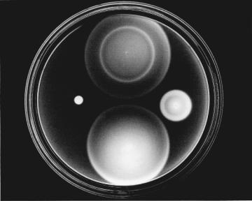
A vivid way of demonstrating motile responses of E. coli to chemical stimuli (chemotaxis) is to deposit a small drop of a cell suspension on a Petri plate containing semisolid agar (~0.2% w/v) in a nutrient medium. Agar is commonly used at higher concentrations (~2%) as a solid matrix on which to grow discrete bacterial colonies. It is like jello but has the advantage of not being digested by ordinary bacteria. The bacteria grow in this medium, so now the robots are self-replicating. The usual nutrient is tryptone, a mixture of amino acids obtained from a pancreatic digest of casein, a protein found in milk. The structures of three such amino acids are shown in Fig. 3.1. When agar is dilute, motile cells swim through the pores in the gel and spread throughout the plate.They do this in a series of expanding “chemotactic rings,” as shown in Fig. 3.2, where clouds of bacteria scatter light and appear white. Adler (1966) found that these rings form as cells consume different nutrients. Wild-type cells, shown at the top, first induce enzymes required for utilization of serine. In front of the outermost ring there is lots of serine; behind it there is practically none. The cells respond to the intervening spatial gradient and move outward. Meanwhile, cells left behind at the point of inoculation induce enzymes required for the utilization of aspartate. In front of the second ring there is lots of aspartate; behind it there is prac-
19
20 3. Cell Populations
FIGURE 3.1. Three amino acids and a dipeptide. In water, the molecules carry positive and negative charges, as shown. Aspartate has a side chain ending in a carboxylic-acid group, serine in a hydroxyl group, and histidine in an imidazole group. The imidazole is a five-membered ring comprising three carbon atoms (indicated by vertices) and two nitrogen atoms. There are 17 other common amino acids that have distinct side chains (not shown). In polypeptides and proteins, amino acids are linked end to end, by the removal of water (H2O) to form peptide bonds, shown within the dashed line. The atoms shown within the dashed line and the adjacent carbon atoms lie in a plane, and this gives polypeptides favored structural motifs, such as the a-helix and the b-pleated sheet.
tically none. Once again, cells respond to the intervening spatial gradient and move outward. In the course of metabolizing serine and aspartate, the cells deplete most of the oxygen, so next, cells near the bottom of the plate consume threonine anaerobically and move outward in a more diffuse ring. And so on. At the right is a mutant that has lost the ability to taste serine. At the bottom is a mutant that has lost the ability to taste aspartate. At the left is a mutant that swims vigorously but is unable to respond to any attractants or repellents. It fails to form any chemotactic rings. This colony is relatively compact. The cells swim, but they are not
Chemotactic Rings |
21 |
FIGURE 3.2. Behavior of four cell types on a tryptone swarm plate. Top: Wild-type cells, showing chemotactic rings for serine and aspartate. Right: Cells missing the serine receptor. Bottom: Cells missing the aspartate receptor. Left: Smooth-swimming cells unable to process information generated by either chemoreceptor. The plate (8.5cm dia.) was inoculated in four places by stabbing the agar with a sterile toothpick dipped in a cell suspension and placed in a humid incubator set at 30°C (86°F). About 8 hours later, it was illumined slantwise from below and photographed against a dark background. (Photograph courtesy of J.S. Parkinson, University of Utah.)
able to change directions, so they get trapped in blind alleys in the agar.
Chemotactic rings can be quite sharp, especially if the bacteria metabolize only a single nutrient. A dramatic example is shown in Fig. 3.3, where one inoculum contained cells that could only metabolize the sugar ribose, the other cells that could only metabolize the sugar galactose, with a plate containing a mixture of the two. The cells of either type do not interfere with one another.
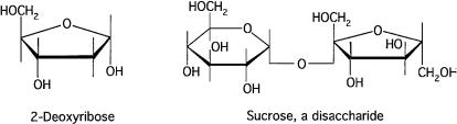
Structures of some sugars are shown in Fig. 3.4. These are ringshaped molecules in which most carbons carry hydroxyl groups. Ribose is a 5-carbon, five-sided ring compound, and galactose is a 6-carbon, six-sided ring compound that differs from glucose only

22 3. Cell Populations
FIGURE 3.3. Behavior of two cell types on a ribose and galactose swarm plate. Both types are chemotactic toward ribose and galactose, but one is unable to metabolize ribose and the other is unable to metabolize galactose. Cells generate a spatial gradient for an attractant only if they consume the attractant. Cells left behind in the original inoculum appear at the center of each ring. (Adler, 1976, and the cover of Nature, 26 July
1979, reprinted with permission).
by the position of some of its hydroxyl groups, i.e., whether they are above or below the plane of the ring.
The swarm assay has been enormously useful for finding mutant cells that are defective for chemotaxis or cells that have regained their ability to respond. In the latter case, a single revertant cell appearing at the edge of a compact colony like that of Fig. 3.2 (left) gives rise to a swarm that moves out into regions of the plate otherwise devoid of bacteria. However, the swarm assay is not simply an assay for a behavioral response, because it requires that the cells take up a substrate, and thus generate a chemical gradient, and multiply, to populate the expanding ring. A mutant that
Capillary Assay |
23 |
FIGURE 3.4. Some sugars. In 2-deoxyribose, the hydroxyl group, present in ribose on carbon 2, is missing. This sugar is part of the backbone of DNA. Sucrose (cane sugar) is a disaccharide: glucose, which has a six-membered ring, is linked to fructose, which has a five-membered ring. The linkage involves removal of water (H2O). The only difference between fructose and ribose is the placement of the hydroxyl groups. E. coli is only weakly chemotactic toward 2-deoxyribose and not chemotactic at all toward sucrose. However, it is chemotactic toward both glucose and fructose. As before, vertices indicate carbon atoms. Hydrogen atoms, located at the end of each unterminated bond, are not shown.
fails to absorb, metabolize, or grow on a substrate will fail to yield a chemotactic ring, even though it might still be able to taste and respond to gradients of that substrate.
Capillary Assay
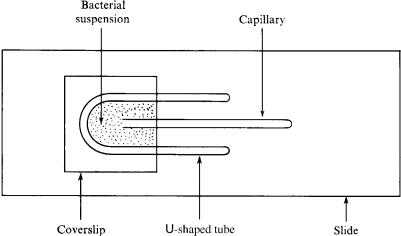
This led Adler to modernize an assay originally developed by Pfeffer, in which the stimulus is a gradient generated by diffusion of a chemical from the mouth of a capillary tube (Adler, 1969, 1973). But first he needed to find conditions that would support vigorous motility without growth: a chelating agent to protect cells from traces of heavy metals, a buffer to keep the pH between 6 and 7.5 (to keep the acidity close to neutral), and, if the cells were grown aerobically, oxygen to allow utilization of an endogenous energy reserve (Adler and Templeton, 1967). It was found that the presence of glucose or growth above 37°C prevented synthesis of flagella. While Pfeffer looked at the cloud of bacteria that formed near the capillary mouth (Fig. 2.3a), Adler counted the number of bacteria that swam inside (Fig. 2.3b). He did this by making serial dilutions of the contents of the tube, plating aliquots on nutrient agar, and counting colonies (Adler, 1973).
24 3. Cell Populations
FIGURE 3.5. The apparatus used in Adler’s version of the capillary assay. The drawing is to scale (microscope slide 1≤ ¥ 3≤), except for the width of the capillary tube (200 mm inside diameter). (Adler, 1973, Fig. 1, reprinted with permission.)
A drawing of Adler’s version of the capillary assay is shown in Fig. 3.5. A U-shaped spacer made from a glass tube about 1.5mm in diameter is placed on a glass microscope slide. A coverslip is added as a roof. The space in between is filled with a bacterial suspension. Finally, a capillary tube (200 mm inside diameter) containing a few millimeters of attractant medium is slid along the glass into the pond. It is withdrawn 30 to 60 minutes later.
Chemicals Sensed
Using the capillary assay, Adler was able to show that E. coli responds to chemicals that it can neither transport (take up from the surrounding medium) nor metabolize (utilize as a source of energy or raw material). Therefore, the cells must recognize the chemicals per se. Taste will do. Consumption is not necessary. An example is shown in Fig. 3.6.
Some of the different kinds of chemicals to which E. coli can respond are listed in Table 3.1. E. coli pays attention to things of low molecular weight, among them oxygen, acids and bases, salts, sugars, amino acids, and dipeptides.

Chemicals Sensed |
25 |
FIGURE 3.6. Numbers of cells entering capillary tubes containing chemicals at different concentrations. Wild-type cells respond strongly to the sugar galactose. Mutant cells also do so, even when defective for uptake and metabolism of galactose.The difference in response is due to the fact that the mutant cells cannot modify the gradient. (Adler, 1987, Fig. 8, reprinted with permission.).
TABLE 3.1. Some chemicals whose gradients strongly affect the motile behavior of wild-type E. coli.
Attractants
Amino acids: e.g., aspartate, serine Dipeptides
Electron acceptors: oxygen, nitrate, fumarate Membrane-permeant bases
Salts at low concentrations
Sugars and sugar alcohols: e.g., fructose, galacitol, galactose, glucitol, glucose, b-glucosides, maltose, mannitol, mannose, ribose, N-acetylglucosamine
Repellents
Alcohols: e.g., ethanol, isopropanol
Amino acids: e.g., leucine, isoleucine, valine
Chemicals at high osmotic strength
Divalent cations: e.g., cobalt, nickel
Glycerol or ethylene glycol at high concentrations
Indole
Membrane-permeant acids
26 3. Cell Populations
Other Stimuli
E. coli also is sensitive to changes in temperature, and there is evidence to suggest that cells accumulate in spatial gradients near temperatures at which they were grown (Maeda et al., 1976). Chemoreceptors (e.g., those for aspartate or serine) also serve as temperature sensors, under some conditions responding when the temperature rises and under others when it falls (e.g., Nishiyama et al., 1999).
There are other species of bacteria that respond to light and others that respond to magnetic fields. Most of the former are photosynthetic (use the energy available from light to fix carbon), and they co-opt this machinery to generate behavioral signals. Others have specific photoreceptors (Spudich, 1998; Spudich et al., 2000). Remarkably, chimeric fusions of the latter with chemoreceptors in E. coli enable E. coli cells to respond to light (Jung et al., 2001). Magnetic bacteria are equipped with arrays of small particles of protein-coated iron oxides (e.g., magnetite) or iron sulfides (e.g., greigite). These cause the cells to line up with the earth’s magnetic field, so they behave like swimming compass needles (Blakemore, 1975; Frankel, 1984; Frankel and Blakemore, 1991). E. coli (without the photoreceptor transplant) is damaged by light at high intensities; it does not respond to magnetic fields. It is sensitive in the blue, so when working under a microscope at high intensities, one needs to use a cutoff filter that blocks wavelengths shorter than about 500nm. In the presence of a dye that absorbs light and generates singlet oxygen, cells tumble and then stop swimming (Taylor and Koshland, 1975; Taylor et al., 1979).
More Exotic Patterns
The formation of chemotactic rings (Figs. 3.2 and 3.3) involves interactions between cells that influence one another by removing chemoattractants from the growth medium. Rings also form when chemoattractants are absent in the growth medium, provided that cells excrete a chemoattractant. This can occur when cells are inoculated on soft agar plates containing a nutrient that is readily metabolized aerobically (e.g., an intermediate of the citric-acid cycle, such as fumarate). Under these conditions, the cells excrete aspartate. Wild-type cells, or mutants still able to respond to aspartate, migrate slowly outward in a compact band,
More Exotic Patterns |
27 |
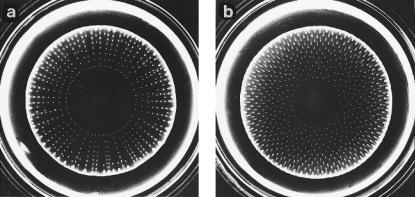
metabolizing the nutrient. This band is unstable, because when cells (by growth) reach a critical density, fluctuations in their number, and thus in aspartate concentration, generate gradients steep enough to cause cells to aggregate. This, in turn, increases the local concentration of aspartate. Therefore, starting at a single point and progressing in both directions, the circular band breaks up into a ring of discrete spots. These spots are left behind as cells continue to migrate outward in a compact band. What happens next depends on the concentration of the nutrient. At relatively low concentrations of nutrient, the cells in a spot begin to run out of fuel and stop excreting aspartate. Those that remain motile leave the spot and move outward, rejoining the band. This raises the concentrations of bacteria at the points where they rejoin, and new spots form there. Thus, one gets radial arrays of spots, as shown in Fig. 3.7a. The spots are frozen in place, because the cells, having run out of nutrient, soon stop swimming. At slightly higher
FIGURE 3.7. (a) Cells of a mutant of E. coli chemotactic to aspartate but not to serine that have spread outward in a soft-agar plate to form radial arrays of spots. (b) Cells of the same kind that have formed a hexagonal array of spots. The carbon source was a-ketoglutarate (2.5mM), which is not a chemoattractant. Plate (a) contained, in addition, 2.5mM hydrogen peroxide, and plate (b) 2.0mM hydrogen peroxide. The plates were inoculated at the center and incubated for 40 hours at 25°C. They were illuminated slantwise from below and photographed against a dark background. The bright ring near the periphery is an illumination artifact. Other conditions were as described in Budrene and Berg (1991). (Adapted from Berg, 1992, Fig. 1.)
28 3. Cell Populations
concentrations of nutrient, cells in the spots continue to excrete aspartate, remaining in place until cells in the advancing band increase in number and aggregate anyway. They do so at points midway in between the earlier spots, where the cell densities are higher (because cells were not removed there when the previous set of spots formed). Thus, one gets hexagonal arrays of spots, as shown in Fig. 3.7b. At even higher concentrations of substrate, the cells remain in spots, as before, but tend to move outward as a group. Cells that are not motile are left behind as a streak, so one gets hexagonal arrays of spots with radial tails. At even higher concentrations of substrate, larger aggregates form that seem to have a life of their own. They move slowly like slugs, with the larger slugs consuming the smaller ones. For a more complete description of such pattern formation, see Budrene and Berg (1995).
The spreading phenomena described thus far are exhibited by cells of normal size swimming through pores of soft agar, responding to chemical gradients that they generate by consumption or excretion. If cells are grown on agar with pore sizes slightly too small for the cells to penetrate (e.g., 0.5%) on a very rich medium, something very different happens. The cells get longer, produce many more flagella, and excrete a lubricant, called slime. They move rapidly outward across the surface of the agar, in parallel arrays in rafts or packs, through coordinated flagellar movement. They appear to “swarm,” like bees (see Harshey, 1994). Near the edge of the swarm, groups of cells rapidly move in swirls, this way and then that, often backing up. At the very edge, they tend to line up, pointing outward. Streams of such cells colonize the entire plate within a few hours. A circularly symmetric swarm is shown in Fig. 3.8a. One in the shape of a four-leaf clover is shown in Fig. 3.8b. The cells used in Fig. 3.8a form chemotactic rings in the presence of aspartate (as in Fig. 3.2, right), but those used in Fig. 3.8b do not. This cloverleaf pattern is reproducible, but the mechanism for its formation is not known. The signals that bring about this swarm transformation are poorly understood. However, cells need to be on a surface, to grow rapidly, to excrete slime, and to be able to swim. They do not need to be chemotactic. Swarming is better known in other flagellated species, especially in Proteus mirabilis, where long swarm cells revert to short vegetative cells, which later develop more swarm cells, generating colonies that are terraced (Rauprich et al., 1996). Unfortunately, the word “swarm” is used in two ways: for the general phenomenon of cells swimming
References 29
FIGURE 3.8. (a) Swarm of an E. coli strain deleted for genes that encode receptors for serine, ribose/galactose, and dipeptides but expressing the gene for the aspartate receptor, tar. (b) As in (a), but for a similar strain expressing a gene for an aspartate receptor unable to bind aspartate, tar(T154I). Cells were inoculated on a Petri plate containing 0.45% Eiken agar (from Japan) and a rich growth medium (peptone, meat extract) and incubated for 16 hours at 30°C. (Burkart et al., 1998, Figs. 4A, B, reprinted with permission.)
through soft agar (as in the formation of Adler’s chemotactic rings), and to denote the particular form of surface translocation just described.
References
Adler, J. 1966. Chemotaxis in bacteria. Science 153:708–716. Adler, J. 1969. Chemoreceptors in bacteria. Science 166:1588–1597.
Adler, J. 1973. A method for measuring chemotaxis and use of the method to determine optimum conditions for chemotaxis by
Escherichia coli. J. Gen. Microbiol. 74:77–91.
Adler, J. 1976. The sensing of chemicals by bacteria. Sci. Am. 234 (4): 40–47.
Adler, J. 1987. How motile bacteria are attracted and repelled by chemicals: an approach to neurobiology. Biol. Chem. Hoppe-Seyler 368: 163–173.
Adler, J., and B. Templeton. 1967. The effect of environmental conditions on the motility of Escherichia coli. J. Gen. Microbiol. 46:175–184.
Berg, H. C. 1992. Response of Escherichia coli to novel gradients. In: Sensory Transduction, Proc. 45th Symp. Soc. Gen. Physiol. Rockefeller University Press, New York, pp. 220–223.
Blakemore, R. 1975. Magnetotactic bacteria. Science 190:377–379.
30 3. Cell Populations
Budrene, E. O., and H. C. Berg. 1991. Complex patterns formed by motile cells of Escherichia coli. Nature 349:630–633.
Budrene, E. O., and H. C. Berg. 1995. Dynamics of formation of symmetrical patterns by chemotactic bacteria. Nature 376:49–53.
Burkart, M., A. Toguchi, and R. M. Harshey. 1998. The chemotaxis system, but not chemotaxis, is essential for swarming motility in
Escherichia coli. Proc. Natl. Acad. Sci. USA 95:2568–2573.
Frankel, R. B. 1984. Magnetic guidance of organisms. Annu. Rev. Biophys. Bioeng. 13:85–103.
Frankel, R. B., and R. P. Blakemore. 1991. Iron Biomaterials. Plenum, New York.
Harshey, R. M. 1994. Bees aren’t the only ones: swarming in Gramnegative bacteria. Mol. Microbiol. 13:389–394.
Jung, K-H., Spudich, E. M., Trivedi, V. D., and Spudich, J. L. 2001. An archael photosignal-transducing module mediates phototaxis in
Escherichia coli. J. Bacteriol. 183:6365–6371.
Maeda, K., Y. Imae, J.-I. Shioi, and F. Oosawa. 1976. Effect of temperature on motility and chemotaxis of Escherichia coli. J. Bacteriol. 127:1039–1046.
Nishiyama, S., I. N. Maruyama, M. Homma, and I. Kawagishi. 1999. Inversion of thermosensing property of the bacterial receptor Tar by mutations in the second transmembrane region. J. Mol. Biol. 286:1275–1284.
Rauprich, O., M. Matsushita, C. J. Weijer, F. Siegert, S. E. Esipov, and J. A. Shapiro. 1996. Periodic phenomena in Proteus mirabilis swarm colony development. J. Bacteriol. 178:6525–6538.
Spudich, J. L. 1998. Variations on a molecular switch: transport and sensory signalling by archaeal rhodopsins. Mol. Microbiol. 28:1051– 1058.
Spudich, J. L., C.-H. Yang, K.-H. Jung, and E. N. Spudich. 2000. Retinylidene proteins: structures and functions from archaea to humans. Annu. Rev. Cell Dev. Biol. 16:365–392.
Taylor, B. L., and D. E. Koshland, Jr. 1975. Intrinsic and extrinsic light responses of Salmonella typhimurium and Escherichia coli. J. Bacteriol. 123:557–569.
Taylor, B. L., J. B. Miller, H. M. Warrick, and D. E. Koshland, Jr. 1979. Electron acceptor taxis and blue light effect on bacterial chemotaxis.
J. Bacteriol. 140:567–573.