

8
Cellular Hardware
Before we look at the biochemical machinery that enables cells to count molecules, to compare counts made at different times, and to use these results to control the direction of flagellar rotation, we need to know more about cell architecture.
Body Plan
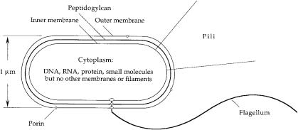
E. coli is a single-celled organism with a multilayered wall (Fig. 8.1). First, there is a thin outer membrane made of lipopolysaccharide, with the sugar chains pointing outward, penetrated by holes due to proteins, called porins. This membrane blocks the passage of most lipid-soluble molecules, but it allows the passage of water-soluble things up to about twice the size of sucrose. Next, there is a porous gauze-like layer of peptidoglycan that gives the cell its rigidity and cylindrical shape. This structure resists the turgor pressure generated when the cell finds itself in a medium of low osmotic strength. The peptidoglycan is immersed in an aqueous layer, called the periplasm, containing a variety of smaller molecules, including a number of proteins that either bind molecules that interest the cell or destroy molecules that pose a threat. Finally, there is a cytoplasmic (phospholipid) membrane similar to the membranes that enclose human cells, spanned by proteins involved in generation of energy, transport of materials, and sensory transduction. This is the main permeability barrier that enables the cell to retain chemicals—DNA, RNA, protein, and a variety of water-soluble molecules of lower molecular weight— essential for life. The multilayered cell wall is on the order of 0.03 mm thick. Unlike most human cells, which contain a number of membranous organelles, including a nucleus, and a variety of rope-like and tubular cytoskeletal structures, the cytoplasm of E. coli is a quasi-homogeneous soup.
69
70 8. Cellular Hardware
FIGURE 8.1. A schematic diagram of E. coli. One flagellum, two type 1 pili, and three porin molecules are shown. A typical cells has four flagella, either zero or a hundred or so pili, and thousands of porins. The flagellum comprises a long helical filament, a short proximal hook, and a basal body. The basal body is embedded in the cell wall. The spacing between the inner and outer membranes is shown larger than scale by a factor of about 4.
Phospholipids are molecules with polar (water-loving) head groups containing phosphate and long hydrocarbon (oily) tails. They form bilayer membranes, with the oily tails on the inside and the head groups on the outside. The oily layer blocks the passage of water-soluble molecules. In bacteria, there also are a variety of polymers in which phospholipids are combined with sugars to form lipopolysaccharide, and peptides are combined with sugars to form peptidoglycan (also called murein). In both cases the sugars appear in long chains. Some of the most effective antibiotics (e.g., penicillin) interfere with the synthesis of these components (e.g., peptidoglycan). Since human cells do not have such walls, we are not harmed by these antibiotics (unless we happen to be allergic). To learn more, see Seltmann and Holst (2002).
The most remarkable molecule in the cytoplasm of E. coli is DNA, a double-helical chain about 1.4mm long (nearly 1000 times longer than the cell) in the form of a closed loop. When cells grow rapidly, there is more than one copy per cell, because the molecule is being replicated at more than one place. The DNA has about 4.6 ¥ 106 base pairs. Since 3 base pairs specify one amino acid, it can code for 1.5 ¥ 106 amino acids (ignoring regions of DNA that specify RNA or are required to bind proteins that turn genes on and off). A typical polypeptide has a molecular weight
Why Cells? 71
of about 50,000, comprising 400 amino acids. So the DNA can code for about 4000 polypeptides. The coding region for each polypeptide is called a gene. The DNA of E. coli K-12 has been sequenced and found to code for 4288 polypeptides (Blattner et al., 1997). Surprisingly, the pathogenic strain O157:H7 has been found to code for 32% more (Perna et al., 2001). The functions of only about 60% of the original set of gene products are known. Fewer than 2% are involved in bacterial chemotaxis.
When a polypeptide is made, the relevant DNA sequence is copied as a messenger RNA (mRNA, often short-lived), which is read by a large RNA-protein particle called a ribosome. This links specific amino acids end to end. These are supplied by transfer RNAs (tRNA) that recognize successive 3-base codons in the mRNA. In an electron micrograph of a sectioned E. coli, the cytoplasm appears granular, because there are many ribosomes, each about 0.03 mm in diameter. Regions in which the DNA is more concentrated appear less granular, because ribosomes tend to be excluded. Dissolved in a finer matrix are mRNA, tRNA, a variety of proteins, and chemicals of low molecular weight.
Why Cells?
It is worth pausing to consider why all free-living things, including E. coli, are cells or are made up of cells. A cell is a relatively small isolated device that can import foodstuffs and export wastes, grow, and replicate. The isolation is essential both for chemistry and for genetics.
For reactions to occur, chemicals need to find one another. The rate of a reaction such as A + B Æ C is proportional to the product of the concentrations of A and B. If A and B are both diluted by a factor of 1000, the reaction rate goes down by a factor of one million. So one needs a concentrated medium in which to do biochemistry. Bacteria are the earliest cells that we know anything about, and they are relatively small. The time required for a small molecule to diffuse across a cell 1 mm in diameter is a matter of milliseconds (see Chapter 6). So early cells did not need any specialized machinery for moving goods from one place to another. Thermal agitation would do.
Specific chemical reactions are catalyzed by enzymes. In the earliest forms of life, these probably were made from ribonucleic acid (RNA). Now they are made from proteins, large molecules
72 8. Cellular Hardware
that also serve as structural elements (see below). Every time a cell divides, the DNA that encodes these proteins replicates. Occasionally, mistakes are made, and a different structure is specified. If the change is beneficial, so that the new cell is more likely to survive, then the mistake can propagate. But this can happen only if the molecule that carries the genetic information is packaged together with the product that it specifies. So evolution works because DNA is able to reap the rewards imposed by natural selection. In an earlier world, before cells were invented, the RNA that catalyzed essential reactions must have been self-replicating.
More on Proteins
By weight, E. coli is about 70% water, 1% inorganic ions, and the balance organic molecules, most of high molecular weight. Proteins are polymers made up of precise linear sequences of 20 different kinds of amino acids—amino acids have a molecular weight averaging about 120, an amino group (as in ammonia), distinctive side chains, and a carboxylic-acid group (as in vinegar) that can form peptide bonds linking one subunit to another, as we saw in Fig. 3.1. Proteins contain one or more long polypeptide chains. For example, hemoglobin, a protein well known to us but not to E. coli—hemoglobin carries oxygen in our blood—has four polypeptide chains. A space-filling model is shown in Fig. 8.2.
Polypeptide chains tend to wind up in a helix, called an a-helix, or line up side by side (parallel or antiparallel) in a sheet, called a b-pleated sheet. Thus, polypeptides also can be represented by ribbon diagrams, in which the shape of the ribbon indicates the local conformation of the chain, as shown in Fig. 8.3.
Growth
When E. coli grows, it gets longer and then divides in the middle. In a sense, it is immortal, because the mother is replaced by two daughters, essentially identical to the daughters of the previous generation. E. coli is haploid; it has only one chromosome. The fate of each cell is determined by a single DNA double helix. Except for mutations, which occur spontaneously for a given gene at the rate of about 10-6 per generation, all the molecules of DNA in a given set of descendents are identical. If fed well and held at

Growth 73
FIGURE 8.2. Hemoglobin, the protein that carries oxygen in our blood, and the sugar glucose, shown on the same scale. This is a space-filling illustration; bonds between individual atoms are not shown. Hemoglobin is a compact globular structure of diameter 5.5nm and molecular weight about 65,000 in which four polypeptide chains are packed together in a tetrahedral array. (From Goodsell, 1993, reprinted with permission.)
the temperature of the human gut (37°C), E. coli divides every 20 minutes. In a medium with only one carbon source (e.g., glucose), it takes longer, about 2 hours. The extra time is required for the cell to synthesize all of the other organic molecules that it needs. A generation time of 20 minutes is prodigious. If we start with one cell at noon today, there will be 23 = 8 cells at 1:00 o’clock, and 272 = 4.7 ¥ 1021 cells at noon tomorrow. Since each cell has a volume of about 10-18 m3, the volume of cells at noon tomorrow will be 4.7 ¥ 103 m3, i.e., a cube about 17 meters = 55 feet on a side! In practice, this does not happen, because the cells are not provided with enough food. However, it explains why, when, say, 100 cells are dispersed on the surface of hard nutrient agar, one soon obtains 100 mounds of cells (colonies) each a millimeter or so in diameter, or why, on soft agar, the progeny of a single cell soon populate the entire plate. It is this speed of replication that makes

74 8. Cellular Hardware
FIGURE 8.3. A ribbon diagram of CheY, a small protein that couples receptors to the flagella (see Chapter 9). CheY becomes active when phosphorylated. The phosphorylation site, aspartate-57, is shown at the top in space-filling format, with the side-chain oxygens in black.A central five-stranded parallel b-sheet is sandwiched between five a-helices. (Falke et al., 1997, Fig. 14A, reprinted with permission.)
bacterial genetics such a pleasure. Start your experiment today and get an answer tomorrow.
External Organelles
E. coli has at least three kinds of extracellular organelles. These include two kinds of fibers, called pili, that extend out from the cytoplasmic membrane.The first, very thin and straight, called type 1 pili (or fimbriae), enable the bacteria to adhere to a variety of surfaces, including cells of the intestinal epithelium. There can be hundreds per cell. A second, called the sex pilus, can bind to other cells and retract, drawing the cells together. This enables cells
References |
75 |
carrying F plasmids (small autonomously replicating circular pieces of DNA) to transfer part of their DNA to other cells. Cells with F plasmids make one such pilus. Some cells used for studies of bacterial behavior have type 1 pili and others do not; few have sex pili.
The extracellular organelle of greatest interest here is the bacterial flagellum. This organelle has three parts, a basal body (a reversible rotary motor) embedded in the cell wall (beginning within the cytoplasm and ending at the outer membrane), a short proximal hook (a flexible coupling or universal joint), and a long helical filament (a propeller). The filament is about 0.02 mm in diameter. Normally it is shaped as a left-handed helix with a wavelength (pitch) of about 2.3 mm and a diameter of 0.4 mm. If a cell is not subjected to viscous shear—filaments are easily broken—its filaments can be up to 10 mm long. The number of flagella vary from cell to cell. The average number is about four.
To learn more about basic molecular and cellular biology, see Alberts et al. (2003). For an earlier more succinct view, see Kendrew (1966).
References
Alberts, B., D. Bray, K. Hopkin, A. Johnson, J. Lewis, M. Raff, K. Roberts, and P. Walter. 2003. Essential Cell Biology. An Introduction to the Molecular Biology of the Cell, Second Edition. Garland, New York.
Blattner, F. R., G. Plunkett, C. A. Bloch, et al. 1997. The complete genome sequence of Escherichia coli K-12. Science 277:1453–1462.
Falke, J. J., R. B. Bass, S. L. Butler, S. A. Chervitz, and M. A. Danielson. 1997. The two-component signaling pathway of bacterial chemotaxis: a molecular view of signal transduction by receptors,kinases,and adaptation enzymes. Annu. Rev. Cell Dev. Biol. 13:457–512.
Goodsell, D. S. 1993. The Machinery of Life. Springer-Verlag, New York. Kendrew, J. C. 1966. The Thread of Life. G. Bell & Sons, London. Perna, N. T., G. Plunkett III, V. Burland, et al. 2001. Genome sequence of
enterohaemorrhagic Escherichia coli O157:H7. Nature 409:529–533. Seltmann, G., and O. Holst. 2002. The Bacterial Cell Wall. Springer,
Berlin.
This page intentionally left blank