

11
Gain Paradox
Receptor Sensitivity
Data obtained early on suggested that the chemotactic response is proportional to the change in receptor occupancy, with that occupancy characterized by a fixed dissociation constant, Kd, the concentration of ligand at which the probability of receptor occupancy is 1/2 (Berg and Tedesco, 1975; Mesibov et al., 1973). Then it became evident that the dissociation constant increases (i.e., cells become less sensitive) at higher concentrations of ligand, as receptors are methylated (Borkovich et al., 1992; Bornhorst and Falke, 2000; Dunten and Koshland 1991; Li and Weis, 2000). However, even at these higher concentrations (e.g., for the nonmetabolizable aspartate analog a-methylaspartate at an ambient concentration of 0.16mM) the gain is prodigious: a step increase in concentration from 0.16 to 0.16 + 0.0027mM (a change of about 1.7%) transiently increases the probability that the motor spins counterclockwise (CCW) by 0.23 (Segall et al., 1986). Computer simulations of the chemotaxis system (e.g., Bray et al., 1993; reviewed by Bray, 2002) fail to predict the necessary gain. Two recent findings appear to resolve the paradox. First, there is an amplification step at the beginning of the signaling pathway: the fractional change in kinase activity is some 35 times larger than the fractional change in receptor occupancy (Sourjik and Berg, 2002a). Second, there is another amplification step at the end of the signaling pathway: the motor is ultrasensitive (Cluzel et al., 2000); see below.
Evidence for the first amplification step was obtained by using fluorescence resonance energy transfer (FRET) to monitor the interaction between the response regulator, CheY-P, and its phosphatase, CheZ. At steady state, the concentration of CheY-P is constant: CheY is phosphorylated at the same rate that it is dephosphorylated. The dephosphorylation rate is proportional to
97
98 11. Gain Paradox
the concentration of the CheY-P/CheZ complex, so from that concentration one can deduce the relative activity of the kinase. The receptor occupancy can be estimated from values for the Kd measured in vitro. One makes a fusion protein between cyan fluorescent protein (CFP) and CheZ, and another fusion protein between yellow fluorescent protein (YFP) and CheY, excites the CFP, and measures the fluorescence emission from both CFP and YFP. If the fluorophores of CFP and YFP are closer to one another than about 10nm, which is the case for the CheY-P/CheZ complex, energy is transferred from CFP to YFP. As a result, the CFP emis-
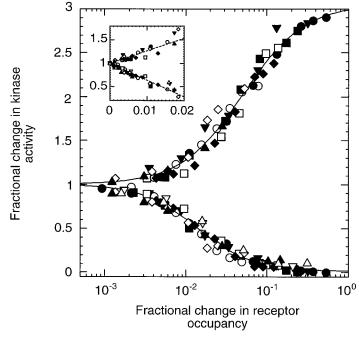
FIGURE 11.1. Fractional change in the activity of the kinase, CheA, upon addition and removal of a nonmetabolizable aspartate analog, a- methylaspartate. The initial activity is 1, and it falls to zero upon addition of enough attractant to saturate the response (lower curve). Given time (several minutes) the cells adapt, and the activity returns to 1. Then, when the attractant is removed, the activity increases (upper curve). These experiments were done with different ambient concentrations of a-methylaspartate, ranging from 0 (closed circles) to 10mM (open triangles). The insert shows the same data plotted on a linear scale. (From Sourjik and Berg, 2002a, Fig. 3C.)
Receptor Clustering |
99 |
sion goes down and the YFP emission goes up. Results from this kind of analysis are shown is Fig. 11.1. Data obtained over a wide range of ambient concentrations, indicated by the different symbols, collapse into a single set of curves. The inset shows these data plotted with a linear abscissa. The slopes of these plots are not ± 1, as expected, but about ± 35. The change in receptor occupancy that occurs during chemotaxis is relatively small. Cells swimming up spatial gradients of aspartate operate near the left end of the lower curve: substantial extensions of run length occur for fractional changes in receptor occupancy as small as 0.002. How is this amplification achieved?
Receptor Clustering
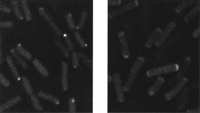
The answer appears to be via receptor–receptor interactions. Receptors tend to cluster, usually at one pole, as shown in the electron microscope by immunogold labeling (Maddock and Shapiro, 1993). Clustering also is evident from the distribution of fluorescent fusion proteins (Fig. 11.2). In addition to receptors, the clusters include CheA, CheB, CheR, CheW, CheY, and CheZ. CheA and CheW bind to the receptor signaling domain, CheB and CheY to CheA, CheR to the receptor C-terminal peptide, and CheZ to the short form of CheA, CheAS. If either CheA or CheW are missing, receptors still appear at the poles, but as diffuse caps, and the other components normally associated with clusters (except CheR) spread throughout the cytoplasm.
The presence of receptor clusters at one pole led to the suggestion that E. coli has a nose. However, when a cell tumbles and chooses a new run direction, either end goes first (Berg and Turner, 1995). If there is some reason for clustering, it does not have to do with how ligands in the external medium interact with the cell body, since the best that one can do is to disperse the receptors over the cell surface, and thus increase the size of the detector (Berg and Purcell, 1977). An alternative is to put receptors in clusters so that they can activate one another, and hence improve sensitivity, as argued by Duke and Bray (1999). Molecular models have been constructed to show what these clusters might look like (Shimizu et al., 2000). There is now direct genetic evidence that defects in a receptor of one type, for example, Tsr (in a region of receptor–receptor contact, identified by x-ray crystallography) can be cured by interaction with a receptor of
100 11. Gain Paradox
FIGURE 11.2. Images of cells expressing a fluorescent fusion protein, YFP-CheR. CheR, the methyltransferase, binds to receptors at their C- terminal pentapeptide, as shown in Fig. 9.2. It does this whether or not the receptors are clustered. The cells at the left are wild-type and show receptor clusters at their poles as diffraction-limited spots. The cells at the right are missing the CheA kinase, and their receptors appear, instead, as diffuse polar caps. That the clusters and caps contain receptors has been verified by labeling with anti-Tsr rabbit antibody (images not shown). Photographs courtesy of Victor Sourjik.
another type, for example, Tar (Ames et al., 2002). In addition, response to a give attractant (e.g., serine) can be enhanced by de novo receptor clustering, forced by the addition of a chemical bearing multiple copies of a different ligand (e.g., galactose) that is sensed by a different receptor (Gestwicki and Kiessling, 2002). Other evidence for cooperativity between receptors is reviewed by Falke (2002). But precisely how receptors activate one another remains to be determined.
Motor Response
The other amplification step comes from the highly cooperative response of the motor to changes in the concentration of CheY-P. The concentration of CheY-P (actually, of a fusion between CheY-P and green fluorescent protein) was measured in single cells by fluorescence correlation spectroscopy. Every cell behaved
Precise Adaptation |
101 |
identically: a shift in concentration of CheY-P from 2.7 to 3.5 mM was enough to change the probability of clockwise (CW) rotation from 0.2 to 0.8 (Cluzel et al., 2000). These data are shown in Fig. 11.3, along with data for the binding of CheY-P to FliM obtained by FRET (Sourjik and Berg, 2002b). The binding curves are not as good as they might be, because a substantial fraction of CFPFliM was free in the cytoplasm. But both sets of data can be fit by the two-state allosteric model of Monod et al. (1965). A more general allosteric model for motor switching has been developed by Duke et al. (2001) in which FliM molecules in a ring of 34 bind CheY-P and interact with their neighbors. Each protein can adopt a CW or a CCW configuration, and the direction of rotation depends on how many proteins are in either state. Given a largeenough interaction energy between adjacent molecules, the ensemble switches from a state in which nearly all are in the CW configuration to one in which nearly all are in the CCW configuration. But once again, the details of the mechanism remain to be determined.
Precise Adaptation
For the system to operate on such a steep response curve (Fig. 11.3), the adaptation mechanism must be precise. Is this accomplished by an as yet unknown feedback mechanism, or is adaptation intrinsically exact (Barkai and Leibler, 1997)? Under the conditions of the tracking experiments (see Chapter 4), it was found that adaptation to aspartate was exact, while that to serine was not: the mean run length in 1mM serine was about three times longer than the mean run length in the absence of serine. However, cells drifted up gradients of either attractant perfectly well. So adaptation need not be exact, but it has to be sufficiently precise to keep cells somewhere near the middle of the motor response curve. In the model of Barkai and Leibler, receptors are in either of two states: active or inactive. In one embodiment, perfect adaptation is achieved by allowing only methylated receptors to be active, specifying that CheR works at saturation, and letting CheB-P act only on receptors that are active. In this scheme, adaptation is robust, in the sense that return to the initial steady state occurs even when the concentrations of system components vary widely. This proposition has been confirmed experimentally (Alon et al., 1999).

102 11. Gain Paradox
FIGURE 11.3. Comparison of the dependence of motor bias ( ) and FliM occupancy (
 ) on concentration of free cytoplasmic CheY-P. Data for the motor bias are from Cluzel et al. (2000) and for the FliM binding from Sourjik and Berg (2002b). Dashed lines are fits to an allosteric model, showing a highly cooperative state function and a nearly linear binding function; see the text. (Adapted from Sourjik and Berg, 2002b, Fig. 2b.)
A Modelers’ Era
We are entering a new phase in the study of chemotaxis in which enough is known about the detailed properties of the signaling network that its behavior can be treated analytically and simulated numerically. It is becoming a subject more in tune with the physical sciences, with constructive interplay between theory and experiment.
References
Alon, U., M. G. Surette, N. Barkai, and S. Leibler. 1999. Robustness in bacterial chemotaxis. Nature 397:168–171.
Ames, P., C. A. Studdert, R. H. Reiser, and J. S. Parkinson. 2002. Collaborative signaling by mixed chemoreceptor teams in Escherichia coli.
Proc. Natl. Acad. Sci. USA 99:7060–7065.
References 103
Barkai, N., and S. Leibler. 1997. Robustness in simple biochemical networks. Nature 387:913–917.
Berg, H. C., and E. M. Purcell. 1977. Physics of chemoreception. Biophys. J. 20:193–219.
Berg, H. C., and P. M. Tedesco. 1975. Transient response to chemotactic stimuli in Escherichia coli. Proc. Natl. Acad. Sci. USA 72:3235–3239.
Berg, H. C., and L. Turner. 1995. Cells of Escherichia coli swim either end forward. Proc. Natl. Acad. Sci. USA 92:477–479.
Borkovich, K. A., L. A. Alex, and M. I. Simon. 1992. Attenuation of sensory receptor signaling by covalent modification. Proc. Natl. Acad. Sci. USA 89:6756–6760.
Bornhorst, J. A., and J. J. Falke. 2000. Attractant regulation of the aspartate receptor-kinase complex: limited cooperative interactions between receptors and effects of the receptor modification state. Biochemistry 39:9486–9493.
Bray, D. 2002. Bacterial chemotaxis and the question of gain. Proc. Natl. Acad. Sci. USA 99:7–9.
Bray, D., R. B. Bourret, and M. I. Simon. 1993. Computer simulation of the phosphorylation cascade controlling bacterial chemotaxis. Mol. Biol. Cell 4:469–482.
Cluzel, P., M. Surette, and S. Leibler. 2000. An ultrasensitive bacterial motor revealed by monitoring signaling proteins in single cells. Science 287:1652–1655.
Duke, T. A. J., and D. Bray. 1999. Heightened sensitivity of a lattice of membrane receptors. Proc. Natl. Acad. Sci. USA 96:10104–10108.
Duke, T. A. J., N. Le Novère, and D. Bray. 2001. Conformational spread in a ring of proteins: a stochastic approach to allostery. J. Mol. Biol. 308:541–553.
Dunten, P., and D. E. Koshland, Jr. 1991. Tuning the responsiveness of a sensory receptor via covalent modification. J. Biol. Chem. 266:
1491–1496.
Falke, J. L. 2002. Cooperactivity between bacterial chemotaxis receptors.
Proc. Natl. Acad. Sci. USA 99:6530–6532.
Gestwicki, J. E., and L. L. Kiessling. 2002. Inter-receptor communication through arrays of bacterial chemoreceptors. Nature 415:81–84.
Li, G., and R. M. Weis. 2000. Covalent modification regulates ligand binding to receptor complexes in the chemosensory system of
Escherichia coli. Cell 100:357–365.
Maddock, J. R., and L. Shapiro. 1993. Polar location of the chemoreceptor complex in the Escherichia coli cell. Science 259:1717–1723.
Mesibov, R., G. W. Ordal, and J. Adler. 1973. The range of attractant concentrations for bacterial chemotaxis and the threshold and size of response over this range. J. Gen. Physiol. 62:203–223.
Monod, J., J.Wyman, and J.-P. Changeux. 1965. On the nature of allosteric transitions: a plausible model. J. Mol. Biol. 12:88–118.
104 11. Gain Paradox
Segall, J. E., S. M. Block, and H. C. Berg. 1986. Temporal comparisons in bacterial chemotaxis. Proc. Natl. Acad. Sci. USA 83:8987–8991.
Shimizu, T. S., N. Le Novère, M. D. Levin, A. J. Beavil, B. J. Sutton, and D. Bray. 2000. Molecular model of a lattice of signalling proteins involved in bacterial chemotaxis. Nature Cell Biol. 2:1–5.
Sourjik, V., and H. C. Berg. 2000. Localization of components of the chemotaxis machinery of Escherichia coli using fluorescent protein fusions. Mol. Microbiol. 37:740–751.
Sourjik, V., and H. C. Berg. 2002a. Receptor sensitivity in bacterial chemotaxis. Proc. Natl. Acad. Sci. USA 99:123–127.
Sourjik, V., and H. C. Berg. 2002b. Binding of the Escherichia coli response regulator CheY to its target measured in vivo by fluorescence resonance energy transfer. Proc. Natl. Acad. Sci. USA 99:12669–12674.