

2
Larger Organisms
Seventeenth Century
Antony van Leeuwenhoek was the first person to see bacteria, and it was their motion that captured his attention. However, he was not the first person to use a microscope or to describe cells. But the single-lens instruments that he made himself had fewer aberrations than the compound instruments of his day, so he was able to see more. And his curiosity was insatiable. His work on little animals—he called them animalcules—is available to the modern reader through translations from the archaic Dutch by the British microbiologist Clifford Dobell (1932), who published them on the 300th anniversary of van Leeuwenhoek’s birth.Van Leeuwenhoek described what he saw in letters written in ink, still jet black, sealed with red wax, and sent from Delft to London, to Henry Oldenberg, the secretary of the Royal Society. Oldenberg translated bits and pieces and published them in the Transactions of the Royal Society. My favorite is the 18th letter in which van Leeuwenhoek describes animalcules in water from his well. He was curious about the effect that pepper might have, so he ground up some in a blue porcelain pot and mixed it in.A number of larger animalcules died out, until on 6 August 1676 he saw large numbers of very small ones:
I now saw very plainly that these were little eels, or worms, lying all huddled up together and wriggling; just as if you saw, with the naked eye, a whole tubful of very little eels and water, with the eels a-squirming among one another: and the whole water seemed to be alive with these multifarious animalcules. This was for me, among all the marvels that I have discovered in nature, the most marvellous of all; and I must say, for my part, that no more pleasant sight has ever yet come before my eye than these many thousands of living creatures, seen all alive in a little drop of water, moving among one another, each several creature having its own proper motion.
7
8 2. Larger Organisms
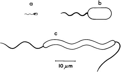
Dobell believed these to be cells of a large spiral organism, Spirillum volutans, shown at the bottom of Fig. 2.1. Van Leeuwenhoek’s first drawings of bacteria came later, in 1683, when he examined the scruff on his teeth. He never saw bacterial flagella, but he marveled at their evident small size. In his 26th letter of 1678 he wrote,
but I see, besides these, other living animalcules which are yet more than a hundred times less, and on which I can make out no paws, though from their structure and the motion of their body I am persuaded that they too are furnished with paws withal: and if their paws be proportioned to their body, like those of the bigger creatures, upon which I can see the paws, then, taking their measure at but a hundred times less, it follows that a million of their paws together make up but the thickness of a hair of my beard; while these paws, besides their organs for motion, must also be furnished with vessels whereby nourishment must pass through them.
FIGURE 2.1. Scale drawings of some flagellated bacteria whose behavior has been studied. (a) E. coli. About four filaments arise at random from the sides of the cell and form a bundle that appears near one pole. The bundle pushes the cell. When one or more filaments transiently changes its direction of rotation, the cell alters course. (b) Chromatium okenii. About 40 filaments arise at one pole. The bundle either pushes or pulls the cell. When the filaments change their direction of rotation, the cell backs up. (c) Spirillum volutans, shown swimming from left to right. The body is helical. About 25 filaments arise at each pole. Those on the left are in the tail configuration; those on the right in the head configuration. When the filaments in either bundle change their directions of rotation and flip over (tail to head, head to tail), the cell swims in the opposite direction, as if reflected in a mirror.
Nineteenth Century |
9 |
Like other early naturalists, van Leeuwenhoek believed that microorganisms were macroorganisms writ small!
Very few of van Leeuwenhoek’s microscopes survive, but they have been studied by modern methods and found to pass muster. There is no doubt that he was able to see what he claimed to see (Ford, 1985, 1991). Robert Brown (1828) used a single-lens instrument in his studies of brownian motion, of which we will hear more later.
Van Leeuwenhoek was born in the same year and christened in the same church as Johannes Vermeer. Vermeer died in his early 40s and van Leeuwenhoek was the executor of his estate. But van Leeuwenhoek lived on to his early 90s. He was buried in the Oude Kerk at Delft, where his daughter Maria erected a monument in his memory.
Nineteenth Century
Bacterial flagella were first seen in 1836, when the German naturalist Christian Ehrenberg (1838) found an enormous bacterium in the brook below the church of Ziegenhayn, near Jena. This organism is shown at the upper right in Fig. 2.1. He called it Monas okenii (now Chromatium okenii), in honor of Oken, the founder of the society then meeting in Jena. C. okenii is a photosynthetic red sulfur bacterium. It converts hydrogen sulfide (H2S) to elementary sulfur (S), which appears as granules in the cell cytoplasm. Ehrenberg took these granules to be stomach cells. He also identified the ovary!
In 1883, Theodor Engelmann, a German physiologist working in Utrecht, found in the waters of a branch of the Rhine a similar organism, which he called Bacterium photometricum. He was amazed by its behavior toward light. If, while looking through the microscope at a population of swimming cells, he passed his hand between the light source and the specimen stage, every cell backed up. This gave Engelmann the impression of fright, so he called it a shock reaction. He then showed that cells accumulate in a spot of light, not because they like the light, but because they are afraid of the dark. They swim into the spot perfectly well, but are not able to get out. When crossing a dark-light boundary from dark to light, they tend to speed up; when crossing in the opposite direction, they back up. Engelmann observed a similar response to carbon dioxide: when cells in a hanging drop were suddenly exposed to this gas, they also backed up.
10 2. Larger Organisms
Engelmann (1881a,b) studied the responses of a variety of other bacteria to oxygen. Some species swam toward higher concentrations of oxygen and others toward lower concentrations. Some were more discriminating, liking some oxygen but not too much. Species of the first kind, when placed in an aqueous suspension under a square coverglass, accumulated at the edge of the coverglass. Species of the second kind fled to the middle. Species of the third kind formed a square array a certain distance away from the edge of the coverglass. When Engelmann blew hydrogen gas at this preparation, the square got larger; when he used oxygen, the square got smaller. Then he had a bright idea: use cells that like oxygen as analytical chemists to indicate where oxygen is generated in a green plant during photosynthesis. The answer proved to be the chloroplast. Engelmann reviewed these experiments in 1894. Figure 2.2 shows one of his drawings of an agal cell called Spirogyra that has a spiral chloroplast. When the cell was illuminated with a spot of light, as shown on the left, the bacteria accu-
FIGURE 2.2. Part of a drawing by Engelmann of oxygen-loving bacteria (possibly Bacillus subtilis) responding to the illumination of Spirogyra, an algal cell with a spiral chloroplast. The original figure is in color.
The Golden Age of Microbiology |
11 |
mulated near its surface, but only if the spot impinged on the chloroplast. If the cell was illuminated uniformly, the bacteria accumulated in a spiral array.
Engelmann had an interesting career. He spent 30 years as a physiologist in Utrecht. He also was a cellist, and his second wife, Emma, a pianist. They were friends of Brahms: to wit, the Engelmann Quartet (Kamen, 1986). He ended his days in Berlin. He was best known for work on excitation of muscle in the heart.
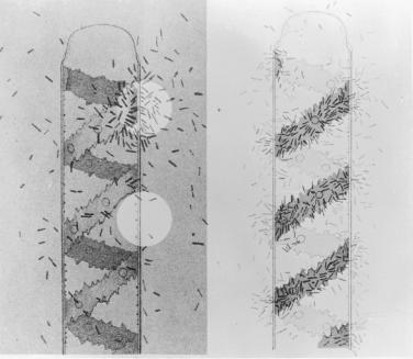
Some beautiful sketches of Chromatium okenii were published by Manabu Miyoshi (1898).While soaking in the Yumoto Spa near Nikko, he became interested in tufts of sulfur in water from the volcanic springs. In ditches and pools nearby he found reddish blooms that proved to be nearly pure cultures of C. okenii. His drawings of their responses to gradients of chemical attractants or repellents are shown in Fig. 2.3. Miyoshi’s reddish blooms are still there. I visited the Yumoto Spa in the spring of 1999 with Chi Aizawa, a colleague then at Teikyo University. We took samples back to his laboratory in Utsunomiya, put them under the microscope, and passed our hands between the light source and the specimen stage. The shock reaction is alive and well.
The capillary assay used by Miyoshi had been developed in the 1880s by Wilhelm Pfeffer (1884), a botanist working in Tübingen. He used it to study the responses of a number of different species of bacteria to a variety of chemicals. Initially, he thought that bacteria could steer toward the mouth of a capillary tube containing a chemical attractant. So he coined the term “chemotaxis.” As we shall see later, bacteria are not able to steer. E. coli uses a strategy called “klinokinesis with adaptation.” But molecular biologists are not fond of nomenclature, so the term chemotaxis has stuck. Pfeffer’s capillary assay was perfected by Julius Adler, a biochemist at the University of Wisconsin in Madison, who began the modern work on chemotaxis in E. coli. We will hear about this shortly. Pfeffer ended his days in Leipzig. He was best known for his work on osmotic pressure (measured with porous porcelain pots impregnated with copper ferrocyanide).
The Golden Age of Microbiology
One does not read about work on the motile behavior of bacteria in books on the history of microbiology.Their emphasis, understandably, is on bacteria that cause disease. Robert Koch isolated
12 2. Larger Organisms
FIGURE 2.3. Drawings by Miyoshi of the responses of C. okenii to a chemical attractant (ammonium nitrate, 0.3% w/v) or a chemical repellent (malic acid, 0.5% w/v) diffusing from the tip of a capillary tube. In the response to the attractant, the bacteria accumulate near the mouth of the capillary tube and then swim inside. The original figures are in color.
the anthrax bacillus in 1876, the tubercle bacillus in 1882, and the cholera vibrio in 1883. He published his postulates specifying the criteria for proof of the cause of infectious disease in 1884. Beginning in 1880, Louis Pasteur demonstrated immunization by atten-

The Golden Age of Microbiology |
13 |
uated bacteria (or virus) for cholera in birds, anthrax in sheep, erysipelas in pigs, and rabies in dogs. The use of agar as a bacteriological medium appeared in 1882, the Gram stain in 1884, the Petri plate in 1887, and the Institut Pasteur in 1888. This was the golden age of medical microbiology. So why bother about bacterial motility? One exception was Koch’s (1877) photographs of stained flagella that appeared as part of a discourse on technical methods for bacterial examination.
Some pathogenic organisms are motile and others are not, as shown in Table 2.1. The importance of motility and chemotaxis
TABLE 2.1. Motility of the main bacterial pathogens discovered between
1877 and 1906.
Pathogen, disease |
Bacterial genus or species |
Cells motile |
|
Typhoid fever |
Salmonella typhi |
Cholera |
Vibrio cholerae |
Tetanus |
Clostridium tetani |
Diarrhea |
Escherichia coli |
Food poisoning |
Salmonella enteritidis |
Botulism |
Clostridium botulinum |
Paratyphoid |
Salmonella paratyphi |
Syphilis |
Treponema pallidum |
Cells nonmotile |
|
Anthrax |
Bacillus anthracis |
Suppuration |
Staphylococcus |
Gonorrhea |
Neisseria gonorrhoeae |
Suppuration |
Streptococcus |
Tuberculosis |
Mycobacterium tuberculosis |
Diphtheria |
Corynebacterium diphtheriae |
Pneumonia |
Streptococcus pneumoniae |
Meningitis |
Neisseria meningitidis |
Gas gangrene |
Clostridium perfringens |
Plague |
Yersinia pestis |
Dysentery |
Shigella dysenteriae |
Whooping cough |
Bordetella pertusssis |
|
|
Note: The organisms in each category, motile or nonmotile, are listed in the order of discovery. The motile pathogens are all peritrichously flagellated rods (peri, around; trichos, hair), except V. cholerae, which has a single, polar, sheathed flagellum, and T. pallidum, which is a spirochete. Some species closely related to the nonmotile pathogens are motile, e.g., Bacillus cereus, the Clostridium species of the motile group, Yersinia pseudotuberculosis and Bordetella bronchiseptica. Motile streptococci are rare, but they do exist (members of the group D, or enterococcus, group).

14 2. Larger Organisms
for pathogenicity is still a matter of active study (Ottemann and Miller, 1997), although the common belief is that for a given species, motile cells are more virulent than nonmotile ones.As will be discussed later, chemotaxis offers a cell an enormous advantage for long-range migration, which might be expected to play an important role in invasiveness.
FIGURE 2.4. A sketch of the forward and backward motion of Chromatium okenii, according to Buder (1915). During forward motion (left), the flagellar bundle turns rapidly counterclockwise (as seen by an observer behind the cell) and the cell body rolls more slowly clockwise.
During backward motion (right) these motions are reversed. Buder analyzed the forces acting on the bundle at an arbitrary point A, assuming (left) that it pressed on the fluid with a force represented by the vector AC and experienced a resistance AB, having components parallel and normal to the helical axis. Scale: the cell body is about 6 mm in diameter.
Early Twentieth Century |
15 |
Early Twentieth Century
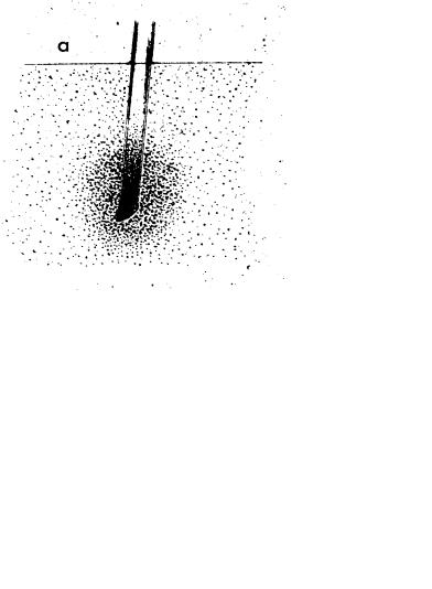
The development of dark-field condensers of high numerical aperture enabled Karl Reichert (1909) to determine optimum conditions for visualizing bacterial flagella. An intense cone of light is focussed on the specimen stage in such a way that none directly enters the objective of the microscope. One sees only light scattered from objects in the field of view. Thus, one can visualize such objects even though their dimensions are below the resolving power of the microscope (its ability to distinguish two objects that are next to one another). A single flagellar filament, for example, blooms up to that resolving power (about 0.2 mm) and looks about 10 times thicker than it actually is. The main problem with the method is that the cell body scatters so much light that it is impossible to distinguish faint objects nearby. However, much of the early work was done with very large organisms, such as C. okenii and S. volutans, with large flagellar bundles that were easy to see. Figure 2.4 shows an example from the studies of Johannes Buder (1915) on the forward and backward motion of
C. okenii.
The most extensive work of this kind was done by Peter Metzner (1920) on the behavioral responses of S. volutans. The most striking thing about this organism is that reversals of its flagellar bundles are synchronized (unless cells are damaged), even though these bundles are relatively far apart (about 50 mm). So this organism is capable of rapid long-range intracellular communication, probably electrical. E. coli does not have this talent.
Late Twentieth Century
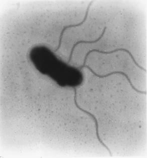
The modern era of work on bacterial behavior began in the 1960s when Julius Adler demonstrated that E. coli has a sense of taste, that is, that bacterial chemotaxis is a matter of aesthetics rather than material gain, and when Tetsuo Iino, Sho Asakura, and Goro Eguchi published a series of papers on the assembly of flagellar filaments. The latter work was done with a close relative of E. coli, now named Salmonella enterica serovar Typhimurium, that I will simply call Salmonella. The first picture of E. coli published by Adler (an electron micrograph) is shown in Fig. 2.5. For a closer look at the classic literature on bacterial chemotaxis, see Berg (1975).
16 2. Larger Organisms
FIGURE 2.5. Electron micrograph of E. coli negatively stained (by exposure of a dried sample to the salt of an element of high atomic number, usually tungsten or uranium). Scale: the cell body is about 1 mm in diameter (2 wavelengths of green light). The flagella are extraordinarily thin. (Adler, 1965, Fig. 1, reprinted with permission).
References
Adler, J. 1965. Chemotaxis in Escherichia coli. Cold Spring Harbor Symp. Quant. Biol. 30:289–292.
Berg, H. C. 1975. Chemotaxis in bacteria. Annu. Rev. Biophys. Bioeng. 4:119–136.
Brown, R. 1828. A Brief Account of Microscopical Observations on the Particles Contained in the Pollen of Plants; and on the General Existence of Active Molecules in Organic and Inorganic Bodies. Richard Taylor, London.
Buder, J. 1915. Zur Kenntnis des Thiospirillum jenense und seiner Reaktionen auf Lichtreize. Jahrb. Wiss. Bot. 56:529–584.
Dobell,C. 1932.Antony van Leeuwenhoek and His “Little Animals.”John Bale, Sons & Danielsson, London. Reprinted by Dover, New York, 1960.
Ehrenberg, C. G. 1838. Die Infusionsthierchen als vollkommene Organismen. Leopold Voss, Leipzig, Germany.
References 17
Engelmann, T. W. 1881a. Neue Methode zur Untersuchung der Sauerstoffausscheidung pflanzlicher und thierischer Organismen. Pflügers Arch. Gesamte Physiol. Menschen Thiere 25:285–292.
Engelmann, T. W. 1881b. Zur Biologie der Schizomyceten. Pflügers Arch. Gesamte Physiol. Menschen Thiere 26:537–545.
Engelmann, T. W. 1883. Bacterium photometricum. Ein Beitrag zur vergleichenden Physiologie des Lichtund Farbensinnes. Pflügers Arch. Gesamte Physiol. Menschen Thiere 30:95–124.
Engelmann, T. W. 1894. Die Erscheinungsweise der Sauerstoffausscheidung chromophyllhaltiger Zellen im Licht bei Anwendung der Bacterienmethode. Pflügers Arch. Gesamte Physiol. Menschen Thiere
57:375–386.
Ford, B. J. 1985. Single Lens. The Story of the Simple Microscope. Harper & Row, New York.
Ford, B. J. 1991. The Leeuwenhoek Legacy. Biopress, Bristol, and Farrand Press, London.
Kamen, M. D. 1986. On creativity of eye and ear: a commentary on the career of T. W. Engelmann. Proc. Am. Phil. Soc. 130:232–246.
Koch, R. 1877. Untersuchungen über Bacterien. VI. Verfahren zur Untersuchung, zum Conserviren und Photographiren der Bacterien.
Beitr. Biol. Pflanz. 2:399–434.
Metzner, P. 1920. Die Bewegung und Reizbeantwortung der bipolar begeißelten Spirillen. Jahrb. Wiss. Bot. 59:325–412.
Miyoshi, M. 1898. Studien über die Schwefelrasenbildung und die Schwefelbacterien der Thermen von Yumoto bei Nikko. J. Coll. Sci. Imp. Univ. Jap. 10:143–173.
Ottemann, K. M., and J. F. Miller. 1997. Roles for motility in bacterialhost interactions. Mol. Microbiol. 24:1109–1117.
Pfeffer, W. 1884. Locomotorische Richtungsbewegungen durch chemische Reize. Unters. Bot. Inst. Tübingen 1:363–482.
Reichert, K. 1909. Über die Sichtbarmachung der Geisseln und die Geisselbewegung der Bakterien. Zentralbl. Bakteriol. Parasitenk. Infektionskr. Abt. 1 Orig. 51:14–94.
This page intentionally left blank