

Полезные материалы за все 6 курсов / Учебники, методички, pdf / INBDEBooster Head and Neck Anatomy Notes
.pdfHead and Neck Anatomy |
81 |
Cricothyroid
•Origin: cricoid cartilage
•Insertion: inferior horn and lamina of the thyroid cartilage
•Action: tenses and elongates vocal folds (raises pitch)
•Innervation: superior laryngeal nerve via CN X
Figure 16.07 A lateral view of the larynx displaying the cricothyroid
Thyroarytenoid
•Origin: thyroid cartilage and cricothyroid ligament
•Insertion: arytenoid cartilage
•Action: relaxes vocal ligament (lowers pitch)
•Innervation: recurrent laryngeal nerve via CN X
Figure 16.08 A lateral view of the larynx displaying the thyroarytenoid and lateral cricoarytenoid
Vocalis
•Origin: thyroid cartilage and cricothyroid ligament
•Insertion: arytenoid cartilage
•Action: relaxes vocal ligament (lowers pitch)
•Innervation: recurrent laryngeal nerve via CN X
Figure 16.09 A superior view of the thyroarytenoid and vocals muscles
INBDE Booster | Booster PrepTM

Head and Neck Anatomy |
82 |
|||
|
|
|
|
|
Intrinsic |
Function |
Innervation |
|
|
laryngeal |
|
|
|
|
muscle |
|
|
|
|
|
|
|
|
|
Posterior crico- |
Abductor |
CN X |
|
|
arytenoid |
|
|
|
|
|
|
|
|
|
Oblique and |
Adductor, sphincter |
CN X |
|
|
transverse |
|
|
|
|
arytenoids |
|
|
|
|
|
|
|
|
|
Lateral crico- |
Adductor |
CN X |
|
|
arytenoid |
|
|
|
|
|
|
|
|
|
Crico-thyroid |
Tenses vocal cord |
CN X |
|
|
|
|
|
|
|
Thyro- |
Relaxes vocal cords |
CN X |
|
|
arytenoid |
|
|
|
|
|
|
|
|
|
Vocalis |
Relaxes vocal cords |
CN X |
|
|
|
|
|
|
|
INBDE Booster | Booster PrepTM
Head and Neck Anatomy |
83 |
Craniofacial Arteries
The craniofacial arteries section is fairly complicated, however the scope of knowledge needed for your board exams requires a more holistic understanding of blood flow than specific details. Candidates will apply their knowledge of the heart, the circulatory system, the branches of the aortic arch, and surrounding anatomical structures to do well on this section. These notes include a good background on the craniofacial arteries to make this topic easier to understand and apply within an anatomical and clinical context.
1 Overview and background
The heart and circulatory system
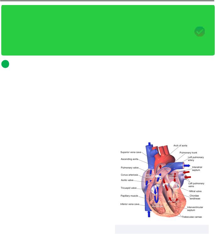
The circulatory system is important for many functions, like protection (clotting and immunological), temperature regulation, gas and nutrient transport, and waste removal to name a few. The heart plays a key role in pumping blood throughout the body to ensure that oxygenated blood reaches organs and tissues to function, while deoxygenated blood and carbon dioxide is brought to the lungs.
Blood flow between the lungs and heart is called the pulmonary circulation whereas blood flow from the heart to the body tissues and back is called the systemic circulation.
Deoxygenated blood:
•enters the heart through the superior and inferior vena cava
•passes into the right atrium
•enters the right ventricle via the right atrioventricular (tricuspid) valve
•enters the pulmonary arteries by passing through the pulmonary semilunar valve
•moves through the pulmonary circulation to reach the lungs to become oxygenated
Oxygenated blood:
•leaves the lungs and enters the heart once more through the pulmonary veins
•enters the left atrium
•passes through the left atrioventricular (bicuspid) valve to reach the left ventricle
•passes into the aorta via the aortic semilunar valve
•moves through the systemic circulation to reach body tissues, becoming deoxygenated
Figure 3.01 The anatomy of the heart
INBDE Booster | Booster PrepTM

Head and Neck Anatomy
Figure 3.01 The classification of blood flow into the body
The vascular system
Although the heart is crucial for pumping blood throughout the body, its vasculature is just as important! There are five main types of vasculature:
•Arteries: contain much smooth muscle and influence blood pressure, carries oxygenated blood
*Exception is the pulmonary arteries (deoxygenated blood)
•Arterioles: primary site of vascular resistance, control total peripheral resistance (TPR)
•Capillaries: site of intermingled arteriole and venule endings at tissue sites to exchange nutrients and oxygen
•Venules: smaller counterpart to veins
•Veins: contain limited smooth muscle, have valves to prevent backflow of blood, often situated near or between skeletal muscles to assist in moving blood towards the heart, carries deoxygenated blood
*Exception is the pulmonary veins (oxygenated blood)
84
2 The aortic arch and major branches
The aortic arch
Oxygenated blood leaves the heart to the body tissues through the largest blood vessel in the body, the aorta. The aorta is a wide vessel, forming the aortic root as it exits the heart. The next region is the ascending aorta, which connects to the aortic arch. The ascending aorta supplies blood to the upper body (such as the head, neck and limbs).
There are three main blood vessels which connect to the aortic arch: the brachiocephalic trunk (which then branches into the right subclavian and common carotid arteries), the left common carotid artery, and the left subclavian artery.
Blood may then pass through the aortic isthmus (embryological constriction between cerebral and placental circulations) and the descending aorta, which supplies blood to the rest of the body.
The aorta "arches" towards the left-hand side of the body, where the descending aorta courses inferiorly along the left side of the vertebral column. Since the aortic arch is already situated more to the left, there is no need for a brachiocephalic trunk on this side. There are some anatomical variations for how the common carotid, subclavian, and brachiocephalic trunks branch off from the aortic arch (and even the number of branches present), but you should just be familiar with the most common arrangement.
INBDE Booster | Booster PrepTM

Head and Neck Anatomy
Figure 3.03 The aortic arch and major branches
Figure 3.04 More detailed diagram if aortic arch and its extended branches.
The right costocervical trunk is not present but would be lateral to the right thyrocervical trunk
The brachiocephalic trunk
The first branch of the aortic arch is the brachiocephalic trunk (synonymous with brachiocephalic artery). In most individuals, it is located on the right side of the aortic arch, where it then branches into the right subclavian and right common carotid arteries. Again, there is no need for this vessel to exist on the left because of the natural leftsidedness of the aortic arch's course.
INBDE Pro Tip:
The brachiocephalic trunk "stretches" obliquely (or superolaterally) and has to cross the anterior aspect of the trachea before it divides into its two branches at the right sternoclavicular joint.
85
The subclavian artery
As evident from its name, the subclavian artery is related to the inferior clavicular region bilaterally. It is the major artery sending blood to the upper limbs and contains numerous branches. These include:
•Internal thoracic (mammary) artery o Supplies the anterior chest wall
bilaterally
•Thyrocervical trunk
oSupplies many structures in and around the neck region (thus thyro-cervical) like the (para)thyroid glands, larynx and pharynx, plus cervical and shoulder muscles
•Vertebral artery
oIts name alludes to its function: supplying the spinal column of the cervical region with blood bilaterally
oConnects to brainstem and brain supply via "feet" of the Circle of Willis (more on this later)
•Costcocervical trunk
oSupplies deep cervical and supreme intercostal arteries
At the level of the clavicle, the subclavian artery becomes the axillary artery, which spans the medial shoulder or axillary region. At the level of approximately the third rib, it becomes the brachial artery of the arm. Inferior to the humerus, the brachial artery splits once more to form the radial and ulnar arteries of the forearm.
INBDE Booster | Booster PrepTM

Head and Neck Anatomy
INBDE Pro Tip:
Be sure not to confuse the location of the radial and ulnar arteries! In the anatomical position, the thumbs are pointed outwards, and the palms are facing frontwards. The radius is located laterally, and the ulna is located medially.
To remember this, the radius is associated with the thumb, so when you rotate your forearm (pronation or supination), your thumb rotates and "draws" a radius of the movement.
Figure 17.05 Summary of the
aortic arch branches
The common carotid artery
The common carotid artery is "common" to its two branches. Recall that this vessel is an immediate branch from the arch of the aorta on the left side, but branches indirectly off the aortic arch on the right side, since it passes through the brachiocephalic trunk first.
This artery bifurcates into the external and internal carotid arteries between the level of C3 and C4. These two branches pass through the carotid sheath and are crucial contributors of the head's blood supply.
86
The internal carotid artery is responsible for supplying the anterior aspect of the brain and ocular region with blood. It enters the carotid canal of the temporal bone and possesses seven segments as described by Allan Bouthillier et al. in 1996.
There are many different classifications used to describe the course and segments of the internal carotid artery, though the Bouthillier classification is frequently used. The segments include:
•C1: cervical segment
•C2: petrous segment
•C3: lacerum segment
•C4: cavernous segment
•C5: clinoid segment
•C6: ophthalmic segment
oOpthalmic branch supplies the orbit, upper nose, and forehead
oForm anastomoses with branches of the external carotid arteries
•C7: communicating segment
These segments of the internal carotid artery are here to give you a better understanding of this important vessel's anatomical course, but they are not crucial for you to know.
Figure 17.06 The internal and external carotid arteries
INBDE Booster | Booster PrepTM
Head and Neck Anatomy
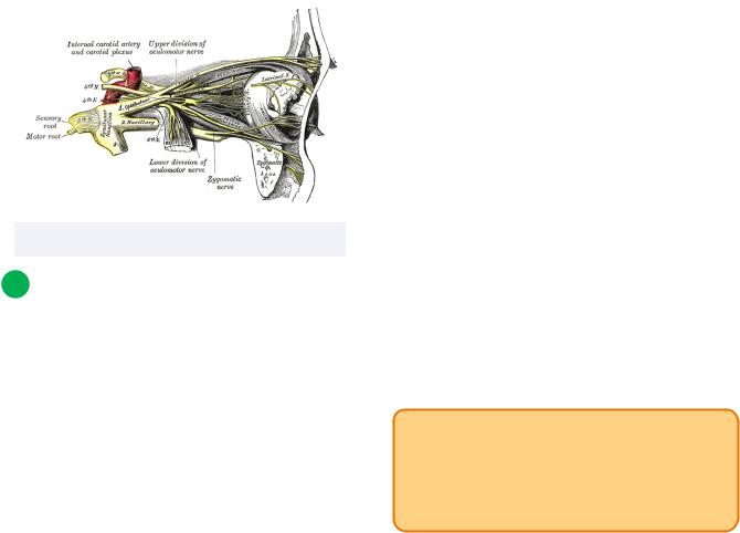
The internal carotid plexus is formed by postganglionic sympathetic fibers from the superior cervical ganglion which surround the internal carotid artery. Fibers from the plexus form the deep petrosal nerve or communicate with the trigeminal ganglion, abducens nerve, or pterygopalatine ganglion.
Figure 17.07 The internal carotid plexus
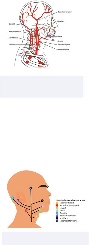
3 The external carotid artery
Overview
After the common carotid artery bifurcates, the external carotid artery differs in its anatomical course from the internal carotid artery. Rather than immediately pass into the skull, the external carotid artery branches into 8 different vessels which supply various regions of the head and neck region. These branches provide a combination of deep and superficial blood supply; this will depend on the vessel and its respective region. Thankfully, the regions and the organs that are supplied by these branches can be inferred by their names.
87
The eight branches, from inferior to superior position, are as follows:
•Superior thyroid artery
Supplies thyroid gland, infrahyoid muscles, sternocleidomastoid
•Ascending pharyngeal artery
Supplies pharynx
•Lingual artery
Supplies intrinsic tongue muscles
•Facial artery
Supplies submandibular gland, tonsils, and palate
•Occipital artery
Supplies posterior scalp
•Posterior auricular artery
Supplies ear and scalp near ear, parotid gland and facial nerve
•*Maxillary artery
Very important branch within the dental field
Supplies external acoustic meatus and tympanic membrane, mandible, gingivae, teeth, all muscles of mastication
•Superficial temporal artery
Temporal aspect of scalp
INBDE Pro Tip:
There is a well-known mnemonic to remember that will make it easier to recall the branches of the external carotid artery: Some Anatomists Like Freaking Fut, Poor Medical Students.
INBDE Booster | Booster PrepTM
Head and Neck Anatomy
Figure 17.08 The branches of the external carotid artery.
Note that the superior thyroid and posterior auricular arteries are not shown
The branches of the external carotid artery can be thought of being organized like a Christmas tree with ornaments. When looking at the right side of the head and neck, there are three branches on the left that move posterolaterally, and four branches on the right that move anteromedially. The external carotid artery can be thought of as the trunk of the tree, where the "end" at the top is the superficial temporal branch.
Figure 17.09 A “Christmas tree” diagram of the branches of the external carotid
88
The maxillary artery
The maxillary artery is important within the field of dentistry because it supplies blood to many key structures of the oral cavity. The maxillary artery branches off from the external carotid artery at the neck of the mandible. Here it will course between the neck of the mandible and the sphenomandibular ligament. It will then pass between the heads of the lateral pterygoid muscle and towards the pterygopalatine fossa where it will end in a series of other small branches.
There are three main parts of the maxillary artery which have numerous branches that are categorized in relation to surrounding structures and organs that are supplied with blood. It is not crucial for you to memorize the branches of each part of the maxillary artery, but a general idea of the vasculature and key vessels relevant to dentistry will be helpful.
•First (mandibular or bony) part
Deep auricular
Anterior tympanic
Middle meningeal
Inferior alveolar
-Supplies mandibular teeth
-Forms mental branch
Accessory meningeal
•Second (pterygoid or muscular) part
Masseteric
Pterygoid branches
Deep temporal
Buccal
•Third (pterygopalatine) part
Sphenopalatine
Descending palatine
-Forms greater and lesser palatine arteries which supply hard and soft palates
Infraorbital
Posterior superior alveolar
-Supplies maxillary teeth
INBDE Booster | Booster PrepTM

Head and Neck Anatomy
Pterygoid canal
Pharyngeal
Middle superior alveolar
-Branch of the infraorbital artery
Anterior superior alveolar
-Branch of the infraorbital artery
Figure 17.10 The three parts of the maxillary artery and its branches
Figure 17.11 Illustration of the maxillary artery, its course, and its branches.
Note that not all branches are depicted here
89
INBDE Pro Tip:
The first part of the maxillary artery has branches that are generally associated with the ear. The second part has branches associated with the muscles of mastication. The third part has branches associated with the infraorbital/ maxillary/palatal regions.
There are some mnemonics that can be useful if you wish to remember all of the maxillary branches.
First part
•Does Audio Make Interesting Allies?
Second part
•Munch Peanuts, Drink Beer!
Third part
•Some Dancing In Paint Presents Positive Mood Adjustments
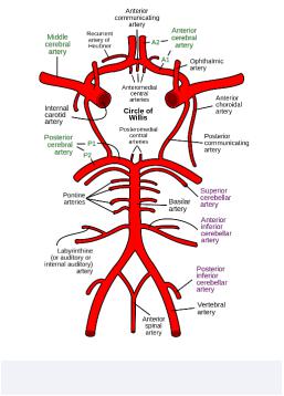
Overview
The blood flow to the brain is supported by an important structure called the Circle of Willis. This circle is an anastomoses of major blood vessels at the base of the brain and brainstem, and connect with other branches that supply surrounding neural tissues.
The Circle of Willis is a part of the cerebral circulation and is important for collateral circulation. In the event that any aspect of the circle is blocked or encounters stenosis (narrowing), blood can often bypass the affected area by passing through other vessels in the circle. This is clinically relevant as it can prevent ischemia to the brain and surrounding tissues.
INBDE Booster | Booster PrepTM
Head and Neck Anatomy
There are 10 major vessels (arteries) that contribute to the Circle of Willis, but this number is not absolute. You should be familiar with them but pay particular attention to the vessels with an asterisk (*) by them. Generally, from inferior to superior they are:
•Anterior spinal
Supplies the anterior aspect of the spinal cord
•*Vertebral
Supplies posterior brain, spinal cord, brainstem and cerebellum
Posterior inferior cerebellar (PICA)
-Supplies this aspect of the cerebellum
•Anterior inferior cerebellar (AICA)
Supplies this aspect of the cerebellum
•Basilar
Supplies the posterior circulation of the brain
Pontine
-Supplies the pons
•Superior cerebellar
Supplies superior cerebellum
•Posterior cerebral
Supplies posterior cerebrum (ie. occipital and temporal lobes)
•Posterior communicating
Key point of connection between the anterior and posterior cerebral circulations via the internal carotid arteries and posterior cerebral arteries, respectively
•*Middle cerebral
Has many branches that supply much of the brain (combination of frontal, parietal, temporal, occipital lobes and surfaces)
Clinically important since it is most frequently occluded; its major blood supply to the lobes of the brains means that middle cerebral artery stroke is very dangerous
-Effects depend on the lobe or region of the brain that experiences a stroke
90
•*Internal carotid
-See earlier information
Opthalmic
-Supplies the orbit, upper nose, and forehead
•Anterior cerebral
Medial cerebral hemisphere surfaces such as the corpus callosum
•Anterior communicating
Point of connection between the two anterior cerebral arteries
Supplies optic chiasma
Recall from earlier in this section when the subclavian artery was discussed. One of the branches from this artery includes the right and left vertebral arteries, from the left and right subclavian arteries, respectively. The vertebral arteries form the inferior portion of the Circle of Willis.
Figure 17.12 The Circle of Willis
INBDE Booster | Booster PrepTM