

Полезные материалы за все 6 курсов / Учебники, методички, pdf / INBDEBooster Head and Neck Anatomy Notes
.pdfHead and Neck Anatomy
2 Features
Fontanelles
The fontanelles are membranous spaces that result from the incomplete fusion of skull bones. Within 18 months after birth, these spaces fuse together. There are two types:
•Anterior/Frontal fontanelle: between sagittal and coronal suture
•Posterior/Occipital fontanelle: between sagittal and lambdoid suture
Sutures
When the fontanelles fuse, sutures form. Unlike the cartilaginous tissue of the fontanelles, the sutures are made up of fibrous tissue, and form immovable joints. Sutures usually close within 20 years after birth, and there are 5 different types:
•Frontal/metopic suture: found at the midline of the frontal bone
Closes 3-9 months following birth
•Sagittal suture: fusion of two parietal bones (one for each side)
•Coronal suture: fusion of the frontal and two parietal bones
•Lambdoid suture: fusion of occipital bone with both parietal bones
•Squamous suture: fusion of parietal bone and temporal bone
Additionally, there are two different anatomical terms that apply to the intersection of two different sutures. These features are remnants from the fontanelles. They include:
•Bregma: intersection of sagittal and coronal sutures
Remnant of frontal fontanelle
•Lambda: intersection of sagittal and lambdoid sutures
Remnant of occipital fontanelle
11
*Note: Do not confuse lambda with the lambdoid suture. The lambdoid suture is a physical suture, but the lambda is a term referring to a specific point. There is a bregma point, but no bregma suture.
Figure 2.02 The fontanelles and sutures of the developing skull
INBDE Pro Tip:
The names of each of these sutures gives away their appearance and location. It also makes it much easier to remember!
•Frontal/metopic suture: is the fusion of the frontal bone; metopic means forehead
•Coronal suture: a cut down this suture provides a coronal view of the skull
•Sagittal suture: a cut down this suture provides a sagittal view of the skull
•Squamous suture: is squished at the side of the head
•Lambdoid suture: remember the lambda symbol? From a superior view, this suture looks like "λ"
For the intersections:
•Lambda: if you look at the intersection points of the lambda symbol, it’s the center of all its points" "
•Bregma: the inverse of lambda; turn lambda upside down and look at the intersection points " "
INBDE Booster | Booster PrepTM
Head and Neck Anatomy
There are a series of birth defects that may arise when the fontanelles prematurely close. This is called craniosynostosis, where brain growth exceeds growth of the skull. Craniosynostosis may present as three different types:
•Scaphocephaly: sagittal suture prematurely closes leading to anterior-posterior skull elongation
•Brachycephaly: coronal and lambdoid sutures prematurely close, causing superior-inferior or vertical elongation of the skull
•Plagiocephaly: one-sided premature closure of coronal and lambdoid sutures, causing asymmetry of the skull
Lines
There exist various imaginary, anatomical lines known as principle lines of the skull. These lines are useful in a clinical setting when treating facial injuries or when oral surgeons are performing procedures.
•Principles lines of force are vertically oriented lines which represent the strongest regions of the skull
Formed due to adaptions resulting from local vertical mechanical stressors such as mastication
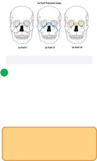
•Principle lines of fracture (Le Fort fracture lines) are generally more horizontally oriented lines which represent the weakest regions of the skull
Limited horizontal mechanical stressors mean this region is more commonly susceptible to fracture
12
One area of clinical Importance is the pterion. This region can be found on the lateral skull where the frontal, parietal, sphenoid and temporal bones articulate.
•Since there are many articulations in this one place, it is considered the weakest area of the calvaria
•Below this area runs the middle meningeal artery, which may be lacerated during injury of the pterion
Results in an epidural hematoma
Figure 2.03 The principle lines of fracture
2 Bones and Regions of Interest
Orbital bones
There are a total of 7 bones that comprise the orbit. These include the frontal, palatine, maxillary, lacrimal, ethmoid, sphenoid, and zygomatic bones. Note that the nasal bone is NOT included here.
INBDE Pro Tip:
The orbital bones are a high yield topic to know for your board exams. One way to remember them is by using the mnemonics “Z, SLEeP For Me,” since we’r talking about the eve after all!
INBDE Booster | Booster PrepTM
Head and Neck Anatomy |
13 |
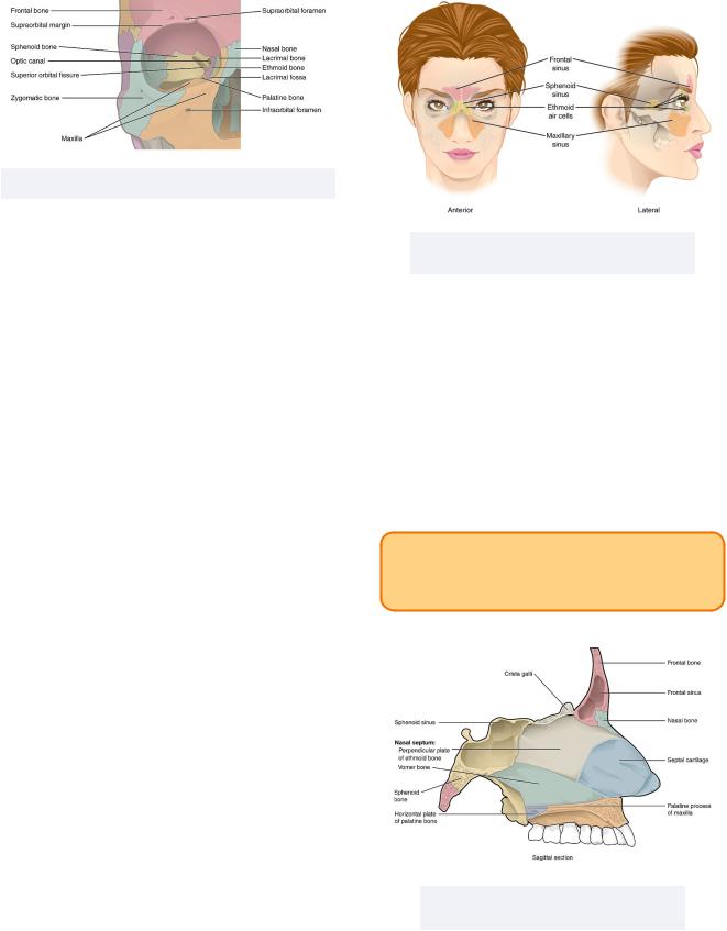
Figure 2.04 The bones that comprise the orbit
Sinuses
The sinuses are an important supporting structure of the skull. They branch off from the lateral nasal wall and into select bones in the surrounding area. They have numerous functions:
•Function as "air pockets" to reduce weight of skull
•Impact voice resonance
•Dampen intranasal pressure
•Humidifying and warming air during inspiration
•Increase surface area for olfaction
Specifically, the frontal, ethmoid, sphenoid and maxilla bones give rise to their respective sinuses. Initially, these sinuses begin as small pockets, and become larger as a person develops.
•Frontal and maxillary sinuses develop after birth
Experience vertical growth
•Ethmoid sinuses begin as ethmoid cells filled with air at birth
Figure 2.05 The 4 different sinuses and their locations
The nasal septum
The nasal septum acts as a physical barrier between the left and right nasal passageways. It is comprised of three main parts, the septal cartilage, the perpendicular plate of the ethmoid bone, and the vomer bone.
Clinically, displacement of the nasal septum into the right or left nasal passageways is called a deviated septum.
INBDE Pro Tip:
The components of the nasal septum are highyield topics for your board exam
Figure 2.06 The 3 main parts of the nasal septum
INBDE Booster | Booster PrepTM

Head and Neck Anatomy
The nasal meatuses
Within the lateral nasal cavity can be found the three air passages, known as the nasal meatuses. These meatuses are located on both sides of the nasal cavity and are inferior to their three respective nasal conchae.
•Superior meatus: contains the opening to the sphenoid sinus, posterior ethmoid sinus, and sphenopalatine foramen
•Middle meatus: houses the opening to the anterior and middle ethmoid sinuses, maxillary sinus, and frontal sinus through the semilunar hiatus
•Inferior meatus: houses the opening to the nasolacrimal duct
INBDE Pro Tip:
The semilunar hiatus as it relates to the middle meatus is an important topic for your board exam.
The ethmoid bone
The ethmoid bone is located within the skull and is known for its unique appearance. It has a series of important features:
•Crista galli: serves as an attachment point for the falx cerebri of the dura mater
•Cribriform (horizontal) plate: olfactory nerve endings from CN I pass through the many, tiny holes here
•Superior and middle nasal conchae: scrolllike shelves of bone that function to control airflow during breathing
•Uncinate process: a downward vertical projection with a wing-like appearance, important for directing drainage of the frontal sinus
14
Figure 1.07 A posterior view of the ethmoid bone and its features
The sphenoid bone
The sphenoid bone is a large, wing-shaped bone from a superior perspective. It also has numerous features of note:
•Sella turcica: means "Turkish saddle" due to the bone's appearance, and encases the pituitary gland
•Greater wing: appears to be a large winglike structure and is a component of the middle cranial fossa
•Lesser wing: appears to be a small-shaped wing structure and is a component of the anterior cranial fossa
•Clinoid process: 4 fang-like bony processes located centrally in the sphenoid bone and surround the sella turcica
Serve as an attachment point for the dura mater
Figure 2.08 A superoposteriar view of the sphenoid bone
INBDE Booster | Booster PrepTM
Head and Neck Anatomy
The mandible
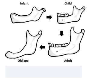
The mandible is considered an adaptive bone due to its ability to make changes in the angle of the mandible based on alterations in the alveolar process and its relation to the teeth.
The angle of the mandible experiences changes as individuals age over time, due to changes in their tooth structure and number.
•No teeth are found in the mandible at birth
Obtuse angle of mandible due to unformed alveolar process
About 150 degrees
•Small deciduous teeth are found in children
Obtuse angle of mandible and mostly unformed alveolar process
About 140 degrees
•Permanent teeth are in adults
Less obtuse angle of mandible as the alveolar process is fully developed
About 120 to 130 degrees
•Edentulous individuals (ie. those lacking teeth) may occur at any age, but is more common as an individual ages
Experience resorption of alveolar process and changes in mental foramen
Mandible more closely resembles the mandible at birth
-Obtuse angle of mandible at about 140 to 150 degrees
Figure 2.09 The age-related changes to the mandibles
15
INBDE Booster | Booster PrepTM
Head and Neck Anatomy |
16 |
Foramina of the Skull
The foramina of the skull is a good topic to know for your board exam, as it helps piece together information studied from other chapters. Using knowledge of the cranial nerves, blood vessels, and nervous system tissues, candidates will apply this information to various foramina and regions of the skull. These notes will simplify and connect information that will help boost confidence for the INBDE.
1 Foramina of the face
A foramen (plural: foramina) is an opening that allows for the passage of key nerves, arteries and veins from one region of the body to another. Often, foramina allow for external nerves and vasculature to connect with internal structures or sources.
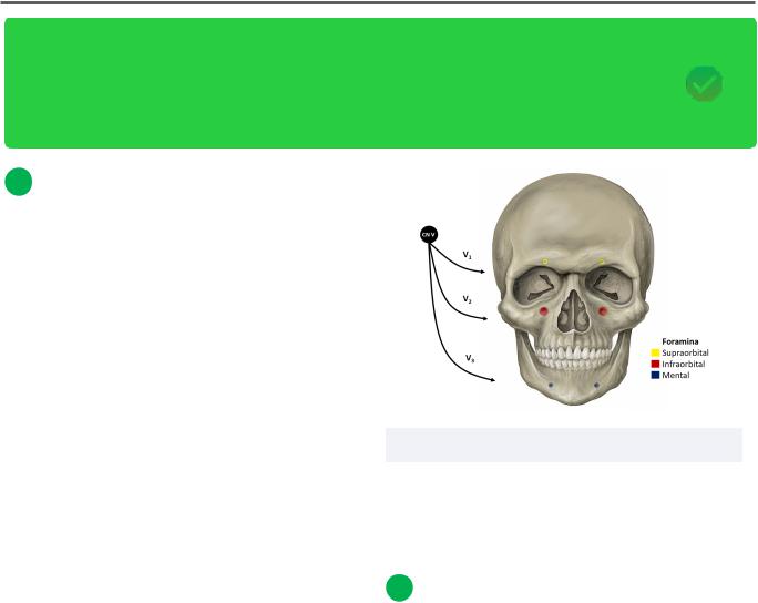
There are three main regions of the face that contain superficial foramina of importance:
•The frontal bone: contains the supraorbital foramen located above each orbit
Supraorbital nerve from CN V1, artery and vein
•The maxilla: contains the infraorbital foramen located below each orbit
Infraorbital nerve from CN V2, artery and vein
•The mandible: contains the mental foramen located below the mandibular gumlines and medially located on either side of the chin
Mental nerve from CN V3, artery and vein
Figure 3.01 Three major foramina of the face
Notice that for each region of the face discussed, there are foramina for each branch of the trigeminal nerve (CN V).
2 Foramina of the Skull Base
Ethmoid bone
Located anteriorly at the base of the skull and surrounded anteriorly and laterally by the frontal bone.
•Contains the cribriform plate, which contains many tiny holes that allow the olfactory fibers from CN I, the olfactory nerve, to pass into the nasal cavity
INBDE Booster | Booster PrepTM
Head and Neck Anatomy
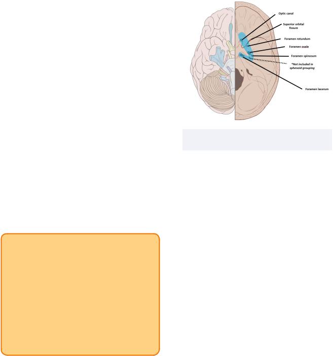
Sphenoid bone
Located posteriorly to the ethmoid and frontal bones. Considered an important structure with regards to foramina of the skull since many reside within this bone.
Generally, from anterior to posterior they include:
•Optic canal: contains CN II and ophthalmic artery
•Superior orbital fissure: contains CN III, CN IV, CN V1, and CN VI
Note: important since it contains all nerves relevant to eye movements
•Foramen rotundum: contains CN V2
•Foramen ovale: contains CN V3, lesser petrosal nerve
•Foramen spinosum: contains middle meningeal artery
•Foramen lacerum: contains greater and deep petrosal nerves
INBDE Pro Tip:
You can remember the order of the foramina by using a mnemonic during your studying "Observing ROSs's League."
•The first two foramina have to do with the optic nerve (thus, observing)
•The next three foramina nearly spell ROSs (your friend's name)
•The last one is foramen lacerum; since Ross plays in a hockey league, this foramen alongside the others looks like a backwards "L" or hockey stick
17
Figure 3.02 The foramina of the skull within the sphenoid bone
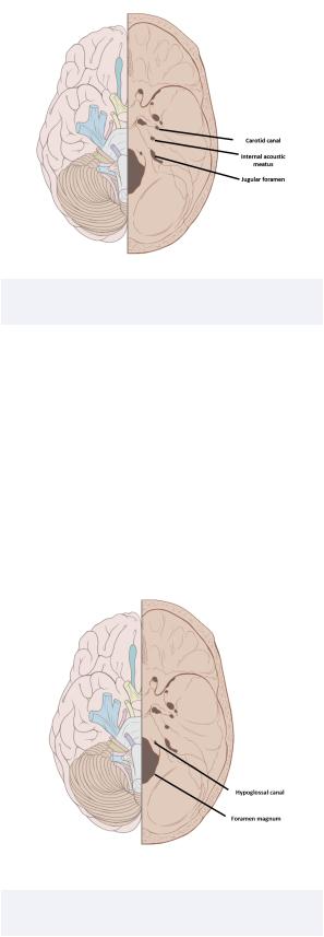
Temporal bone
Located roughly in the middle of the base of the skull, between the sphenoid and occipital bones.
•Internal auditory meatus: contains CN VII and CN VIII
Note: In contrast to the external auditory meatus which we know is the auditory canal
-Since this foramen deals with hearing, it makes sense CN VIII is involved
•Carotid canal: contains the internal carotid artery
•Jugular foramen: contains CN IX, CN X, CN XI
Note: When you think of the word "jugular" you often think of the neck; the above cranial nerves all have some association with the neck region!
INBDE Booster | Booster PrepTM
Head and Neck Anatomy |
18 |
Figure 3.03 The foramina of the skull within the temporal bone
Occipital bone
Located posteriorly at the base of the skull.
•Foramen magnum: communicates with the vertebral column, thus containing the spinal cord, spinal and vertebral arteries)
Note: The Latin name quite literally means "big hole," which describes its appearance
•Hypoglossal canal: contains CN XII (the hypoglossal nerve)
Figure 3.04 The foramina of the skull within the occipital bone
INBDE Booster | Booster PrepTM
Head and Neck Anatomy |
19 |
Fossae of the Skull
The fossae of the skull can be a challenging topic to know for your board exam, since it tests many different bony, nervous, and circulatory structures. Candidates will apply their knowledge of anatomical structures, borders, and regions of communication to gain a more holistic anatomical understanding. These notes will simplify information to help boost confidence for the INBDE.
1 External fossae
Introduction
A fossa (plural: fossae) can be described as a depression, pit, or groove on the surface of an organ or structure. Often, this term is applied to bony structures, but may also be applied to other tissues, such as the heart (ex. the fossa ovalis). A fossa is not to be confused with a foramen, which is a complete opening of the bone through which supporting nerves and vessels pass.
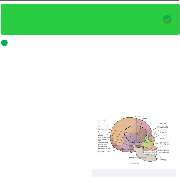
Temporal fossa
The temporal fossa is a hollow area of the temporal region of the skull. It is comprised of five main bones: frontal, parietal, sphenoid, temporal, and zygomatic. There are five main borders that outline this fossa, though more attention should be drawn to the contents of this region.
•Inferior: zygomatic arch and greater wing of sphenoid (infratemporal crest)
•Superior: superior temporal line
•Anterior: zygomatic bone (frontal process) and frontal bone (zygomatic process)
•Posterior: superior temporal line
•Floor: frontal and greater wings of sphenoid, parietal bone, temporal bone (squamous portion)
•Contains: temporalis muscle, middle and deep temporal arteries and veins, superficial temporal artery and vein, auriculotemporal nerve zygomaticotemporal nerve, temporal branches of facial nerve
Clinically, this region contains the pterion, which is the articulation between the frontal, sphenoid, parietal, and temporal bones. As a result of its numerous articulating surfaces, it is a considered a weaker region of the skull and is prone to injury.
Figure 4.01 The temporal fossa
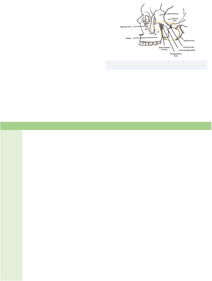
Infratemporal fossa
The infratemporal fossa can be found below the temporal fossa.
INBDE Booster | Booster PrepTM
Head and Neck Anatomy
•Superior/roof: greater wing of sphenoid
Also contains foramen spinosum and foramen ovale
•Inferior/floor: medial pterygoid muscle
•Anterior: posterolateral border of the maxillary sinus
•Posterior: carotid sheath
•Medial: lateral pterygoid plate; tensor veli palatini and levator veli palatini muscles, and superior constrictor muscle
•Lateral: coronoid process and ramus of the mandible bone
•Contains: medial and lateral pterygoids, temporalis muscle, mandibular nerve, auriculotemporal nerve, buccal nerve, lingual nerve, inferior alveolar nerve, chorda tympani, otic ganglion, maxillary artery and vein, pterygoid venous plexus, and middle meningeal vein
20
Figure 4.02 The infra temporal fossa
Pterygopalatine fossa
The pterygopalatine fossa is fairly complex in comparison to the others discussed in this section. It is shaped like an inverted cone and is deeper in relation to the infratemporal fossa. It both communicates with and houses numerous structures.
Surface |
Border |
Opening |
Communica3on |
Contents |
|
|
|
|
|
Superior/ |
Body of sphenoid bone |
Inferior orbital |
Orbit |
Zygomatic branch of CN V2 and |
roof |
and orbital process of |
fissure |
|
infraorbital artery and vein |
|
palatine bone |
|
|
|
|
|
|
|
|
Inferior/ |
Palatine bone |
Greater and |
Palate |
Greater and lesser palatine |
floor |
(pyramidal process) |
lesser palatine |
|
artery, vein, and nerve |
|
and canals |
canals |
|
|
|
|
|
|
|
Anterior |
Posterior wall of |
Inferior orbital |
Orbit |
Zygomatic branch of CN V2 and |
|
maxillary sinus |
fissure |
|
infraorbital artery and vein |
|
|
|
|
|
Posterior |
Sphenoid bone |
Foramen |
Middle cranial fossa |
CN V2 |
|
(pterygoid process) |
rotundum |
(Meckel cave and |
|
|
|
|
cavernous sinus) |
Pterygoid canal's artery, vein |
|
|
Pterygoid canal |
Middle cranial fossa |
and nerve |
|
|
|
to medial pterygoid |
|
|
|
|
plate |
Pharyngeal branches of CN V2 |
|
|
Pharyngeal canal |
Nasopharynx |
and artery |
|
|
|
|
|
Medial |
Palatine bone |
Sphenopalatine |
Nasal cavity |
Sphenopalatine artery and vein, |
|
(perpendicular plate) |
foramen |
|
plus the nasopalatine nerve |
|
|
|
|
|
Lateral |
N/A |
Pterygomaxillary |
Infratemporal fossa, |
Posterior superior alveolar |
|
|
fissure |
masticator space |
nerve and terminal aspect of |
|
|
|
|
maxillary artery |
|
|
|
|
|
INBDE Booster | Booster PrepTM