

Полезные материалы за все 6 курсов / Учебники, методички, pdf / INBDEBooster Head and Neck Anatomy Notes
.pdfHead and Neck Anatomy
3 Extrinsic Auricular Muscles
Quick review
The extrinsic auricular muscles were discussed in the muscles of facial expression section. As a quick summary, this group of muscles includes the superior, posterior, and anterior auricular muscles. They move the ear superiorly, posteriorly and anteriorly, respectively. The superior auricular muscle is the largest, and the anterior auricular muscle is the smallest.
61
There are 6 main intrinsic auricular muscles: the helicis major and minor, tragicus, antitragicus, transverse and oblique muscles. The transverse and oblique muscles are located on the posterior surface of the ear. Each muscle name is easy to remember as it corresponds to its respective region of the ear.
Figure 11.04 The intrinsic muscle of the ear
Figure 11.03 The extrinsic muscle of the ear
4 Intrinsic Auricular Muscles
Overview
The intrinsic auricular muscles are limited in terms of their uses. As opposed to other animals which make greater use of intrinsic muscles to move their ears and gain a better sense of their environment through hearing, humans do not rely on these muscles in day-to- day life. These muscles are found within the cartilage of the ear.
INBDE Booster | Booster PrepTM

Head and Neck Anatomy |
62 |
Salivary Glands
The salivary glands are necessary for mastication and digestion, so it will be important for candidates to apply their knowledge from other sections to fully comprehend the function of these exocrine glands. These notes will provide an outline of everything candidates must know with respect to the structure of salivary glands, their function, innervation, anatomical features, and differences. Tips, tricks, and helpful summaries along the way will ensure that this content is easy to understand and remember come time to write the INBDE.
1 The Structure of Salivary Glands
Overview
The salivary glands are an example of exocrine glands in the body, meaning that the contents in the gland are secreted into ducts and released into a body cavity. These glands release saliva, which contain enzymes, antibodies from residing immunological cells, electrolytes, and a combination of water and mucous. Saliva is important for breaking down food into a bolus to assist in mastication and swallowing, initiates the process of digestion, alters the pH of the oral cavity, and "cleans" the mouth.
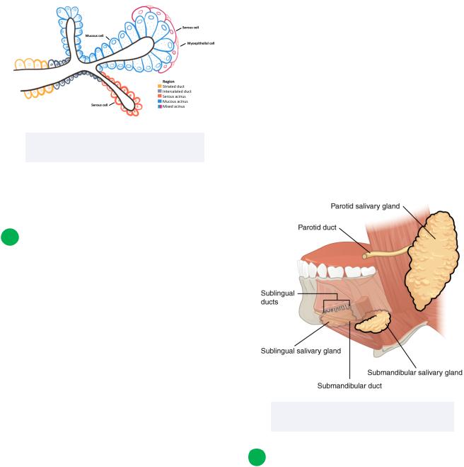
Before looking at the gross anatomical salivary glands, it is important to first understand the histological structure of these glands by looking at cellular components and their organization. There are three main cells that will be discussed:
•Serous cell
Pyramidal in shape produce watery secretions with proteins and tend to stain dark histologically
•Mucous cell
These cells are usually larger than the serous salivary glands, are pyramidal in shape, and contain secretory granules with glycoproteins
Produce thicker, more mucous-like secretions
•Myoepithelial cell
Are thin contractile cells located at the basal end of cells
Contractions cause secretory contents within the acini to enter into the surrounding ducts
There are five main regions that are also relevant:
•Serous acinus
Ball or sac-shaped cavity comprised of many serous cells
•Mucous acinus
Ball or sac-shaped cavity comprised of many mucous cells
•Mixed acinus
Ball or sac-shaped cavity that contains a combination of serous and mucous cells
•Intercalated duct
The region that connects to the acini and are usually cuboidal epithelial cells
•Striated duct
The region that connects to the intercalated ducts where secretions are initially produced, and release contents into interlobular excretory ducts
Tend to contain columnar epithelial cells
INBDE Booster | Booster PrepTM
Head and Neck Anatomy
Figure 12.01 The histological organization of salivary glands
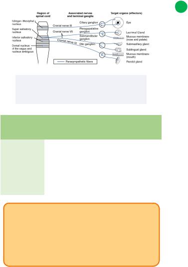
There are three major salivary glands that will be discussed in this section: the parotid, submandibular, and sublingual glands.
2 The Parotid Gland
Overview
The parotid gland is the largest of the three salivary glands. It is located within the retromandibular fossa, inferior to the zygomatic arch and posterior to the masseter.
•Features:
Contains a superficial and deep lobe which is separated by the facial nerve (CN VII)
Contains the facial nerve but does not innervate it!
External carotid artery
Retromandibular vein
•Innervation:
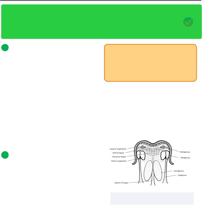
Parasympathetic fibers from inferior salivatory nucleus --> glossopharyngeal nerve (CN IX) lesser petrosal nerve --> otic ganglion --> auriculotemporal nerve (of CN V3)
63
•Secretion:
Contains predominately serous acini resulting in watery secretions
Serous cells of parotid glands secrete
α-amylase causes hydrolysis of carbohydrates
•Duct:
Stenson's duct, which courses from the anterior aspect of the superficial lobe, over the masseter, and penetrates the buccinator muscle
Opens into the oral cavity near the maxillary second molar
Figure 12.02 A simplified diagram of the three salivary glands
3 The submandibular gland
Overview
When compared with the other two salivary glands, the submandibular gland is the second largest. It is located in the submandibular fossa on the medial aspect of the mandible by the mylohyoid line.
INBDE Booster | Booster PrepTM

Head and Neck Anatomy
•Features:
Contains a superficial and deep lobe that is divided by the mylohyoid muscle
Likely to experience sialolithiasis (salivary gland stone)
•Innervation:
Parasympathetic fibres from superior salivatory nucleus --> CN VII --> chorda
tympani --> lingual nerve (of CN V3) --> submandibular ganglion
•Secretion:
Contains a mix of both serous and mucous-secreting acini; slightly more mucous acini means the saliva produced is a bit thicker
Produces most of the saliva
Combination of α-amylase from serous acini and lysozyme (hydrolyses bacterial cell walls) plus mucin from mucous acini
•Duct:
Wharton's duct courses around the posterior aspect of the mylohyoid muscle, then superomedially towards the floor of the mouth on either side of the lingual frenulum
4 The Sublingual Gland
Overview
When compared with the other salivary glands, the smallest is the sublingual gland. It can be found on the floor of the oral cavity between the geniohyoid, hyoglossus, and mylohyoid muscles. It is bordered laterally by the mandible.
•Features:
Contributes minimally to the overall saliva production in the oral cavity
Lacks striated and intercalated ducts
64
•Innervation:
Parasympathetic fibres from superior salivatory nucleus --> CN VII --> chorda
tympani --> lingual nerve (of CN V3) --> submandibular ganglion
•Secretion:
Contains some mucous and serous acini, though is more of a mucous-secreting gland
Amylase, lysozyme and mucins present
•Duct:
Bartholin's duct is responsible for transporting saliva into the oral cavity
-Joins Wharton's duct and thus drains into the oral cavity by the lingual frenulum
Since this gland lacks striated and intercalated ducts, saliva is also excreted near the plica fimbriata of the ventral tongue through the ducts of Rivinus
Figure 12.03 The surrounding structures and associated excretory ducts of the salivary glands
INBDE Booster | Booster PrepTM
Head and Neck Anatomy
Figure 12.04 The parasympathetic innervation of the saliva glands and other visceral motor pathways
Salivary |
Inner- |
Duct |
Secretion |
gland |
vation |
|
|
|
|
|
|
Parotid |
CN IX |
Stenson's |
Serous |
|
|
|
|
Sub- |
CN VII |
Wharton's |
Both |
mandibular |
|
|
|
|
|
|
|
Sublingual |
CN VII |
Bartholin's |
Mucous |
|
|
|
|
INBDE Pro Tip:
To remember the information in the table above, a mnemonic can be used.
•Perry's 9 Salty Sandwiches
•Manny's 7 Weird Bananas
•Lucy's 7 Baked Macaroons
65
5 Other Minor Salivary Glands
Overview
There are hundreds of small salivary glands that are accessory to the three major glands discussed above. They contribute minimally to the overall salivary production and are of lesser importance. Nonetheless, they still have a role to play in salivary production, and are mentioned here for completeness. They can be found in the submucosa of the:
•Sinonasal cavity
•Cheeks
•Soft and hard palate
•Tongue
•Nasopharynx
•Larynx
•Trachea
•Lips
INBDE Booster | Booster PrepTM
Head and Neck Anatomy |
66 |
Muscles of the Tongue
The muscles of the tongue is a topic you should be familiar with for the INBDE. Candidates should know the origin and insertion of these muscles, but should focus particular attention to their innervation and action. These notes will provide a good basis for this section of the board exam with simplified, clear descriptions for each muscle group.
1 Muscles of the Tongue overview
The muscles of the tongue are crucial to the functions of the tongue, which include speech, manipulating food during mastication, and swallowing. Though not related to the muscles of the tongue specifically, the tongue is also involved in taste.
Embryologically, this group of muscles is derived from the paraxial mesoderm which later form the occipital somites. These muscles are innervated by the either the vagus nerve (CN X) or the hypoglossal nerve (CN XII).
There are two main groups of muscles for this organ, intrinsic and extrinsic.
2 Intrinsic Muscles
Overview
These muscles can be found within the tongue. Unlike the extrinsic muscles, these are found within the tongue, and are responsible for finer, more complex movements. Generally, they can change the shape of the tongue, rather than provide mass movement of the organ. These muscles include the superior and inferior longitudinal, plus the transverse and vertical lingual muscles.
These muscles are all innervated by the hypoglossal nerve (CN XII).
INBDE Pro Tip:
To remember which muscles are intrinsic, pay attention to their names. Each have to do with a specific plane, which tells us something about the fibers, and thus the action, of that particular muscle.
Superior longitudinal
Located most superficially to the other intrinsic muscles of the tongue, below the lingual submucosa. Oriented longitudinally, or in a postero-anterior fashion.
•Origin: submucosa of posterior tongue
•Insertion: anterolateral aspect of tongue
•Action: shortens tongue, curls it upward
Figure 13.01 The intrinsic and extrinsic muscles of the tongue (coronal view)
This muscle is located inferior to all other intrinsic muscles of the tongue. Runs posteroanteriorly.
•Origin: root of tongue, body of hyoid bone
•Insertion: apex of tongue
•Action: shortens tongue, curls it downward
INBDE Booster | Booster PrepTM
Head and Neck Anatomy
Transverse lingual
Fibers run laterally from the medial aspect of the tongue.
•Origin: lingual septum
•Insertion: lateral aspect of tongue
•Action: elongates and narrows tongue
Vertical lingual
Fibers run vertically in an infero-superior fashion.
•Origin: root of tongue, genioglossus
•Insertion: lingual aponeurosis
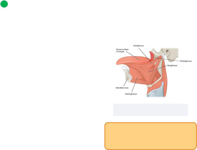
3Extrinsic Muscles
•Action: flattens tongue
Overview
These muscles are located externally to the tongue. They are responsible for mass movements of the tongue, and are primarily involved in swallowing and mastication. These muscles include the genioglossus, styloglossus, hyoglossus, and palatoglossus.
Genioglossus
The word "genio" essentially means "chin," which is the general location for this muscle's origin.
•Origin: Mandible (superior mental spine)
•Insertion: lingual aponeurosis, body of hyoid bone, dorsum of tongue
•Action: protrusion of tongue
Styloglossus
"Stylo" refers to the styloid process, which is where the origin of this muscle is found.
•Origin: styloid process, stylomandibular ligament
•Insertion: inferior longitudinal muscle, hyoglossus muscle
•Action: retraction of tongue bilaterally
67
Hyoglossus
The word "hyo" is associated with the hyoid bone, which is where this muscle originates.
•Origin: body and greater horn of hyoid bone
•Insertion: lateral tongue (inferior aspect)
•Action: depression and retraction of tongue
Palatoglossus
"Palato" is related to the word "palate," which points to this muscle’s origin.
•Origin: soft palate (palatine aponeurosis)
•Insertion: lateral aspect of tongue
•Action: elevation of posterior tongue, closure of oropharyngeal Isthmus, maintains palatoglossal arch
•*Innervation: The only muscle of the tongue innervated by the vagus nerve (CN X)
Figure 13.02 The extrinsic muscles of the tongue
INBDE Pro Tip:
All the extrinsic muscles of the tongue have a prefix that alludes to its origin, and a suffix that points to its insertion (though non-specific).
INBDE Booster | Booster PrepTM

Head and Neck Anatomy |
68 |
Muscles of the Soft Palate
The muscles of the soft palate are relevant for the INBDE, as it relates to the process of swallowing, and even air pressure equalization. Candidates should know the origin and insertion of these muscles, with more emphasis on innervation and function.
These notes will clearly outline the muscles of the soft palate, so you're ready to face any question on your board exam that tests this knowledge.
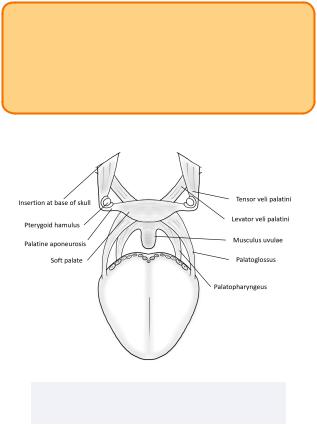
1 Muscles of the Soft Palate Overview
The muscles of the soft palate combine aponeurotic and muscular tissue to form the soft palate. The soft palate is considered an extension of the hard palate, which is comprised of bone, or more specifically, the palatine and maxillary bones. Generally, the soft palate is important for swallowing and breathing, but also contains mucous glands necessary for lubrication of the oral cavity.
These muscles are innervated by the vagus nerve (CN X), though the tensor veli palatini is innervated by the trigeminal nerve (CN V3).
There are 5 muscles that will be discussed, including the levator veli palatini, tensor veli palatini, palatoglossus, palatopharyngeus, and musculus uvulae.
INBDE Pro Tip:
The names of these muscles will tell you something important about them!
•For the palatoglossus and palatopharyngeus, the prefix is the origin, and the suffix is the insertion
•"Levator" means "to raise," "tensor" means "to tense," and veli means a "curtain" or a "veil", and "palatini" refers to the palate
2 Muscles of the Soft Palate
Levator veli palatini
•Origin: medial eustachian tube and temporal bone (petrous part)
•Insertion: palatine aponeurosis
•Action: elevates soft palate during swallowing (prevents food from entering nasopharynx); opens auditory tube opening during swallowing to equalize pressure between the middle ear and pharynx
Tensor veli palatini
•Origin: medial pterygoid plate of sphenoid, lateral cartilage of eustachian tube
•Insertion: palatine aponeurosis
•Action: tenses soft palate during swallowing (prevents food from entering nasopharynx); opens auditory tube opening during swallowing to equalize pressure between middle ear and pharynx
Palatoglossus
•Origin: palatine aponeurosis
•Insertion: posterolateral aspect of tongue
•Action: depresses soft palate, elevates posterior tongue to facilitate swallowing
INBDE Booster | Booster PrepTM
Head and Neck Anatomy |
69 |
Palatopharyngeus
•Origin: hard palate and palatine aponeurosis
•Insertion: thyroid cartilage (superior aspect)
•Action: tenses soft palate, moves pharynx superoanteriorly and closes larynx during swallowing
Musculus uvulae
•Origin: posterior nasal spine (palatine bone) and palatine aponeurosis
•Insertion: mucosa of uvula, palatine aponeurosis
•Action: shortens uvula, ipsilateral contraction causes ipsilateral elevation
INBDE Pro Tip:
During swallowing, the nasal passages are blocked by the levator and tensor veli palatini. The palatopharyngeus blocks the larynx during swallowing.
Figure 14.01 The muscles of the soft palate (frontal view)
INBDE Booster | Booster PrepTM

Head and Neck Anatomy |
70 |
Muscles and Triangles of the Neck
The muscles of the neck and their associated triangles can be challenging to understand, however they are very useful when locating key structures in a clinical setting. For the board exam, candidates should know the various muscles of the neck, their function and innervation, plus their relationship to the triangles of the neck, their borders, and key contents. These notes include diagrams, tips, and tricks that will outline everything you need to know for this section so that you are well-prepared for any potential question.
1 Introduction to Muscles of the Neck
Collectively, the muscles of the neck are important for supporting and protecting the head and neck region, in addition to providing their movement. In total, there are three main types of neck muscle categories: anterior, lateral, and posterior. Muscles are organized into these categories based on their position, though it is also related to their function in most cases.
There are many muscles that can be discussed for this region, though more focus will be placed on the anterior neck muscles for the board exam.
2 Superficial Neck Muscles
Introduction
As the name suggests, these muscles will be found in the most superficial layer of the neck.
Platysma
The platysma is a thin muscle often referred to as the "shaving muscle" because its activation tenses the skin of the neck to facilitate shaving.
•Origin: skin/fascia of superior thoracic and shoulder regions (clavicle, acromial region, pectoralis major and deltoid muscles)
•Insertion:
Medial fibers: lower border of the mandible and the skin of the lower lip
Lateral fibers: skin of the perioral region, modiolus
•Action: lateral fiber contraction causes corners of mouth and lip to lower while medial fiber contraction may depress mandible and open the mouth
Sternocleidomastoid
This muscle of the neck conveys its origin and insertion points by its name alone.
•Origin:
Clavicular head: clavicle (medial third)
Sternal head: sternum (manubrium)
•Insertion: temporal bone (mastoid process)
•Action:
Bilateral contraction: lateral neck flexion ipsilaterally, lateral head rotation contralaterally
Unilateral contraction: neck flexion
•Innervation: spinal accessory nerve (CN IX), spinal nerves C2 and C3
INBDE Booster | Booster PrepTM