

Полезные материалы за все 6 курсов / Учебники, методички, pdf / INBDEBooster Head and Neck Anatomy Notes
.pdfHead and Neck Anatomy |
91 |
Figure 17.13 The position of the Circle of Willis relative to the brain and brainstem
The vessels of the Circle of Willis can be remembered by its distinctive shape, which looks like a person.
•Anterior spinal
The tail
•Vertebral
The legs
Posterior inferior cerebellar (PICA)
-The feet
•Anterior inferior cerebellar (AICA)
A belt
•Basilar
The spine
Pontine
-The ribs
•Superior cerebellar
The arms
•Posterior cerebral
A scarf
•Posterior communicating
The cheeks
•Middle cerebral
The glasses (temple)
Internal carotid
-The eyes
Opthalmic
-The eyebrows
•Anterior cerebral
The top of the head
•Anterior communicating
The hair
Figure 17.14 The simplified diagram of the Circle of Willis
INBDE Booster | Booster PrepTM

Head and Neck Anatomy |
92 |
Craniofacial Veins
The craniofacial veins are comparable to the craniofacial arteries, with some differences in structure and organization. For this section, candidates will apply their understanding of the head, neck, upper limbs, and thorax to gain a complete understanding of how these structures drain venous blood into surrounding vasculature. This section will provide a solid basis on the craniofacial veins to ensure complete understanding, boost confidence and provide clinical relevance in preparation for the board exam.
1The branches of the superior vena cava
The superior vena cava
Deoxygenated blood enters the heart from the body tissues through the superior and inferior vena cava. For the board exam and the field of dentistry, the superior vena cava is more relevant, as it drains the head, neck, upper thorax and limb regions. Generally, the superior vena cava drains structures above the diaphragm, whereas structures below the diaphragm drain into the inferior vena cava.
The brachiocephalic (innominate) vein
The first branch of the superior vena cava is the brachiocephalic or innominate vein. The superior vena cava will bifurcate at the level of the first right costal cartilage; thus, there are two brachiocephalic veins - one for each side of the thorax. This is unlike the brachiocephalic trunk/artery which is only present on the right side of the thorax.
The superior vena cava is situated anterior and slightly to the right of the vertebral column. Due to its position, the left brachiocephalic vein is longer than the right brachiocephalic vein, as it needs to pass anterior to the trachea to reach the left side.
Figure 18.01 The superior vena cava and its branches
Figure 18.02 The position of the superior vena cava and brachiocephalic veins relative to the heart and aortic arch
INBDE Booster | Booster PrepTM

Head and Neck Anatomy |
93 |
There are five main branches that drain into |
|
the brachiocephalic vein: |
|
• Inferior thyroid vein |
|
Drains inferior thyroid |
|
• Internal thoracic vein |
|
Drains the chest wall and the breasts |
|
• Vertebral vein |
|
Drains cervical spine, prevertebral and |
|
suboccipital muscles |
|
• Accessory vertebral vein |
|
Supports drainage of the vertebral vein |
|
• Internal jugular vein |
|
Important vessel since it contributes to |
Figure 18.03 Indirect venous drainage of |
much of the head and neck's venous |
the upper limb into the subclavian vein |
drainage |
|
Drains the brain and venous sinuses |
There are two main blood vessels that drain |
Exits via jugular foramen |
into the subclavian vein: |
|
• Dorsal scapular vein |
The subclavian vein also drains into the |
Drains dorsal aspect of scapula |
brachiocephalic vein from the clavicular and |
• External jugular vein |
axillary regions. |
The base of this vein drains the |
|
suprascapular and transverse cervical |
The subclavian vein |
veins laterally, and the anterior jugular |
The subclavian vein is located inferior to the |
vein medially |
clavicle bilaterally and receives drainage from |
Moving superiorly, branches into the |
the axillary vein. The distinction between these |
posterior auricular vein and |
two veins is the level of the first rib, where the |
retromandibular vein |
axillary vein becomes the subclavian vein. |
|
The axillary vein drains blood from the brachial |
|
vein, in addition to the basilic (medial) and |
|
cephalic (lateral) veins. The latter two veins are |
|
connected via the median cubital vein, which |
|
is clinically important for drawing blood or IV |
|
injections. |
|
Figure 18.04 Venous drainage of the neck depicting the anterior, internal, and external jugular vein
INBDE Booster | Booster PrepTM

Head and Neck Anatomy |
94 |
Figure 18.05 Summary of the venous drainage from the inferior vena cava to the neck and upper limbs
INBDE Pro Tip:
Unlike the arterial system, there is nothing analogous to the common carotid artery for the venous system. The internal carotid artery branches off the brachiocephalic vein, and the external carotid vein branches off the subclavian vein, respectively.
2 Drainage of the head and face
The facial vein
The facial vein branches indirectly from the internal jugular vein and drains from the angular vein. It is positioned obliquely across the face alongside the facial artery. It is an important site of drainage for numerous facial structures, as it communicates with the cavernous sinus indirectly, pterygoid plexus, deep facial vein, infraorbital vein indirectly, and ophthalmic vein indirectly.
The facial vein is responsible for draining the anterior scalp and forehead, the eyes and nose, the lips, chin, cheeks, submandibular gland, and thyroid gland.
Figure 18.06 Venous drainage of the head and face.
The course of the facial vein can be
The pterygoid plexus
The pterygoid plexus is a web of veins within the infratemporal fossa and is affiliated with the pterygoid muscles. It drains blood from the nasal cavity, paranasal sinuses, palate, nasopharynx, and the auditory tube. The plexus anastomoses with the maxillary vein posteriorly, the facial vein anterolaterally, and the cavernous sinus superiorly.
Important for dentistry, the pterygoid plexus collects venous blood from the inferior alveolar vein and superior (both posterior and anterior) alveolar veins, which supply the mandibular and maxillary teeth, respectively.
INBDE Booster | Booster PrepTM
Head and Neck Anatomy
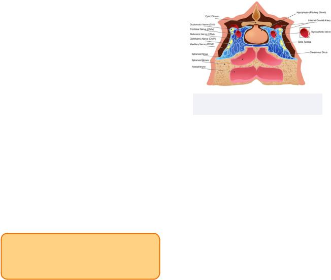
The cavernous sinus
The cavernous sinus is found resting on the sphenoid bone and encircling the pituitary gland. It is a bilateral pair of venous plexuses responsible for draining blood from the brain and select regions of the face (ex. the superior and inferior ophthalmic veins, superficial middle cerebral vein).
Namely, the cavernous sinus contains some key neurovascular structures that pass through it such as:
•Internal carotid artery
•CN III (Oculomotor)
•CN IV (Trochlear)
•CN V1 (Opthalmic)
•CN V2 (Maxillary)
•CN VI (Abducens)
Note: the maxillary nerve may sometimes be found outside the sinus and may therefore not be included.
INBDE Pro Tip:
To remember the contents of the cavernous sinus, a mnemonic can be used: Interesting Onions Turn Odorous, Mostly.
Clinically, a condition known as cavernous sinus thrombosis may result from facial infections. It is generally a rare condition and may be caused by dental infections approximately 10% of the time and sinusitis about 30% of the time. The most common cause is nasal boils (or furuncles) about half the time.
Since the aforementioned neurovascular structures pass through this sinus, an infection may impede their functions, which could lead to a stroke, mydriasis (pupil dilation), vision loss, and ophthalmoplegia (extraocular muscle paralysis) to name a few.
95
Figure 18.07 The cavernous sinus and surrounding structures
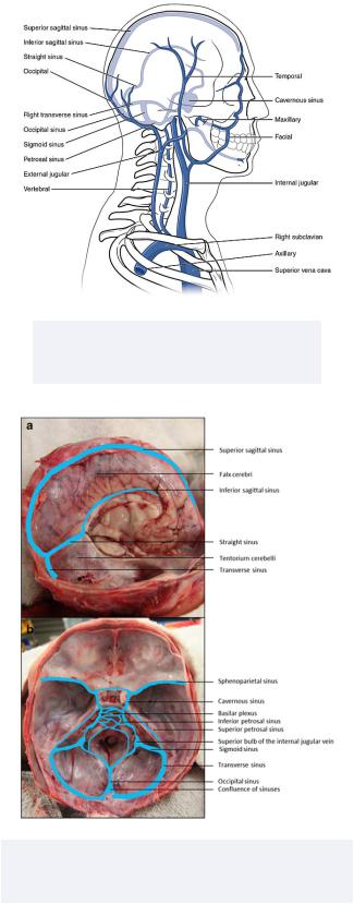
The superior sagittal sinus
The deep venous portions of head are important for drainage of the brain and supporting tissues. One key vessel is the superior sagittal sinus, which is an unpaired vein which lacks valves and courses along the falx cerebri (sheet-like fold of dura separating hemispheres of the brain) along the midline of the superior brain.
The cerebrospinal fluid is able to drain into the superior sagittal sinus via arachnoid villi and granulations. This is because the meningeal dura mater separates the superior sagittal sinus from the subarachnoid space. Due to this vessels' relation to the brain, its interconnectivity with many surrounding vessels, and its size, injury or infection may have serious effects.
INBDE Booster | Booster PrepTM
Head and Neck Anatomy |
96 |
Figure 18.08 The superior sagittal sinus, in addition to other sinuses and major veins of the head and neck
Figure 18.09 A cadaveric image displaying the venous drainage of the brain, with the brain partially intact in the first image
INBDE Booster | Booster PrepTM
Head and Neck Anatomy |
97 |
The Lymphatic System
The lymphatic system is not often discussed, but a solid understanding of this system can be applied in a clinical setting when assessing infections in patients. Candidates will apply their knowledge of immune cells, the circulatory system, and surrounding anatomical structures when studying for this section. These notes will cover the basics of the lymphatic system and the major lymph node groups so that you are prepared for any question that may apply this knowledge on your board exam.
1 The Lymphatic System
Overview
The circulatory system is not the only system with vasculature that spans the body and transports fluids to various organs. The lymphatic system is responsible for moving lymph throughout the body and for its involvement in the body's immune response. The lymphatic system has three main roles:
1.Drains fluids
Interstitial fluid drained from capillaries is returned to the blood
2.Immunological protection
Creates and distributes lymphocytes
Antigen uptake via white blood cells can assist in mounting an immune response
3.Lipid transport
Lipids absorbed by the gastrointestinal tract are transferred throughout the body
-Blood contains water (a polar substance) which does not properly carry non-polar lipids
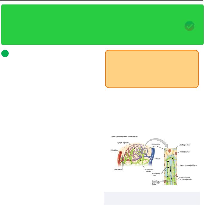
The lymphatic system takes excess fluid, such as plasma, from the circulatory and transports it to nearby lymph nodes via lymphatic vessels. Lymph is created by interstitial fluid that is released into surrounding tissue spaces via a combination of capillary filtration and leakage caused by blood pressure.
INBDE Pro Tip:
When between cells, fluid lost from the circulatory system is called interstitial fluid. However, when it is later taken up by surrounding lymphatic vessels, it becomes lymph.
Components
Lymphatic capillaries will be found between cells, which provides an optimal position for them to absorb interstitial fluid. These capillaries will converge to create larger lymphatic vessels.
Figure 19.01 The anatomical and histological structure of lymphatic capillaries and vessels
INBDE Booster | Booster PrepTM
Head and Neck Anatomy
Lymph is able to move around the body through two main methods:
•Skeletal muscle
Similar to how veins are strategically placed near or between skeletal muscles throughout the body, lymphatic vessels are as well
Skeletal muscle contraction causes lymphatic vessels to constrict, forcing lymph throughout the lymphatic circulation
The presence of lymphatic valves also ensures one-way flow throughout the system
•Respiration
Pressure changes during breathing drives lymph flow
Inhalation means abdominal pressure is high, so lymph moves to the thoracic region towards lower pressure
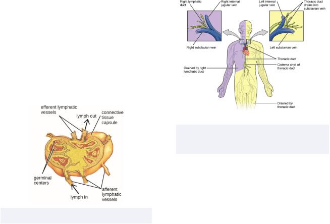
The lymph nodes are ovular clusters of lymphatic tissue that are joined by lymphatic vessels and house lymph in addition to many white blood cells. Lymphocytes in the lymph nodes isolate, sample, and kill pathogens. Clinically, swollen lymph nodes is a sign of infection. A local infection may be indicated by swollen lymph nodes in a particular area.
98
Lymphoid nodules are unlike lymph nodes since they are not encapsulated. These ovular structures can be found in the connective tissue surrounding various organs in the body, often called mucosa-associated lymphatic tissue (MALT). The tonsils are one key example of MALT specific to the oral cavity.
Lymphatic trunks are formed after emerging from lymph nodes. Jugular trunks drain the head and neck. The lymphatic trunks drain into the left and right lymphatic ducts.
•Right lymphatic duct
Drains right side of head and neck, right arm, right thorax
•Left lymphatic (or thoracic) duct
Drains remaining areas of the body (left side and below diaphragm)
Figure 19.03 Lymphatic drainage for the body mapped according to the right and left lymphatic ducts’ responsibilities
Figure 19.02 The anatomy of a lymph node
INBDE Booster | Booster PrepTM
Head and Neck Anatomy
Primary and secondary organs and tissues
There are two main types of lymphatic tissues, primary and secondary. They differ based how they influence lymphocytes.
•Primary: where lymphocytes develop, mature, proliferate, and become immunocompetent
Includes the red bone marrow and thymus
•Secondary: where immune responses are initiated
Includes the lymph nodes, spleen, and lymphatic nodules
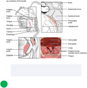
The tonsils
Recall that the tonsils are important lymphoid nodules. They play a key role in providing immunity against pathogens introduced within the oral cavity. Like other nodules, tonsils are not completely encapsulated. As such, they have easy access to sample pathogens passing through the oral cavity.
Recurring tonsilitis may require individuals to have certain tonsils removed (a tonsillectomy) to prevent further infection/inflammation and ease current or future eating/breathing. There are three major types of tonsils:
•Lingual tonsils
Found in submucosa of posterior third of tongue
•Pharyngeal tonsils
Found in mucosa of nasopharyngeal roof
When infected and enlarged, called an adenoid
•Palatine tonsils
Found in tonsillar bed of lateral oropharyngeal wall
opening of the Eustachian tube
Together, all four types of tonsils form
Waldeyer's tonsillar/lymphatic ring, which is one of many regional lymphatic node categorizations in the body.
99
There is one other type of tonsil, which is less commonly discussed.
• Tubal tonsils
These tonsils are located in the lateral wall of the nasopharynx encircling the opening of the Eustachian tube
Together, all four types of tonsils form
Waldeyer's tonsillar/lymphatic ring, which is one of many regional lymphatic node categorizations in the body.
Figure 19.04 The palatine, pharyngeal, and lingual tonsils
2 Superficial Lymph Nodes
Overview
Looking specifically at the head and neck region, there are two major groups of lymph nodes, the superficial and deep cervical.
There are 8 superficial lymph node groups which collect lymph from their associated regions and form a ring-like shape. They drain lymph from the scalp, face, and neck. These groups drain into deep lymph nodes and include:
INBDE Booster | Booster PrepTM

Head and Neck Anatomy
•Pre-auricular nodes
•Occipital nodes
•Mastoid nodes
•Parotid nodes
•Submental nodes
•Submandibular nodes
•Facial nodes
•Superficial cervical (anterior and posterior lateral) nodes
Note: many sources consider different groups or subgroups to be a part of the superficial group.
INBDE Pro Tip:
There are many different ways to remember the superficial lymph nodes of the head and neck region. The first is an acronym "POMPS, So Flipping Special." A pomp is an extravagant display, which would probably be for a very special event. Perhaps it could be you when you celebrate your board exam that you passed!
Now that we have the letters in mind, we can also group the different lymph nodes together in terms of function/location to remember them better.
•Special senses: Occipital and pre-auricular
•Glands: Parotid, submandibular
•Bone/Muscle: Mastoid and facial, respectively
•Other: Superficial cervical, submental
100
Overview
Once lymph has passed through the superficial nodes of the head and neck region, they will drain into the deep lymph nodes. These nodes are vertically oriented and can be found near the internal jugular vein. Once lymph has passed through the deep lymph nodes, it will move through the jugular lymphatic trunks.
The deep lymph nodes drain to the left venous angle through the thoracic duct and the right venous angle through the right lymphatic duct.
There are two main groups of deep lymph nodes, superior and inferior deep lymph nodes. There are many lymph nodes in this area, though they are not well categorized. For your interest, they include the pre-laryngeal, pre-tracheal, para-tracheal, thyroid, lateral and anterior jugular, jugulo-omohyoid, and supraclavicular nodes.
INBDE Booster | Booster PrepTM