

Полезные материалы за все 6 курсов / Учебники, методички, pdf / INBDEBooster Head and Neck Anatomy Notes
.pdfHead and Neck Anatomy
Subclavius
This muscle is small and triangular in shape. By its name alone, its position beneath the clavicle can be inferred.
•Origin: first rib
•Insertion: inferior aspect of clavicle (middle third)
•Action: stabilizes and depresses clavicle
•Innervation: subclavian nerve (C5 and C6)
Figure 15.01 The sternocleidomastoid muscle
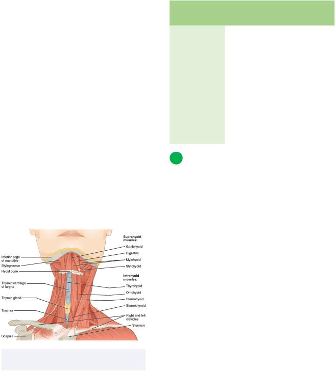
3 Suprahyoid Muscles
Introduction
There are two major groups of muscles that are bordered by the hyoid bone superiorly and inferiorly, called the suprahyoid and infrahyoid muscles respectively. There are four muscles that comprise the suprahyoid group.
71
INBDE Pro Tip:
All muscles relating to the hyoid bone have an easy way to remember their location, group, and, thus, general function.
The prefixes of these muscles are their origin points, whereas their suffixes are their insertion points. Since you know the origin points are either above or below the hyoid bone, you now know its general categorization as a suprahyoid or infrahyoid muscle.
Suprahyoid muscles generally elevate the hyoid bone and depress the mandible, whereas infrahyoid muscles generally depress the hyoid bone and larynx (which opens the airway).
Geniohyoid
The origin of this muscle is associated with the inferior tongue region.
•Origin: mandible (inferior mental spine)
•Insertion: superior aspect of body of hyoid bone
•Action: may elevate hyoid bone or depress mandible
•Innervation: C1 via the hypoglossal nerve (CN XII)
Mylohyoid
The floor of the mouth is predominantly formed by this muscle.
•Origin: mylohyoid line of mandible
•Insertion: mylohyoid raphe and superior aspect of hyoid bone
•Action: elevation of hyoid bone or depression of mandible
•Innervation: mandibular nerve branch (CN V3)
INBDE Booster | Booster PrepTM

Head and Neck Anatomy
Digastric muscle
This muscle's name translates to "two stomachs" or "bellies," which accurately describes its anatomy.
•Origin:
Anterior belly: inferior aspect of mandible at the digastric fossa
Posterior belly: mastoid process of temporal bone at mastoid notch
•Insertion: intermediate tendon by the superior aspect of the body of the hyoid bone
•Action: elevation of hyoid bone and larynx to close epiglottis or depresses mandible
•Innervation:
Anterior belly: branch of CN V3
Posterior belly: branch of CN VII
Stylohyoid
This muscle is as thin and tubular as its bony origin point. "Stylo" means "column" or "pillar."
•Origin: styloid process of temporal bone
•Insertion: body of hyoid bone
•Action: elevates hyoid bone, retracts tongue, assists inspiration by holding pharynx open
•Innervation: facial nerve (CNVII)
Suprahyoid |
Function |
Innervation |
muscle |
|
|
|
|
|
Geniohyoid |
elevates hyoid bone or |
C1 via CN |
|
depresses mandible |
XII |
|
|
|
Mylohyoid |
elevates hyoid bone or |
CN V3 |
|
depresses mandible |
|
|
|
|
Digastric |
elevates hyoid bone or |
Anterior: |
|
depresses mandible, |
CN V3 |
|
closes epiglottis |
Posterior: |
|
|
CN VII |
|
|
|
Stylohyoid |
elevates hyoid bone, |
CN VII |
|
retracts tongue, opens |
|
|
pharynx |
|
|
|
|
72
4 Infrahyoid Muscles
Introduction
This muscle group, also called the "strap muscles" due to their appearance, can be found inferior to the hyoid bone. Another four muscles comprise this group.
Thyrohyoid
This muscle's prefix is related to its association with the thyroid gland region and thyroid cartilage.
•Origin: oblique line of the lamina of thyroid cartilage
•Insertion: inferior aspect of greater horn and body of hyoid bone
•*Action: depresses hyoid bone but elevates larynx
•Innervation: C1 and hypoglossal nerve (CN XII)
Omohyoid
The omohyoid muscle is a very uniquely shaped muscle of the neck. It forms an obtuse J-like shape from its origin point to its insertion point. The prefix "omo" translates to the shoulder region.
•Origin:
Superior belly: intermediate tendon
Inferior belly: superior aspect of scapula
•Insertion:
Superior belly: inferior aspect of body of hyoid bone
Inferior belly: intermediate tendon
•Action: depresses hyoid bone and larynx
•Innervation: the ansa cervicalis formed via spinal nerves C1 to C3 within the cervical plexus
INBDE Booster | Booster PrepTM
Head and Neck Anatomy
Sternothyroid
The prefix and suffix of this muscle conveys its origin and insertion points, respectively.
•Origin: first rib and manubrium of sternum
•Insertion: thyroid cartilage
•Action: depresses hyoid bone and larynx
•Innervation: the ansa cervicalis formed via spinal nerves C1 to C3
Sternohyoid
This muscle spans a longer distance than the sternothyroid muscle.
•Origin: sternum (superior aspect of manubrium) and posteromedial aspect of clavicle
•Insertion: inferior aspect of body of hyoid bone
•Action: depresses hyoid bone larynx
•Innervation: the ansa cervicalis formed via spinal nerves C1 to C3 within the cervical plexus
Figure 15.02 The supra hyoid and infrahyoid muscles of the neck
|
|
73 |
|
|
|
Infrahyoid |
Function |
Innervation |
muscle |
|
|
|
|
|
Thyrohyoid |
*depresses |
*C1 via CN XII |
|
hyoid bone but |
|
|
elevates larynx |
|
|
|
|
Omohyoid |
depresses hyoid |
ansa cervicalis |
|
bone and larynx |
via C1 to C3 |
|
|
|
Sternothyroid |
depresses hyoid |
ansa cervicalis |
|
bone and larynx |
via C1 to C3 |
|
|
|
Sternohyoid |
depresses hyoid |
ansa cervicalis |
|
bone and larynx |
via C1 to C3 |
|
|
|
5 Other Muscles of Importance
Trapezius
This muscle is a part of the posterior muscle group and within the superficial layer subcategory. The trapezius is also considered a muscle of the back because it spans a great width and height, forming a diamond shape.
•Origin:
Descending fibers (superior part): medial third of superior nuchal line
Transverse part: nuchal ligament of C1C6, spinous processes and supraspinous ligaments of vertebrae C7-T3
Ascending fibers (inferior part): supraspinous ligaments and spinous processes of T4-T12
•Insertion:
Descending fibers (superior) part: posterior aspect of lateral third of clavicle
Transverse part: medial acromial margin and superior crest of scapular spine
Ascending fibers (inferior part): medial aspect of scapular spine
INBDE Booster | Booster PrepTM
Head and Neck Anatomy
•Action:
lateral flexion and contralateral head rotation unilaterally, head extension bilaterally
specific fibers/regions support specific movements of the scapula such as rotation, retraction, elevation and depression
•Innervation: spinal accessory nerve (CN XI)
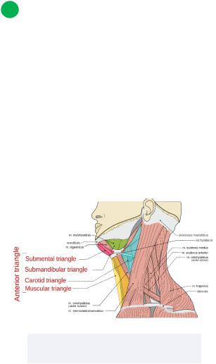
6 Anterior Neck Triangle
The muscles of the neck can be divided into anterior and posterior triangles of the neck via the sternocleidomastoid muscle. The anterior triangle is bound superiorly by the mandible, laterally by the sternocleidomastoid, and medially by the median line of the neck.
There are four main triangles found within this group: the submandibular/digastric, submental/suprahyoid, muscular/ omotracheal/infrahyoid, and carotid.
Figure 15.03 The anterior triangle of the neck and its contents
74
Submandibular/digastric triangle
•Borders:
Superior: mandible
Posterior/Lateral: posterior belly of digastric and stylohyoid
Anterior/Medial: anterior belly of digastric
•Contents:
Lymph nodes, submandibular gland, branches of facial nerve (CN VII), artery and vein, mylohyoid, hyoglossus, middle and superior constrictor muscles,
styloglossus, branches of CN V3, sublingual artery, vein and gland, and CN
XII
Submental triangle
•Borders:
Superior: mandible
Inferior: body of hyoid bone
Medial: midline
Lateral: anterior belly of digastric
•Contents:
Lymph nodes, anterior jugular vein branches, branches of facial artery and vein
Muscular/omotracheal/infrahyoid triangle
•Borders:
Superior: hyoid bone
Lateral: sternocleidomastoid and superior belly of omohyoid
Medial: midline
•Contents:
Infrahyoid muscles, anterior jugular vein, parathyroid and thyroid glands
INBDE Booster | Booster PrepTM
Head and Neck Anatomy
Carotid triangle
•Borders:
Superior: posterior belly of digastric
Anterior/medial: superior belly of omohyoid
Posterior/lateral: sternocleidomastoid
•Contents:
Common, internal, and external carotid arteries, carotid sheath, internal jugular vein, CN X, CN XI, CN XII, and ansa cervicalis
INBDE Pro Tip:
The contents of the anterior triangles of the neck can be easily determined from their names!
Submandibular:
•this triangle has more contents compared to other triangles of the neck
•we are looking at the submandibular area so we will find branches of key cranial nerves associated with the head/face (CN V3 and VII)
•two major glands
•a few muscles of the tongue (hence CN XII) and pharynx
Submental:
•think key vasculature of the neck: anterior jugular and facial artery/vein
Muscular:
•here just remember the infrahyoid group of muscles and (para)thyroid glands!
•this triangle is inferior to the submental
triangle, so the anterior jugular vein will continue here!
Carotid:
•major presence of key neurovasculature!
•carotid vasculature, internal jugular
•CNs X to XII and ansa cervicalis
75
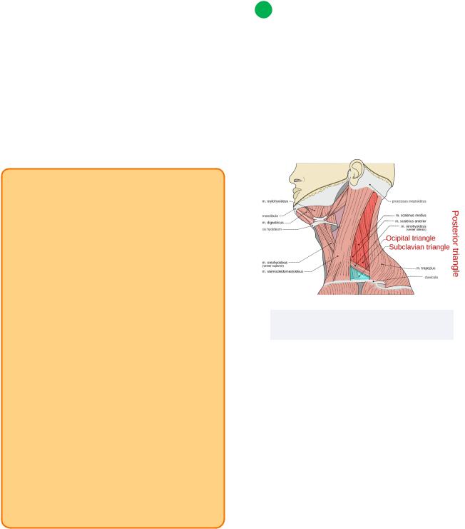
7 Posterior Neck Triangle
The posterior triangle is bound anteriorly by the sternocleidomastoid, posteriorly by the anterior aspect of the trapezius muscle, and inferiorly by the middle third of the clavicle.
There are two main triangles found within this group: the occipital, and supraclavicular/ omoclavicular/subclavian.
Figure 15.04 The posterior triangle and the neck and its contents
Occipital
•Borders:
Anterior: sternocleidomastoid
Posterior: anterior aspect of trapezius muscle
Inferior: inferior belly of omohyoid
•Contents:
CN XI, branches of cervical and brachial plexus
Supraclavicular/omoclavicular/subclavian
•Borders:
Superior: inferior belly of omohyoid
Inferior: clavicle
Anterior: sternocleidomastoid
•Contents:
Brachial plexus trunks, lymph nodes
INBDE Booster | Booster PrepTM
Head and Neck Anatomy |
76 |
INBDE Pro Tip:
The contents of the posterior triangles of the neck can also be easily determined from their names!
Occipital:
•this region is associated with the SCM and trapezius, both of which are innervated by CN XI
•since we are dealing with the posterolateral neck, branches from the brachial and cervical plexuses will be found here
Supraclavicular:
•not much going on here, just a continuation of the brachial plexus (trunks) and some lymph nodes!
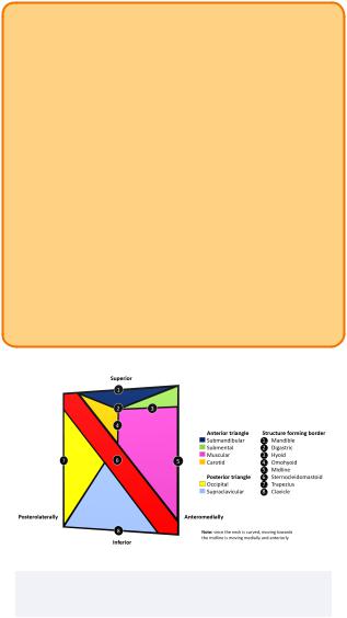
Figure 15.05 The right side of the neck displaying all anatomical triangles
INBDE Booster | Booster PrepTM
Head and Neck Anatomy |
77 |
Muscles of the Pharynx and Larynx
The muscles of the pharynx and larynx can be a bit complicated and daunting at first glance, but they are important in a clinical setting as they relate to both the digestive and respiratory systems. For the board exam, candidates should know the actions of the muscles within these regions and their innervations. These notes include useful diagrams, tips, and tricks that will simplify everything you need to know for this section and help you feel more comfortable if asked a question on this topic.
1 Introduction to the Pharynx
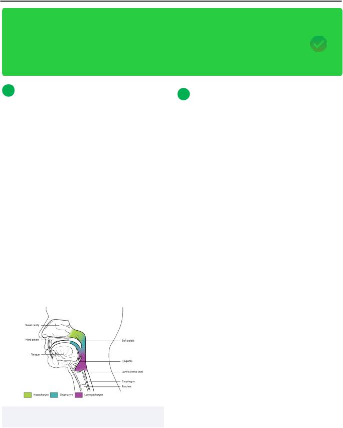
The pharynx is part of both the respiratory tract and digestive system. It can be divided into three main regions: the nasopharynx, oropharynx, and laryngopharynx. These regions are affiliated with the nasal cavity, oral cavity, and the larynx, respectively. Within the nasopharynx, the Eustachian tubes can be found, serving as a connection between the middle ear and nasal cavity to equalize air pressure and remove secretions.
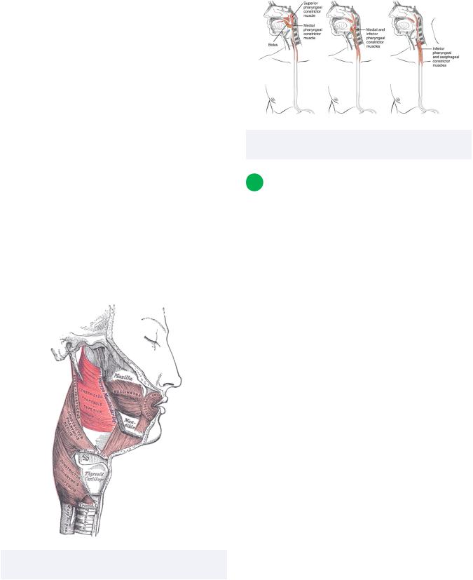
There are two main groups of muscles specific to the pharynx which include the constrictor muscles and longitudinal muscles. Many of these muscles aid in the deglutition, which is the process of swallowing. When food is chewed within the oral cavity, the combination of saliva and food particles forms a ball-like shape called a bolus.
2 Pharyngeal Muscles
Introduction to constrictor group
These muscles will all have a similar function but differ in terms of their anatomical position within the pharynx. They each comprised of involuntary skeletal muscle and act specifically within the scope of the pharynx. Note that these three muscles form the outer circular layer of the pharynx.
Superior constrictor muscle
•Origin: pterygoid hamulus, pterygoid plate, pterygomandibular ligament, mylohyoid line
•Insertion: pharyngeal tubercle of occipital bone, pharyngeal raphe, tongue
•Action: constriction of pharynx, closes soft palate, moves bolus downward
•Innervation: branches of pharyngeal plexus via CN X
Middle constrictor muscle
•Origin: stylohyoid ligament, greater and lesser horn of hyoid bone
•Insertion: pharyngeal raphe
•Action: constriction of pharynx moves bolus downward
•Innervation: branches of pharyngeal plexus via CN X
Figure 16.01 The three main parts of the pharynx: the nano, pro, and laryngopharynx
INBDE Booster | Booster PrepTM
Head and Neck Anatomy
Inferior constrictor muscle
•Origin:
Thyropharyngeal part: lamina of thyroid cartilage
Cricopharyngeal part: cricoid cartilage of larynx
•Insertion:
Thyropharyngeal part: medial pharyngeal raphe
Cricopharyngeal part: esophageal fibers
•Action: constriction of pharynx moves bolus downward
•Innervation: branches of pharyngeal plexus, external and recurrent laryngeal nerves via CN X
Note: some consider the cricopharyngeal part as its own muscle (called the cricopharyngeus) but grouped with the inferior constrictor muscle
Figure 16.02 The pharyngeal constrictor muscles and inner longitudinal group
78
Figure 16.03 The pharyngeal constrictor muscles assisting in deglutition
3 Introduction to the Larynx
Introduction
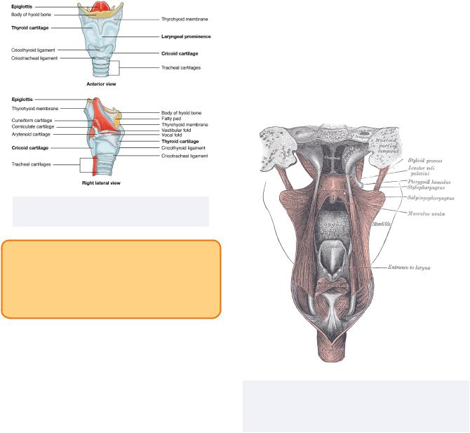
The larynx is important for connecting the upper segment of the gastrointestinal tract (mouth/oral cavity, laryngopharynx) with the respiratory tract. One of the major components here is the epiglottis, which closes off the larynx during swallowing to ensure that food does not enter the trachea and impede breathing. The larynx is important for the ability to speak since it houses the vocal cords.
The larynx is also home to many intricate muscles that support and act on the laryngeal cartilages. The details related to these cartilaginous components are beyond the scope of the board exam. You are welcome to read more about them on your own if you are interested, as it may help you understand the overall relationship between the laryngeal muscles and the laryngeal cartilages.
Many laryngeal muscles have names that allude to their origin and insertion which can be seen by looking at their prefixes and suffixes, respectively. Additionally, many names include the laryngeal cartilages they are associated with, such as the thyroid, arytenoid, an
INBDE Booster | Booster PrepTM
Head and Neck Anatomy |
79 |
Palatopharyngeus
• Origin: hard palate, palatine aponeurosis
• Insertion: thyroid cartilage
• Action: elevates pharynx during swallowing, assists in moving bolus downwards
• Innervation: branches of pharyngeal plexus via CN X
Figure 16.04 The bony and cartilaginous components of the larynx
INBDE Pro Tip:
To ensure you don't confuse the function of the larynx and pharynx remember this:
•The pharynx is for "phood" and air
•The larynx is for language (air)
Introduction to the inner longitudinal group
This section will detail the muscles of the inner longitudinal group, which are associated with both the pharyngeal and laryngeal regions.
This group includes the stylopharyngeus, palatopharyngeus and salpingopharyngeus.
Stylopharyngeus
•Origin: styloid process (temporal bone)
•Insertion: thyroid cartilage, pharyngeal constrictors
•Action: elevates pharynx during swallowing
•Innervation: glossopharyngeal nerve (CN IX)
Figure 16.5 A posterior view of the muscles of the palate, pharynx, and larynx. The palatopharyngeus (labelled pharyngo-palatinus) can be seen posterior to the muscles uvulae
Salpingopharyngeus
•Origin: auditory tube
•Insertion: palatopharyngeus muscle
•Action: elevates pharynx during swallowing
•Innervation: branches of pharyngeal plexus via CN X
INBDE Booster | Booster PrepTM
Head and Neck Anatomy
Inner |
Function |
Innervation |
longitudinal |
|
|
muscle |
|
|
|
|
|
Stylo-pharyngeus |
elevates |
*CN IX |
|
pharynx |
|
|
|
|
Palato- |
elevates |
CN X |
pharyngeus |
pharynx |
|
|
|
|
Salpingo- |
elevates |
CN X |
pharyngeus |
pharynx |
|
|
|
|
Introduction to larynx-specific muscles
The intrinsic laryngeal muscles include various sub-groups based on function:
1.Abductors: posterior cricoarytenoid
2.Adductors: lateral, oblique and transverse arytenoid
3.Sphincters: transverse and oblique arytenoid
4.Vocal cord relaxers: thyroarytenoid, vocalis
5.Vocal cord tensors: cricothyroid
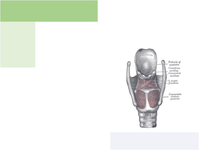
Posterior cricoarytenoid
•Origin: cricoid lamina
•Insertion: muscular process of arytenoid cartilage
•Action: abduction of vocal folds, opens glottis
•Innervation: recurrent laryngeal nerve via CN X
Oblique and transverse arytenoids
•Origin: muscular process of arytenoid cartilage
•Insertion: superior aspect of contralateral arytenoid cartilage
•Action: sphincter of laryngeal inlet which prevents food/liquids from entering trachea, adducts arytenoid cartilage
•Innervation: recurrent laryngeal nerve via CN X
80
Lateral cricoarytenoid
•Origin: cricoid cartilage
•Insertion: muscular process of arytenoid cartilage
•Action: adducts and shortens vocal folds
•Innervation: recurrent laryngeal nerve via CN X
Figure 16.06 A posterior view of posterior cricoarytenoid, oblique (X-like shape) and transverse (horizontal fibers) arytenoids
INBDE Booster | Booster PrepTM