

Полезные материалы за все 6 курсов / Учебники, методички, pdf / INBDEBooster Head and Neck Anatomy Notes
.pdfHead and Neck Anatomy
INBDE Pro Tip:
There are many ways to remember the cranial nerves by grouping:
•CNs I, II, and VIII are grouped together because they are all special sensory cranial nerves (smell, vision, balance, and hearing, respectively)
•CNs III, IV, and VI are all related to muscles that move the eye
•CNs V, VII, IX, and X are all major cranial nerves that have numerous important branches and innervate many key structures; thus, they have both sensory and motor components
•CNs XI and XII are the remaining cranial nerves that only move a few muscles each
The cranial nerves can also be remembered by linking their names to their functions. Their numbers can also be used for the same purpose. This will be described throughout the notes for many of the cranial nerves.
2 Cranial nerves I and II
CN I (Olfactory nerve)
The olfactory nerve is related to the nasal cavity in terms of its course and function.
•Modality:
Sensory
•Location:
Extends from telencephalon
•Function:
Smell
•Pathway:
Olfactory nerve endings (pass through crista galli of ethmoid bone) --> olfactory bulb --> olfactory tract --> primary olfactory cortex in temporal lobe
31
INBDE Pro Tip:
Human beings only have one nose, just like the olfactory nerve is cranial nerve one! The olfactory nerve sounds like "old factory," which probably doesn't smell too good.
Figure 6.03 The olfactory nerve and its path
CN II (Optic nerve)
The optic nerve is related to the eyes and is crucial for vision.
•Modality:
Sensory
•Location:
Extends from diencephalon and exits via the optic canal
•Function:
Vision
•Pathway:
Retina --> optic nerve --> optic chiasma ("crossing" of visual information across hemispheres) --> lateral geniculate bodies --> occipital lobe
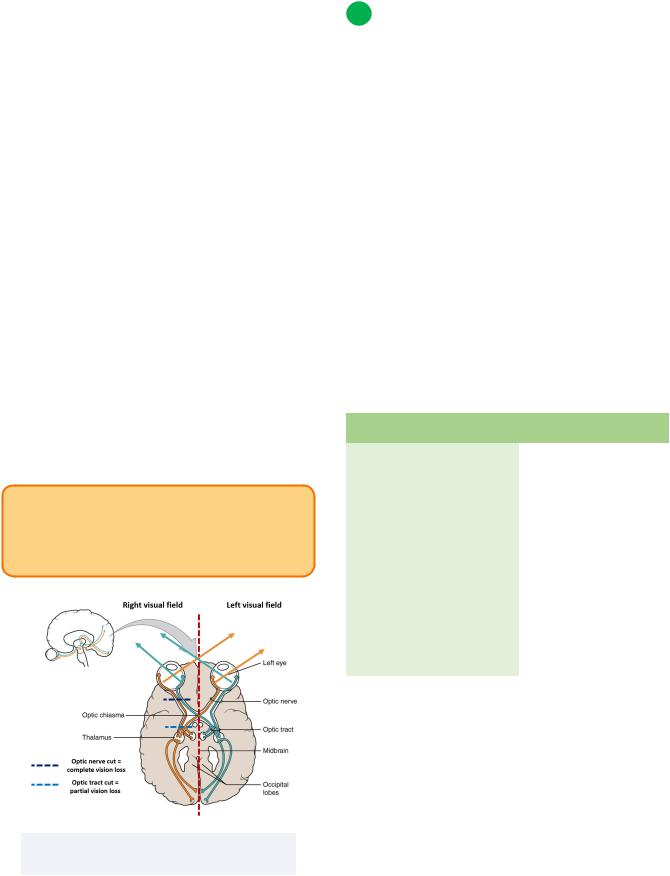
The eyes are able to receive visual information from the medial (or nasal) and lateral (or temporal) aspects of the eyes. The left visual field is sensed by the medial aspect of the eye for both eyes. The right visual field is sensed by the lateral aspect of the eye for both eyes.
INBDE Booster | Booster PrepTM
Head and Neck Anatomy
Depending on where an injury, lesion ,or tumor may arise on the optic nerve pathway, visual outcomes differ. For example:
•Optic nerve cut:
Complete vision loss = unable to see both fields of vision in one eye
-Ex. optic nerve cut on right side means left and right visual fields in that eye are affected
•Optic tract cut:
Partial vision loss = loss of visual field on contralateral (opposite) side of lesion in both eyes
-Ex. a cut of the right optic tract means left visual field on both eyes affected
One important note is that the eyes contain rod and cone cells, which are important for seeing in low light conditions and for distinguishing between colours or shades. Women have more cone cells than men, which is why they are able to distinguish between shades better than men can.
INBDE Pro Tip:
Human beings have two eyes, just like the pic nerve is cranial nerve two! The optic nerve has a name that gives away its functions
32
3 Cranial nerves III, IV, VI
Overview
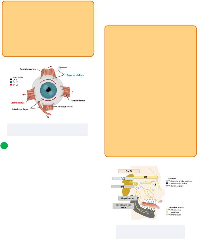
The extraocular eye muscles are controlled by three cranial nerves: the oculomotor (CN III), trochlear (CN IV), and abducens (CN VI).
These muscles are discussed in more detail in their own section of the notes.
•Modality:
Motor
•Location:
Not important for INBDE
•Function:
Moves the eye
-*CN III also constricts the pupils via PSNS fibers to smooth muscles in the eye
•Pathway:
Not important for INBDE
Cranial nerve |
Secretion |
|
|
CN III: Oculomotor |
Levator palpabrae |
|
superioris* |
|
Superior rectus |
|
Inferior rectus |
|
Medial rectus |
|
Inferior oblique |
|
*opens upper eyelid |
|
|
CN IV: Trochlear |
Superior oblique |
|
|
CN VI: Abducens |
Lateral rectus |
|
|
Figure 6.04 The pathway and clinical application of the optic Nerve
INBDE Booster | Booster PrepTM
Head and Neck Anatomy
INBDE Pro Tip:
The trochlear nerve is the fourth cranial nerve. The abducens nerve is the sixth nerve.
One way to remember the innervation of the extraocular muscles is the chemistry formula-like mnemonic LR6SO4O3 where the letters stand for the muscles and the subscript numbers stand for the cranial nerves that innervate them. Note that O3 stands for all other muscles.
Figure 6.05 The extra ocular muscles and their innervation
4 Cranial nerve V
Overview
The trigeminal nerve is very important in the field of dentistry and for the board exam. This is because it has three major branches that are relevant to the face, glands, and muscles of mastication.
•Modality:
Both sensory and motor
•Location:
Lateral pons
•Function:
Various
The three branches of CN V are the ophthalmic (CN V1), maxillary (CN V2), and mandibular (CN V3).
33
These branches course through the superior orbital fissure, the foramen rotundum, and the foramen ovale, respectively.
INBDE Pro Tip:
The branches of the trigeminal nerve differ in their modality. V1, V2, and V3 are sensory, sensory, and mixed.
One way to remember this is by imagining what each branch innervates. On the face, most structures are immovable in the ophthalmic and maxillary regions. Don't worry about the eye or facial muscles, as those are innervated by other cranial nerves. The mandible can be moved, and that's why V3 is mixed; it has both sensory and motor modalities.
Thus, for the branches of CN V in order, "Stationary, Stationary, Moveable" can be a useful mnemonic.
Additionally, to remember the course of the branches through foramina of the skull, use the mnemonic "Sticky Raisin Oatmeal" (Superior orbital fissure, foramen Rotundum, foramen Ovale).
Figure 6.06 The branches of the trigeminal nerve
INBDE Booster | Booster PrepTM

Head and Neck Anatomy
CN V1
The ophthalmic branch is related to the eye and upper scalp regions.
•Modality:
Sensory
•Regions:
Forehead, scalp, upper conjunctiva, frontal and ethmoidal sinuses, upper eyelid, and medial nose
34
•Posterior superior alveolar nerve
Innervates maxillary sinus, gingiva, and cheek
Innervates upper molar teeth (1st, 2nd, 3rd) except mesiobuccal root of 1st molar about 30% of the time
•Middle superior alveolar nerve
Innervates maxillary sinus, gingiva, and cheek
Innervates upper premolar teeth (1st and 2nd) and mesiobuccal root of 1st molar about 30% of the time
•Anterior superior alveolar nerve
Innervates the maxillary sinus
Innervates the upper incisors (central and lateral) and canines
Figure 6.07 The sensory innervation of the trigeminal branches
CN V2
The maxillary branch is related to the infraorbital and maxillary regions.
•Modality:
Sensory
•Regions:
Inferior eyelid, anterior cheek, palate, maxillary teeth, maxillary sinus, lower eyelid, lateral nose, upper lip
The maxillary branch also has three important sub-branches relevant to the teeth. They are named according to their position relative to one another and their general position in the maxillary region. They include the posterior, middle, and anterior superior alveolar nerves.
Figure 6.08 The superior alveolar nerve branches of CN V2
Figure 6.09 The superior alveolar nerve branch innervation of maxillary teeth
INBDE Booster | Booster PrepTM
Head and Neck Anatomy
CN V3
The mandibular branch is related to many important regions of the oral cavity..
•Modality A):
Sensory
•Regions:
Buccal, parotid and temporal regions, anterior 2/3 of tongue, lower lip, mandibular teeth
•Modality B):
Motor
•Regions:
Mylohyoid, anterior belly of digastric, tensor palatini, tensor tympani
The muscles of mastication
-Temporalis, masseter, medial and lateral pterygoids
5 Cranial nerve VII
Overview
The facial nerve is another important cranial nerve to know well.
•Modality:
Both sensory and motor
•Location:
Pontomedullary junction
•Function:
Four main ones that provide sensory info to ear, palate, and tongue
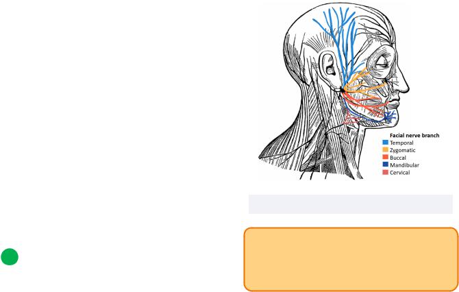
There are five branches of the facial nerve. From superior to inferior they include:
•Temporal
•Zygomatic
•Buccal
•Mandibular
•Cervical
These branches innervate muscles in their respective regions. For example, the zygomatic branch innervates the zygomaticus major and minor muscles, and the cervical branch innervates the platysma.
35
Figure 6.10 The branches of the facial nerve
INBDE Pro Tip:
A useful mnemonic to remember the order of the facial nerve branches from superior to inferior is "Two Zombies Bit My Cat."
Somatic motor innervation:
•Muscles of facial expression*
•Occipital belly of occipitofrontalis
•Stapedius
•Posterior auricular muscles
•Stylohyoid
•Posterior belly of digastric
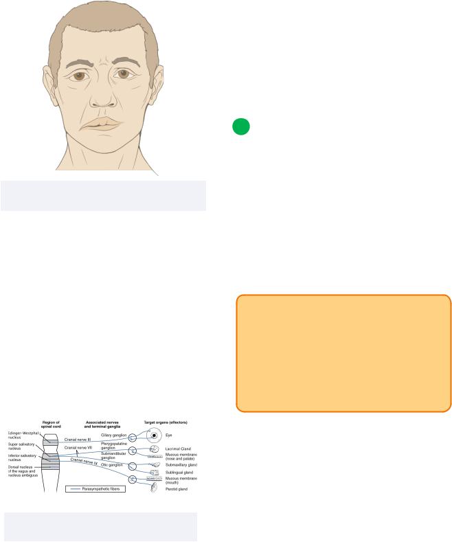
*Clinically, damage to the facial nerve may result in weakness or paralysis on one side of the face. This condition is known as Bell's Palsy. Symptoms include an inability to the close eyes, asymmetrical facial tone, and a sagging face.
INBDE Booster | Booster PrepTM
Head and Neck Anatomy
Figure 6.11 Bell’s palsy demonstrating facial paralysis of the right side of the face
Visceral motor innervation:
Stimulates secretions of the:
•Lacrimal gland
•Sublingual gland
•Submandibular gland
•Nasal, palatine, pharyngeal mucous glands
Damage to the facial nerve can also affect gland secretion. As such, visceral motor function of any of the above glands may be affected. In the case of salivary glands being impacted, xerostomia (dry mouth) may ensue.
Figure 6.12 The innervation of various glands located in the head
36
Special sensory innervation:
Receives information from the:
• Anterior 2/3 of the tongue for taste
Somatic sensory innervation:
Receives information from the:
• External acoustic meatus of ear
6 Cranial nerves VIII and IX
CN VIII
The vestibulocochlear nerve is of lesser importance for the board exam. It provides the body with the special sensory ability to sense equilibrium and hear.
•Modality:
Sensory
•Function:
Balance and hearing
INBDE Pro Tip:
The vestibulocochlear nerve alludes to its function from its name. The vestibular component is related to equilibrium, and the cochlear component is related to hearing. One way to remember the number of this nerve is by flipping the 8 sideways, the infinity sign looks a little bit like a balancing act "∞."
CN IX
The glossopharyngeal nerve is another major cranial nerve in the body because its scope of innervation is quite large. It is involved in numerous pathways, ranging from taste to salivary gland stimulation.
•Modality:
Both sensory and motor
•Location:
Medulla
•Function:
Various (4)
INBDE Booster | Booster PrepTM
Head and Neck Anatomy
There are six main branches of the glossopharyngeal nerve which include:
•Tympanic
•Carotid sinus
•Pharyngeal
•Muscular branch to stylopharyngeus
•Tonsillar
•Lingual
Figure 6.13 The branches of the glossopharyngeal nerve
INBDE Pro Tip:
To remember the glossopharyngeal nerve branches, use the mnemonic "This Could Pull Me To Love.”
Somatic motor innervation:
Stimulates the:
•Stylopharyngeus to widen the pharynx and elevate the larynx during swallowing
Visceral motor innervation:
Stimulates the:
• Parotid gland for salivary secretions
37
Somatic sensory innervation:
Receives information from the:
•Middle ear
•Pharynx
•Soft palate
•Tonsils
•Posterior 1/3 of tongue
Special sensory innervation:
• Posterior 1/3 of the tongue for taste
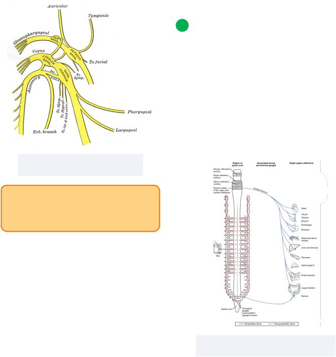
7 Cranial nerve X
Overview
The vagus nerve is also a major cranial nerve in the body because it spans a great distance and is associated with a variety of key organs and organ systems.
•Modality:
Both sensory and motor
•Location:
Medulla
•Function:
Various (4)
Figure 6.14 The overview of the vagus nerve’s parasympathetic innervation
INBDE Booster | Booster PrepTM

Head and Neck Anatomy
INBDE Pro Tip:
To remember the vagus nerve, the word "vagus" means "wandering," which is exactly what this nerve does as it courses and descends throughout the body.
Additionally, the vagus nerve starts with a "V" and if you add those two roman numerals together, the sum is 10 - the number of this cranial nerve!
Special sensory:
Receives information from the:
• Epiglottis for taste
Somatic sensory:
Receives information from the:
•Larynx
•Laryngopharynx
•Superficial external ear
•Epiglottis
Visceral sensory/motor:
Under PSNS control, the following regions receive fibers from the vagus nerve:
•The heart
Influences heart rate
•Lungs
Influences respiratory rate
•Digestive tract
May promote digestive processes via peristalsis and secretions
Somatic motor:
Support swallowing by stimulating the:
•Pharynx
•Larynx
•Soft palate muscles
38
Figure 6.15 The vagus nerve and its course as it descends into the thorax
INBDE Booster | Booster PrepTM
Head and Neck Anatomy
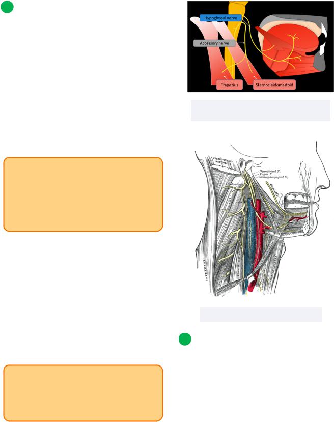
8 Cranial nerves XI and XII
CN XI
The (spinal) accessory nerve is not as important as other cranial nerves for the board exam. Thankfully, it is still very straightforward to understand and remember
•Modality:
Motor
•Location:
Medulla
•Function:
Moves the sternocleidomastoid and trapezius
INBDE Pro Tip:
The (spinal) accessory nerve is the 11th cranial nerve. One way to remember this is that the number 11 looks a bit like the spinal column, which makes sense considering that this cranial nerve innervates two muscles in relation to the neck.
CN XII
This cranial nerve is also not often tested on the board exam, though it is relevant to dentistry due to the select muscles it innervates.
•Modality:
Motor
•Location:
Medulla
•Function:
Moves muscles of the tongue
INBDE Pro Tip:
The hypoglossal nerve tells us its function based on its name. Its prefix and suffix relate to the inferior muscles of the tongue, which is the functional innervation for this cranial nerve.
39
Figure 6.16 The spinal accessory and hypoglossal nerves
Figure 6.17 The hypoglossal nerve
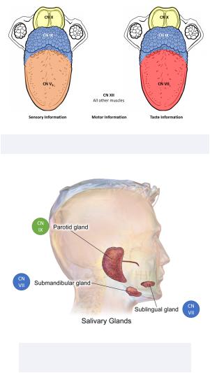
9 Putting it all together
Summary
The cranial nerves may be intimidating at first, but once you start noticing patterns and linking information from other sections together, it becomes much easier to understand.
INBDE Booster | Booster PrepTM
Head and Neck Anatomy |
40 |
Figure 6.18 The innervation of the tongue
Figure 6.19 The innervation of the Three main salivary glands
INBDE Booster | Booster PrepTM