Полезные материалы за все 6 курсов / Учебники, методички, pdf / Kaplan Pediatrics USMLE 2CK 2021
.pdf
USMLE Step 2 CK λ Pediatrics
−Findings (with salt losing):
°Progressive weight loss (through 2 weeks of age), anorexia, vomiting, dehydration, weakness, hypotension
°Affected females—masculinized external genitalia (internal organs normal)
°Males normal at birth; postnatal virilization
•Laboratory evaluation
−Increased 17-OH progesterone
−Low cortisol, increased androstenedione and testosterone
−Hyperkalemia, hyponatremia, low aldosterone
−High plasma renin activity (PRA), particularly the ratio of PRA to aldosterone (markers of impaired mineralocorticoid synthesis)
−Definitive test—measure 17-OH progesterone before and after an intravenous bolus of ACTH
•Treatment
−Hydrocortisone
−Fludrocortisone if salt losing
−Increased doses of both hydrocortisone and fludrocortisone in times of stress
−Corrective surgery for females
Cushing Syndrome
•Exogenous—most common reason is prolonged exogenous glucocorticoid administration.
•Endogenous
−In infants—adrenocortical tumor (malignant)
−Excess ACTH from pituitary adenoma results in Cushing disease (age >7 years)
•Clinical findings
−Moon facies
−Truncal obesity
−Impaired growth
−Striae
−Delayed puberty and amenorrhea
−Hyperglycemia
−Hypertension common
−Masculinization
−Osteoporosis with pathologic fractures
•Laboratory evaluation
−Dexamethasone-suppression test (single best test)
−Determine cause—CT scan (gets most adrenal tumors) and MRI (may not see if microadenoma)
•Treatment—remove tumor; if no response, remove adrenals; other tumor-specific protocols
174

Chapter 16 λ Endocrine Disorders
Clinical Recall
What laboratory abnormality is expected in patients with 21-hydroxylase deficiency?
A.Hyperglycemia
B.Hyponatremia
C.Hypokalemia
D.High cortisol
E.High aldosterone
Answer: B
DIABETES MELLITUS
Type 1
An 8-year-old boy arrives in the emergency department with vomiting and abdominal pain of 2 days’ duration. His mother states he has been drinking a lot of fluids for the past month and has lost weight. Physical examination reveals a low-grade fever and a moderately dehydrated boy who appears acutely ill. He is somnolent but asks for water. Respirations are rapid and deep. Laboratory tests reveal a metabolic acidosis and hyperglycemia.
•Etiology—T-cell−mediated autoimmune destruction of islet cell cytoplasm, insulin autoantibodies (IAA)
•Pathophysiology—low insulin catabolic state
−Increased glucose production and decreased tissue utilization lead to increased serum glucose concentration → osmotic diuresis (hyperosmotic state); result is a loss of fluid and electrolytes, and eventual dehydration
−Activation of renin-angiotensin-aldosterone axis can lead to accelerated potassium loss
−Increased catabolism → cellular loss of Na, K and phosphate
−Increased release of free fatty acids from peripheral fat stores = substrates for hepatic ketoacid production → depleted buffer system → metabolic acidosis
•Clinical presentation
−Polyuria
−Polydipsia
−Polyphagia
−Weight loss
−Most initially present with diabetic ketoacidosis
Published by dr-notes.com |
175 |
|
|
|
|

USMLE Step 2 CK λ Pediatrics
•Diagnostic criteria
−Impaired glucose tolerance test: fasting blood sugar 110–126 mg/dL or 2-hour glucose during OGTT<200 mg/dL but ≥125 mg/dL
−Diabetes: symptoms + random glucose ≥200 mg/dL or fasting blood sugar ≥126 mg/dL or 2 hour OGTT glucose ≥200 mg/dL
−Diabetic ketoacidosis—hyperglycemia, ketonuria, increased anion gap, decreased HCO3 (or total CO2), decreased pH, increased serum osmolality
•Treatment
−Insulin replacement—goal is to provide in as physiologic a manner as possible. Give basal insulin and a preprandial insulin. The basal insulin is either long-acting (glargine or detemir) or intermediate-acting (NPH).
−American Diabetes Association—test blood sugar before meals and snacks, before bed, before exercising or driving, and with suspicion of low blood sugar
−Dietary management—healthy, balanced diet (high in carbohydrates and fiber, low in fat)
−Close patient follow-up
−Diabetic ketoacidosis:
°Most important FIRST step: start insulin infusion to accelerate the movement of glucose into cells → decreases hepatic glucose production + stops the movement of FFA from periphery to liver
°Begin rehydration at the same time; also lowers serum glucose level by improving renal perfusion and renal excretion
°There is a rapid initial decrease in serum glucose; when the glucose level is <180 mg/dL (renal threshold), diuresis stops and rehydration accelerates.
°Rehydration is slow, 24–36 hours depending on severity of DKA. Note: A rapid decline in effective serum osmolality can represent an excess of free water entering the vascular space and increasing risk of cerebral edema.
−Exercise
°All forms of exercise or competitive sports should be encouraged.
°Regular exercise improves glucose control.
°May need additional CHO exchange
Type 2
•Most common cause of insulin resistance is childhood obesity.
•Symptoms more insidious
−Usually excessive weight gain
−Fatigue
−Incidental glycosuria (polydipsia and polyuria uncommon)
•Risk factors
−Age 10-19 years
−Overweight to obese (BMI for age and sex >85%)
−Non-Caucasian
−History of type 2 DM in 1stor 2nd-degree relatives
−Having features of the metabolic syndrome
176

Chapter 16 λ Endocrine Disorders
•Features of the Metabolic Syndrome
−Glucose intolerance leads to L hyperglycemia
−Insulin resistance
−Obesity
−Dyslipidemia
−Hypertension
−Acanthosis nigricans
•Screening and Treatment
−Who: All who meet the BMI criteria + 2 risk factors
−How to screen: fasting blood glucose every 2 years beginning at age 10 years or onset of puberty if above criteria are met
−Diagnosis: same criteria (glucose levels) as adults
−Treatment: first and most important is nutritional education and improved exercise level, but most will eventually need an oral hypoglycemic
Maturity-Onset Diabetes of Youth (MODY)
−Primary autosomal dominant defect in insulin secretion (6 types based on gene mutation)
−Diagnosis: 3 generations of DM with autosomal; dominant transmission and diagnosis of onset age <25 years
−Best test: molecular genetics for mutation (facilitates management and prognosis)
Published by dr-notes.com |
177 |
|
|
|
|


Orthopedic Disorders 17
Chapter Title
Learning Objectives
Recognize and describe treatments for childhood disorders of the hip, knee, foot, spine, and upper limbs
Diagnose and describe treatments for osteomyelitis, septic arthritis, osteogenesis imperfecta, and bone tumors
DISORDERS OF THE HIP
Developmental Dysplasia of the Hip (DDH)
•General ligamental laxity
−Family history
−Significantly more females
−Firstborn
−Breech
−Oligohydramnios
−Multiple gestation
•Physical examination
−Barlow: will dislocate an unstable hip; is easily felt (clunk not a click)
−Ortolani (most important clinical test for detecting infant hip dysplasia): reduces a recently dislocated hip (most at 1–2 months of age), but after 2 months, usually not possible because of soft-tissue contractions
•All infants with positive exams should immediately be referred to an orthopedic surgeon (per standard of practice of the AAP); no radiographic confirmation is needed
•If equivocal, can repeat exam in 2 weeks and if equivocal then a dynamic U/S of the hips is the best test (age <4 months) or hip x-ray (age >4 months)
•Treatment
−Pavlik harness for 1–2 months (highly effective)
−Casting (if needed), surgery (rarely necessary)
•Complications—acetabular dysplasia, leg length discrepancy
Published by dr-notes.com |
179 |
|
|
|
|

USMLE Step 2 CK λ Pediatrics
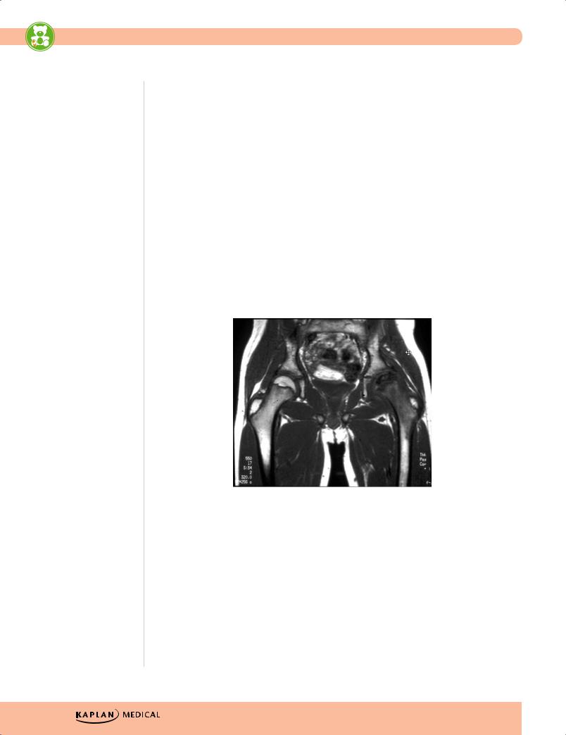
Legg-Calvé-Perthes Disease
A 5-year-old boy has developed progressive limping. At first painless, it now hurts to run and walk. The pain is in the anterior thigh. The pain is relieved by rest. Parents recall no trauma.
•Idiopathic avascular necrosis of the capital femoral epiphysis in immature, growing child
•More in males; 20% bilateral; sometimes after trauma
•Presentation—mild intermittent pain in anterior thigh with painless limp with restriction of motion
•Diagnosis—anterior/posterior and frog leg lateral x-ray shows compression, collapse, and deformity of femoral head
•Treatment
−Containment (femoral head within acetabulum) with orthoses or casting
−Bed rest
−Abduction stretching exercises
−If significant femoral deformity persists, surgical correction
©2007 Kaplan Medical. Reproduced with permission from
Dr. Philip Silberberg, University of California at San Diego
Figure 17-1. MRI Demonstrating Legg-Calve-Perthes Disease
Slipped Capital Femoral Epiphysis (SCFE)
•Most common adolescent hip disorder
•Either obese with delayed skeletal maturation, or thin with a recent growth spurt
•Can occur with an underlying endocrine disorder
•Clinical presentation
−Pre-slip stable; exam normal; mild limp external rotation
−Unstable slip; sudden-onset extreme pain; cannot stand or walk; 20% complain of knee pain with decreased hip rotation on examination
180

Chapter 17 λ Orthopedic Disorders
•Complications—osteonecrosis (avascular necrosis) and chondrolysis (degeneration of cartilage)
•Diagnosis—AP and frog-leg lateral x-ray, earliest finding: widening of physis without slippage (preslip); as slippage occurs, femoral neck rotates anteriorly while head remains in acetabulum
•Treatment—open or closed reduction (pinning)
©2007 Kaplan Medical. Reproduced with permission from
Dr. Philip Silberberg, University of California at San Diego
Figure 17-2. X-ray of the Hips Demonstrating
Slipped Capital Femoral Epiphysis
Transient Synovitis
•Viral; most 7–14 days after a nonspecific upper respiratory infection; most at 3–8 years of age
•Clinical presentation
−Acute mild pain with limp and mild restriction of movement
−Pain in groin, anterior thigh, and knee
•Diagnosis
−Small effusion (±)
−Slight increase in ESR
−Normal x-rays
−No to low-grade fever; non-toxic-appearing
•Treatment—bed rest and no weight-bearing until resolved (usually <1 week), then 1–2 weeks of limited activities
Published by dr-notes.com |
181 |
|
|
|
|

USMLE Step 2 CK λ Pediatrics
Note
In talipes equinovarus, the patient’s heel can’t go flat on the exam surface (as opposed to metatarsus adductus, in which the heel can).
Clinical Recall
A 12-year-old boy presents with a limp. He is overweight. Radiographs are concerning for slipped capital femoral epiphysis. What is the treatment of choice?
A.Pavlik harness
B.Surgical pinning
C.Casting and rest
D.Physical therapy
E.Antibiotics
Answer: B
INTOEING
Metatarsus Adductus
•Most common in firstborn (deformation)
•Treatment—primarily nonsurgical; serial plaster casts before 8 months of age; orthoses, corrective shoes; if still significant in a child age >4 years, may need surgery
Talipes Equinovarus (Clubfoot)
A newborn is noted to have a foot that is stiff and slightly smaller than the other one. The affected foot is medially rotated and very stiff, with medial rotation of the heel.
•Congenital, positional deformation, or associated with neuromuscular disease
•Hindfoot equinus, hindfoot and midfoot varus, forefoot adduction (at talonavicular joint)
•Treatment
−Complete correction should be achieved by 3 months (serial casting, splints, orthoses, corrective shoes); if not, then surgery
Internal Tibial Torsion
•Most common cause of intoeing <2 years of age (also because of in utero positioning); often with metatarsus adductus
•Measure prone thigh/foot angles
•No treatment needed—resolves with normal growth and development; takes 6–12 months (is physiologic)
182

Chapter 17 λ Orthopedic Disorders
Internal Femoral Torsion (Femoral Anteversion)
•Most common cause of intoeing ≥2 years of age; entire leg rotated inwardly at hip during gait
•Most are secondary to abnormal sitting habits (W-sitting).
•Treatment—observation; takes 1–3 years to resolve; surgery only if significant at >10 years of age
DISORDERS OF THE KNEE
Osgood-Schlatter Disease
•Traction apophysitis of tibial tubercle (overuse injury)
•Look for active adolescent (running, jumping)
•Swelling, tenderness, increased prominence of tubercle
•Treatment—rest, restriction of activities, knee immobilization, isometric exercises
•Complete resolution requires 12–24 months
DISORDERS OF THE SPINE
Scoliosis
A 12-year-old girl is seen for routine physical examination. She voices no complaints. Examination is remarkable for asymmetry of the posterior chest wall on bending forward. One shoulder appears higher than the other when she stands up.
•Most are idiopathic
•Others are congenital, with neuromuscular disorders, compensatory, or with intraspinal abnormalities.
•Slightly more females than males; more likely to progress in females
•Adolescent (>11 years) more common
•Adams test bending forward at hips —almost all with >20-degree curvature are identified in school screening programs (but many false positives)
•Diagnosis—x-ray is standard: posterior/anterior and lateral of entire spine gives greatest angle of curvature
•Treatment—trial brace for immature patients with curves 30–45 degrees and surgery for those >45 degrees (permanent internal fixation rods)
Published by dr-notes.com |
183 |
|
|
|
|
