

Полезные материалы за все 6 курсов / Учебники, методички, pdf / Kaplan Pediatrics USMLE 2CK 2021
.pdfUSMLE Step 2 CK λ Pediatrics
•Diagnosis
−Urinalysis shows proteinuria (3–4 +)
−Some with microscopic hematuria
−24-hour urine protein—40 mg/m2/hour in children but now preferred initial test is a spot urine for protein/creatinine ratio >2
−Serum creatinine usually normal but may be increased slightly
−Serum albumin <2.5 g/dL
−Elevated serum cholesterol and triglycerides
Note
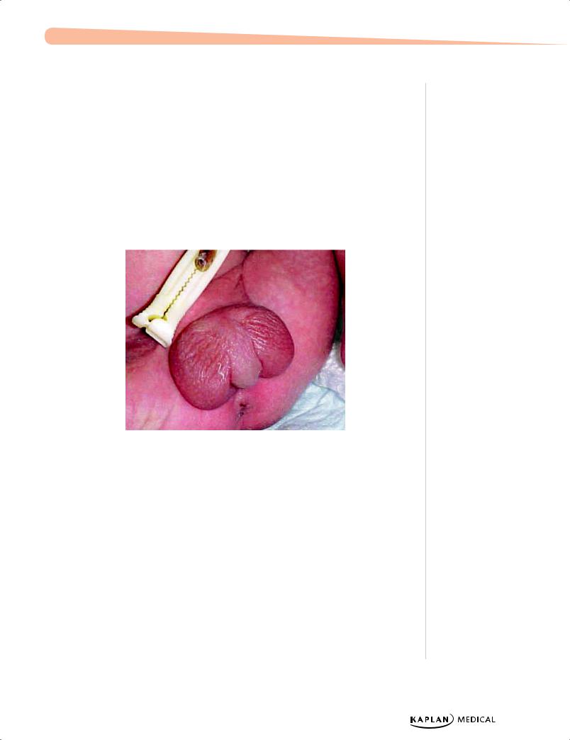
Differentiate undescended testes from retractile testes
(brisk cremasteric reflect age >1 [can manipulate into scrotum]).
−C3 and C4 normal
•Treatment
−Mild—outpatient management; if severe—hospitalize
−Start prednisone for 4–6 weeks, then taper 2–3 months without initial biopsy
−Consider biopsy with hematuria, hypertension, heart failure, or if no response after 8 weeks of prednisone (steroid resistant)
−Sodium restriction
−If severe—fluid restriction, plus intravenous 25% albumin infusion, followed by diuretic to mobilize and eliminate interstitial fluid
−Re-treat relapses (may become steroid-dependent or resistant); may use alternate agents (cyclophosphamide, cyclosporine, high-dose pulsed methylprednisolone); renal biopsy with evidence of steroid dependency
•Complications
−Infection is the major complication; make sure immunized against Pneumococcus and Varicella and check PPD
−Most frequent is spontaneous bacterial peritonitis (S. pneumoniae most common)
−Increased risk of thromboembolism (increased prothrombotic factors and decreased fibrinolytic factors) but really with aggressive diuresis
•Prognosis
−Majority of children have repeated relapses; decrease in number with age
−Those with steroid resistance and who have focal segmental glomerulosclerosis have much poorer prognosis (progressive renal insufficiency).
MALE GENITOURINARY DISORDERS
Undescended Testes
•Most common disorder of sexual differentiation in boys (more in preterm)
•Testes should be descended by 4 months of age or will remain undescended
•Usually in inguinal canal, but some are ectopic
•Prognosis
− Treated: bilateral (50–65% remain fertile), unilateral (85% remain fertile)
–Untreated or delay in treatment: increased risk for malignancy (seminoma most common)
•Surgery (orchiopexy) at 9–15 months
164

Chapter 15 λ Renal and Urologic Disorders
Testicular Torsion
•Most common cause of testicular pain age >12 years
•Clinical presentation—acute pain and swelling; tenderness to palpitation
•Testicle in transverse lie and retracted, no cremasteric reflex
•Diagnosis—Doppler color flow ultrasound (only to determine direction of torsion in order to guide manual detorsion, if urologist decides this is warranted; also to confirm successful detorsion in a completely asymptomatic patient)
•Treatment—emergent surgery (scrotal orchiopexy); if within 6 hours and <360-degree rotation, >90% of testes survive
Torsion of Appendix Testes
•Most common cause of testicular pain age 2–11 years
•Clinical presentation
−Gradual onset
−3–5 mm, tender, inflamed mass at upper pole of testis
−Naturally resolves in 3–10 days (bed rest, analgesia)
•Diagnosis
−Clinical—blue dot seen through scrotal skin
–Ultrasound if concerned with testicular torsion
–Scrotal exploration if diagnosis still uncertain
Epididymitis
•Ascending, retrograde urethral infection → acute scrotal pain and swelling (rare before puberty)
•Main cause of acute painful scrotal swelling in a young, sexually active man
•Urinalysis shows pyuria (can be Chlamydia or N. gonorrhoeae [GC], but organisms mostly undetermined)
•Treatment—bed rest and antibiotics
Testicular Tumors
•65% are malignant
•Palpable, hand mass that does not transilluminate
•Usually painless
•Diagnosis
–Ultrasound
–Serum AFP, beta-HCG (markers for germ cell tumors)
•Treatment—radical orchiectomy
Published by dr-notes.com |
165 |
|
|
|
|

Endocrine Disorders 16
Chapter Title
Learning Objectives
Recognize and describe treatments for thyroid, parathyroid, and adrenal disorders
Describe the epidemiology and treatment of childhood diabetes mellitus
PITUITARY DISORDERS
Hypopituitarism
•Deficiency of growth hormone ± other hormones; also delay in pubertal development is common; results in postnatal growth impairment corrected by growth hormone
•Isolated growth-hormone deficiency or multiple pituitary deficiencies
−Congenital—autosomal dominant, recessive, or X-linked recessive
−Acquired—any lesion that damages the hypothalamus, pituitary stalk, or anterior pituitary (most common is craniopharyngioma)
•Clinical presentation
−Congenital hypopituitarism:
°Normal size and weight at birth; then severe growth failure in first year
°Infants—present with neonatal emergencies, e.g., apnea, hypoglycemic seizures, hypothyroidism, hypoadrenalism in first weeks or boys with microphallus and small testes ± cryptorchidism
°Also have a variety of dysmorphic features; appearance
−Acquired hypopituitarism:
°Findings appear gradually and progress: growth failure; pubertal failure, amenorrhea; symptoms of both decreased thyroid and adrenal function; possible DI
°If there is an expanding tumor: headache, vomiting; visual changes, decreased school performance; papilledema, cranial nerve palsies
•Laboratory evaluation
−Screen for low serum insulin-like growth factor (IGF)-1 and IGF-binding protein-3 (IGF-BP3)
−Definitive test—growth-hormone stimulation test
−Examine other pituitary function:
°Thyroid-stimulating hormone (TSH), T4
°Adrenocorticotropic hormone (ACTH), cortisol, dehydroepiandrosterone (DHEA) sulfate, gonadotropins, and gonadal steroids
Published by dr-notes.com |
167 |
|
|
|
|

USMLE Step 2 CK
Note
If there is a normal response to hypothalamic-releasing hormones, the pathology
is located within the hypothalamus.
λPediatrics
•Other studies
−X-ray most helpful with destructive lesions (enlargement of sella, erosions)
−Calcification
−Bone age—skeletal maturation markedly delayed (BA 75% of CA)
−MRI is indicated in all patients with hypopituitarism (superior to CT scan)
•Differential diagnoses (the major ones)
−Systemic conditions (Weight is often proportionally much less than height.)
−Constitutional delay (delayed BA, delayed adolescent growth spurt, and pubertal development)
−Familial short stature (BA = CA, short parents)
−Primary hypothyroidism
−Emotional deprivation (psychosocial dwarfism)
•Treatment
−Classic growth-hormone deficiency—recombinant growth hormone
−Need periodic thyroid evaluation—develop reversible hypothyroidism
•Indications—growth hormone currently approved in United States for
−Documented growth-hormone deficiency
−Turner syndrome
−End-stage renal disease before transplant
−Prader-Willi syndrome
−Intrauterine growth retardation (IUGR) without catch-up growth by 2 years of age
– Idiopathic pathologic short stature
Note
If the history suggests anything other than familial tall stature or obesity, or if there are positive physical findings, then the patient needs laboratory evaluation.
Hyperpituitarism
•Primary—rare; most are hormone-secreting adenomas
•Majority are deficiencies of target organs and because of negative feedback, there are increases in hypothalamus and pituitary hormones
•Laboratory evaluation
−Screen—IGF-1 and IGF-BP3 for growth hormone excess; confirm with a glucose suppression test
−Need MRI of pituitary
−Chromosomes especially in tall males (decreased upperto lower-body segment ratio suggests XXY; intellectual disability suggests fragile X)
−Thyroid tests
•Management
−Treatment only if prediction of adult height (based on BA) >3 SD above the mean or if there is evidence of severe psychosocial impairment
−Trial of sex steroids (accelerates puberty and epiphyseal fusion)
168
Chapter 16 λ Endocrine Disorders
Prolactinoma
•Most common pituitary disorder of adolescents; more common in girls
•Headache, visual disturbances (with large tumors), galactorrhea, amenorrhea ± findings of hypopituitarism (again with large tumors)
•Diagnosis: increased serum prolactin level then best test, MRI
•Treatment: bromocriptine (still the only dopamine-agonist approved for children)
Physiologic Gynecomastia
•Breast tissue in the male: common (estrogen: androgen imbalance)
•Distinguish from pseudogynecomastia: adipose tissue in an overweight male
•May occur in newborns (estrogen effect) or adolescents (most common)
•Symmetric or asymmetric; may be tender
•Usually up to age 2 years
•If significant with psychological impairment, consider danazol (anti-estrogen) or surgery (rare)
Precocious Puberty
•Definition
−Girls—sexual development age <8 years
−Boys—sexual development age <9 years
•Most common etiologies
−Sporadic and familial in girls
−Hamartomas in boys
•Clinical presentation—advanced height, weight, and bone age; early epiphyseal closure and early/fast advancement of Tanner stages
•Evaluation
−Screen—significant increase in luteinizing hormone
−Definitive—GnRH stimulation test; give intravenous GnRH analog for a brisk, luteinizing hormone response
−If positive, then order MRI
•Treatment—stop sexual advancement and maintain open epiphyses (stops BA advancement) with leuprolide
Incomplete Precocious Puberty
•Premature thelarche
−Usually isolated, transient (from birth due to maternal estrogens)
−May be first sign of true precocious puberty
•Premature adrenarche—early adrenal androgen production (variation of normal)— axillary, inguinal, and genital hair. It is familial.
•Premature menarche—very rare (other causes of bleeding much more common)
Published by dr-notes.com |
169 |
|
|
|
|

USMLE Step 2 CK λ Pediatrics
Clinical Recall
A 7-year-old boy is seen by his pediatrician and noted to be Tanner Stage 3. Initial work-up reveals no oncologic process. What is the treatment of choice?
A.Growth hormone
B.Bromocriptine
C.Leuprolide
D.Thyroid hormone
E.Surgical resection of the testicles
Answer: C
THYROID DISORDERS
Hypothyroidism
A 2-month-old patient appears to be having inadequate weight gain. His mother states he is constipated. On examination, he has decreased muscle tone, a large fontanel, a large tongue, and an umbilical hernia.
•Congenital hypothyroidism—most are primary (i.e., from thyroid gland)
−Sporadic or familial; with or without a goiter
°Most common is thyroid dysgenesis (hypoplasia, aplasia, ectopia); no goiter
°Defect in thyroid hormone synthesis—goitrous; autosomal recessive
°Transplacental passage of maternal thyrotropin (transient)
°Exposure to maternal antithyroid drugs
°Radioiodine exposure/fetal exposure to excessive iodine (topical iodine antiseptics) (now rare in U.S.)
°Iodine deficiency or endemic goiter
°Central hypopituitarism
−Clinical presentation is known as “cretinism.”
°Prolonged jaundice
°Large tongue
°Umbilical hernia
°Edema
°Intellectual disability; developmental delay
°Anterior and posterior fontanels wide
°Mouth open
°Hypotonia
−Other findings—weight and length normal at birth, feeding difficulties, apnea, sluggish, decreased appetite, increased sleep, constipation, decreased temperature, skin cold and mottled, peripheral anemia; apathetic appearance
170
Chapter 16 λ Endocrine Disorders
−Laboratory evaluation:
° Low serum T4 or free T4; increased TSH
−Treatment—sodium thyroxine
•Acquired hypothyroidism
−Hashimoto; thyroiditis is most common cause; may be part of autoimmune polyglandular syndrome
−Typically presents in adolescence
−Other causes—iatrogenic (medications, irradiation, surgery, radioiodine); systemic disease (cystinosis, histiocytic infiltration)
•Clinical presentation
−Many more girls than boys
−First sign usually deceleration of growth
−Then myxedema, constipation, cold intolerance, decreased energy, increased sleep, delayed osseous maturation, delayed puberty, headache, visual problems
−Diffusely increased, firm, nontender thyroid; but may be atrophic so can be nongoitrous
−Laboratory and treatment—same as congenital
Hyperthyroidism
A 12-year-old girl has a 6-month history of hyperactivity and declining school performance. Appetite is increased, but she shows no weight gain. Physical examination reveals a slight tremor of the fingers, mild exophthalmos, and a neck mass.
•Almost all cases are Graves disease
•Peak at age 11–15 years; girls > boys
•Most with family history of some form of autoimmune thyroid disease
•Findings
−Infiltration of thyroid and retro-orbital tissue with lymphocytes and plasma cells
→ exophthalmos
−Lymphadenopathy and splenomegaly
−Thymic hyperplasia
•In whites, association with HLA-B8 and DR3 is also seen with other DR3-related disorders (Addison disease, diabetes mellitus, myasthenia gravis, celiac disease).
•Clinical
−Most signs and symptoms appear gradually
−Earliest usually emotional lability and motor hyperactivity
−Decreased school performance, tremor, increased appetite with weight loss, skin flushed with increased sweating, muscle weakness, tachycardia, palpitations, arrhythmias, hypertension
−Goiter, exophthalmos
−Thyroid storm—acute onset of hyperthermia, severe tachycardia, restlessness → rapid progression to delirium, coma, and death
•Laboratory evaluation
−Increased T4, T3, free T4
−Decreased TSH
−Measurable TRS-AB (and may have thyroid peroxidase antibodies)
Note
Autoimmune Polyglandular
Disease
Type I
•Hypoparathyroidism
•Addison disease
•Mucocutaneous candidiasis
•Small number with autoimmune thyroiditis
Type II (Schmidt syndrome)
•Addison disease, plus:
•Insulin-dependent DM
•With or without thyroiditis
Note
Thyroid cancer in children is uncommon, but you should know about medullary carcinoma (parafollicular cells), seen in 2 of the multiple endocrine neoplasias (MEN):
•MEN IIA: hyperplasia or cancer of thyroid plus adrenal medullary hyperplasia or
pheochromocytoma plus parathyroid hyperplasia
•MEN IIB (mucosal neuroma syndrome): multiple neuromas plus medullary thyroid cancer plus pheochromocytoma
Published by dr-notes.com |
171 |
|
|
|
|
USMLE Step 2 CK λ Pediatrics
•Treatment
−Propylthiouracil (PTU) or methimazole
−Beta blockers for acute symptoms (thyroid storm)
−If medical treatment not adequate, radioablation or surgery; then treat as hypothyroid (daily thyroxine replacement)
PARATHYROID DISORDERS
Hypoparathyroidism
•Parathyroid hormone (PTH) deficiency
•Etiologies—most due to DiGeorge syndrome or velocardiofacial syndrome (aplasia/ hypoplasia); remainder are defects involving production of parathyroid hormone (X-linked recessive, autosomal dominant) and postsurgical, autoimmune, and idiopathic defects
•Clinical presentation—early onset of muscle pain/cramps, numbness, and tingling; then laryngeal and/or carpopedal spasm; and finally seizures, with very low calcium (common initial presentation of DiGeorge)
•Laboratory evaluation
−Decreased serum calcium (5–7 mg/dL)
−Increased serum phosphorus (7–12 mg/dL)
−Normal or low alkaline phosphatase
−Low 1,25 [OH]2D3 (calcitriol)
−Normal magnesium
−Low parathyroid hormone (immunometric assay)
−EKG: prolongation of QT
•Treatment
−Emergency for neonatal tetany → intravenous 10% calcium gluconate and then 1,25[OH]2D3 (calcitriol); this normalizes the calcium
−Chronic treatment with calcitriol or vitamin D2 (less expensive) plus adequate calcium intake (daily elemental calcium)
−Decrease foods high in phosphorus (milk, eggs, cheese)
Table 16-1. Lab Diagnosis of Parathyroid Disease
|
|
PTH |
|
|
Calcium |
|
|
Phosphate |
|
|
Alkaline Phosphatase |
|
|
|
|
|
|
|
|
|
|
|
|
|
|
Primary Hypo |
Decreased |
Low |
High |
Normal |
||||||||
|
|
|
|
|
|
|
|
|
|
|
|
|
Pseudo Hypo |
Increased |
Low |
High |
NL or SL increased |
||||||||
|
|
|
|
|
|
|
|
|
|
|
|
|
Primary Hyper |
Increased |
High |
Low |
Increased |
||||||||
|
|
|
|
|
|
|
|
|
|
|
|
|
Secondary Hyper |
Increased |
NL to SL decreased |
Low |
Huge increase |
||||||||
|
|
|
|
|
|
|
|
|
|
|
|
|
172
Chapter 16 λ Endocrine Disorders
Vitamin D Deficiency
•Most common cause of rickets
•Poor intake, inadequate cutaneous synthesis
•Low serum phosphate, normal to low serum calcium lead to increased PTH and increased alkaline phosphatase
•Increased 25-hydroxy vitamin D
•Fractures, rachitic rosary, craniotabe bone deformities
•Treatment: initial vitamin D replacement and calcium, then adequate dietary calcium and phosphate
ADRENAL DISORDERS
TheFetus.net.
Figure 16-1. Ambiguous Genitalia Seen in Congenital Adrenal Hyperplasia
Congenital Adrenal Hyperplasia (CAH)
A 1-month-old infant is seen with vomiting and severe dehydration. Physical examination reveals ambiguous genitalia.
•21-Hydroxylase deficiency (most common)
−Autosomal-recessive enzyme deficiency
−Decreased production of cortisol → increased ACTH → adrenal hyperplasia
−Salt losing (not in all cases; some may have normal mineralocorticoid synthesis)
−Precursor steroids (17-OH progesterone) accumulate → pathway shunts to androgen synthesis → masculinized female external genitalia
Note
Other 3 Main Defects in CAH
•3-beta-hydroxysteroid deficiency: salt-wasting, male and female pseudohermaphrodites, precocious pubarche; increased 17-OH pregnenolone and DHEA
•11-beta-hydroxylase deficiency: female pseudohermaphroditism, postnatal virilization, hypertension; increased compound S, DOC, serum androgens, and hypokalemia
•17-alpha hydroxyl/17,20 lyase deficiency: male pseudohermaphroditism, sexual infantilism, hypertension; increased DOC, 18-OH DOC, 18-OH corticosterone, and 17-alpha- hydroxylated steroids; hypokalemia
Published by dr-notes.com |
173 |
|
|
|
|