

Review Test
1.A 1-week-old male infant is brought to the emergency department because of vomiting and decreased feeding for 3 days. On examination, you find that he is jaundiced and has hepatomegaly. The infant is admitted to the neonatal intensive care unit, but later he dies of Escherichia coli sepsis. Urine-reducing substances are positive. Which of the following is the most likely diagnosis?
A.Gaucher disease
B.Galactosemia
C.Glycogen storage disease type 1
D.Hurler disease
E.Niemann–Pick disease
2.The parents of a 15-year-old boy consult you because they are concerned that their son has been acting strangely for the past 6 months. The boy has developed ataxia, tremors, and seizures. His father reports that his son seems to have developed a different personality. On examination, you note that the boy has brown rings at the edge of his corneas bilaterally. Which of the following is the best serum laboratory screen for the most likely diagnosis?
A.Aluminum
B.Lead
C.Porphobilinogen
D.Zinc
E.Ceruloplasmin
3.A 6-year-old girl is brought to the clinic for a routine health maintenance visit. Her growth was normal until 2 years of age, when her height started to fall off the growth curve, steadily decreasing to below the fifth percentile. On examination, she has a webbed neck with a normal range of motion, a shield chest with widely spaced nipples, and scoliosis. Past medical history is significant for repaired coarctation of the aorta at 3 years of age. Which of the following is the most likely diagnosis?
A.Noonan syndrome
B.Achondroplasia
C.Turner syndrome
D.Russell–Silver syndrome
E.Marfan syndrome
4.The mother of a 6-year-old boy brings her son to the office for a second opinion regarding her child’s developmental delay. Your nurse takes the initial history and reports that he was adopted from another country. He was born at term by normal spontaneous vaginal delivery and seemed normal at birth, but has not been meeting his developmental milestones. After assessing his vital signs, your nurse pulls you aside and states, “That mother has no control over her child! He is hyperactive, still in diapers, and he stinks!” You walk into the examination room and immediately notice a mousy, musty smell. Which of the following is the most likely diagnosis?
A.Phenylketonuria
B.Tyrosinemia type I
C.Maple syrup urine disease
D.Homocystinuria
E.Cystinuria
5.During a health maintenance visit, the parents of a 9-month-old infant note that their infant is unable to sit alone or roll over and startles very easily to sound. On examination, you note a cherry-red macula and poor eye contact. Which of the following is correct regarding the likely
194
diagnosis?
A.Hearing loss is a common complication of this condition.
B.Radiography of the lower extremity reveals an Erlenmeyer flask–shaped distal femur.
C.Radiography of the extremities reveals dysostosis multiplex.
D.Mild developmental delay is expected.
E.Microcephaly should be apparent.
6.A 5-day-old male infant is brought to the emergency department in coma. The infant’s diaper has a sickly sweet smell. Laboratory results reveal no acidosis, hypoglycemia, or hyperammonemia, but there are significant amounts of ketones in the urine. Maple syrup urine disease is in the differential diagnosis. Which of the following statements regarding the acute management of this patient is correct?
A.Antibiotics are not indicated because the infant likely has an inborn error of metabolism.
B.Intravenous glucose should be administered.
C.Total parenteral nutrition with protein and lipids should be started.
D.Sodium benzoate should be administered.
E.Enteral feeds with a soy-based formula should be given.
7.A 14-year-old boy with mental retardation has been referred to you. The underlying cause of his mental retardation has never been identified. On physical examination, you note that he has large ears and large testes. Which of the following syndromes is associated with mental retardation and these physical findings?
A.Klinefelter syndrome
B.Down syndrome
C.Prader–Willi syndrome
D.Williams syndrome
E.Fragile X syndrome
8.A 4-month-old male infant has been brought to your office for a routine health maintenance evaluation. You note that his height is below the third percentile, yet his head circumference is at the 75th percentile. Facial findings include a prominent forehead and hypoplasia of the
midface region. He also has trident-shaped hands and bilateral short femurs and upper arms. Which of the following is a potential complication of this patient’s likely disorder?
A.Lumbar kyphosis in late childhood
B.Atlantoaxial instability
C.Spinal cord compression
D.Aortic dissection
E.Delayed puberty
9.The mother of a 5-year-old boy is very concerned and shows you a note from his kindergarten teacher. The boy is very active, easily distracted, and unable to perform skills at the same level as others in his class, and the teacher expresses concern and wonders whether the boy could have a severe learning problem. On examination, you note that the boy’s height and weight are at the 50th percentile, but his head appears very small. In addition, he has short palpebral fissures and a long, smooth philtrum with a thin upper lip. The remainder of the examination is normal. Which of the following is the most likely diagnosis?
A.Angelman syndrome
B.Down syndrome
C.Fetal phenytoin syndrome
D.Fetal alcohol syndrome
E.Prader–Willi syndrome
10.A 12-year-old boy is brought to the office for a routine health maintenance visit by his parents. They report that he has struggled in school, requiring some special educational assistance since kindergarten, but has been otherwise healthy. Examination reveals that the boy is very
195
tall (>95th percentile) with long, thin arms with fingers of normal size. Mild scoliosis is evident. On auscultation, a murmur consistent with mitral regurgitation is audible. Based on the likely diagnosis, which of the following would you also expect to find on further evaluation?
A.Increased upper-to-lower segment ratio
B.Downward lens subluxation
C.Aortic root dilatation
D.Joint laxity
E.Hypogonadism
11.You are called to the newborn nursery to evaluate a 1-day-old female infant with unusual physical findings. On examination, you note that the neonate’s hands are clenched with overlapping digits and her lower extremities are extended and crossed. You also note the presence of rocker bottom feet and delicate, small facial features. Which of the following chromosomal abnormalities is the most likely cause of the patient’s features?
A.Trisomy 13
B.Trisomy 18
C.Trisomy 21
D.Deletion on chromosome 7
E.Absence of a region on paternally derived chromosome 15
The response options for statements 12–14 are the same. You will be required to select one answer for each statement in the following set.
A.Ehlers–Danlos syndrome
B.Cri du chat syndrome
C.Osteogenesis imperfecta
D.Williams syndrome
E.Angelman syndrome
F.Hurler syndrome
G.Hunter syndrome
H.Tay–Sachs disease
I.Gaucher disease
J.Homocystinuria
For each patient, select the most likely genetic condition.
1.Six-year-old boy with coarsened facial features, stiff joints, and a cloudy cornea
2.Four-year-old boy with microcephaly, hypertelorism, mental retardation, and a deletion on the short arm of chromosome 5
3.Fourteen-year-old girl with joint laxity, easily bruisable skin, and a defect in type V collagen
196
Answers and Explanations
1.The answer is B [VI.A]. Galactosemia should always be considered in the differential diagnosis of any newborn who develops hypoglycemia and has hepatomegaly. Infants with galactosemia develop vomiting and diarrhea after feeding with either breast milk or cow’s milk–based formulas, because both types of feeding contain galactose. Soy milk does not contain galactose, which means that an infant who is fed a soy formula will not be symptomatic, which delays the diagnosis. Infants with galactosemia are vulnerable to Escherichia coli sepsis, and if the condition is not diagnosed, they may die in early infancy. Children with Gaucher disease present with neurodegeneration, splenomegaly, and bony changes (the most characteristic of which is an Erlenmeyer flask–shaped distal femur). After the first year of life, individuals with Hurler disease present with developmental delay, coarse facies, corneal clouding, and dysostosis multiplex. Individuals with with Niemann–Pick disease may present with hepatomegaly and seizures, but hypoglycemia and E. coli sepsis are not typical features of these diseases. Patients with glycogen storage disease type 1 may have hepatomegaly and hypoglycemia, but they will not have urine-reducing substances, and they do not have a special propensity to get E. coli sepsis.
2.The answer is E [XI.A.2]. Wilson disease should always be considered in a patient with personality changes, ataxia, and seizures. The patient’s signs and symptoms are suggestive of Wilson disease, which is caused by a defect in copper excretion leading to copper deposition in the brain, eyes, and liver. Kayser–Fleischer rings, which represent copper deposition in Descemet’s membrane, are pathognomonic for Wilson disease. The most commonly used screening test for Wilson disease is a low serum ceruloplasmin, which is very suggestive of the disorder. None of the other answer choices are associated with the signs and symptoms present in this patient. Aluminum, zinc, or lead does not cause the signs and symptoms seen in this patient. Serum porphobilinogen, while not addressed given the scope of this text, is elevated in acute intermittent porphyria, which may present with weakness, abdominal pain and autonomic instabilty.
3.The answer is C [III.C.1]. Girls with Turner syndrome are usually diagnosed in childhood after an evaluation for short stature, or during adolescence after an evaluation for delayed puberty. Patients with Turner syndrome classically have a webbed neck with a low posterior hairline, a shield chest with widely spaced nipples, and transient swelling of the hands and feet during the newborn period. Noonan syndrome can occur in females and has similar physical findings, but affected patients usually have right-sided heart lesions (e.g., pulmonary stenosis). Patients with Turner syndrome have left-sided heart lesions (e.g., coarctation of the aorta). Patients with achondroplasia are short from birth with shortening of the proximal long bones (rhizomelic short stature), those with Russell–Silver syndrome have short stature with skeletal asymmetry, and those with Marfan syndrome are tall, not short.
4.The answer is A [Table 5-5]. Patients with mild phenylketonuria (PKU) may present in childhood with developmental delay, hyperactivity, and a classic mousy or musty odor. Although newborn screening identifies the vast majority of cases, patients who are born in countries without newborn screening can present with untreated PKU. Patients with tyrosinemia type I present with peripheral neuropathy and renal and liver disease, and may produce an odor of rotten fish or cabbage. Children with mild maple syrup urine disease may also present with developmental delay, but their urine has a sweet maple syrup odor. Neither homocystinuria nor cystinuria has a peculiar odor as a feature; however, patients with homocystinuria may have developmental delay as a result of strokes from their hypercoagulable state.
5.The answer is A [IX.A.1]. The infant has infantile Tay–Sachs disease, a devastating
197
progressively neurodegenerative disease caused by hexosaminidase A deficiency. The onset of disease is in early infancy when the infant presents with a hyperactive startle and loses eye contact. Classic features include a cherry-red macula, enlarging head circumference, neurodegeneration with severe developmental delay, progressive blindness, and seizures. Death usually occurs by 5 years of age. An Erlenmeyer flask–shaped distal femur is a feature of Gaucher disease and not Tay–Sachs disease. Dysostosis multiplex (bony abnormalities that include a thickened skull, malformed vertebrae, and abnormal ribs and clavicle) is found in patients with mucopolysaccharidoses (e.g., Hunter and Hurler syndromes).
6.The answer is B [IV.C]. In general, the acute management of an inborn error of metabolism involves supplying a source of energy that can be used, removing toxic metabolites, and preventing continued exposure to the offending substance. Glucose is a basic energy source that can be used in any patient, regardless of the inborn error of metabolism, and should be administered intravenously in a patient who is markedly hypoglycemic and in coma or shock. Sodium benzoate is a scavenger agent that would be useful to facilitate ammonia excretion, but this patient does not have hyperammonemia. Although the patient’s condition is likely to be an inborn error of metabolism (maple syrup urine disease, given the diaper odor consistent with the odor of maple syrup), sepsis is more common and can present in a similar fashion. Therefore, initial management should include intravenous antibiotics. Patients with maple syrup urine disease cannot adequately metabolize branched-chain amino acids; therefore, offering parenteral nutrition with protein would continue the toxic exposure. Soy formulas, while appropriate for galactosemia, still contain a full amount of branch-chain amino acids, and similar to regular formulas would overwhelm the patient’s ability to metabolize the branched chain amino acids, resulting in toxic exposure as well.
7.The answer is E [III.C.2]. The characteristic physical features of fragile X syndrome include large ears, macrocephaly, and large testes. Klinefelter syndrome is characterized by tall stature, gynecomastia, and a small penis and testes; Down syndrome, by characteristic facial features, endocardial cushion defects, duodenal atresia, mental retardation, single palmar creases, and a wide space between the first and second toes; Prader–Willi syndrome, by infantile hypotonia, hypogonadism, short stature, and obesity and hyperphagia later in childhood; and Williams syndrome, by a loquacious very friendly personality, supravalvular aortic stenosis, and hypercalcemia. Testes are unaffected in Down syndrome and in Williams syndrome.
8.The answer is C [III.D.2.c]. This patient’s physical features are consistent with achondroplasia, the most common skeletal dysplasia. Potential complications of this disorder include cord compression caused by foramen magnum stenosis, obstructive sleep apnea, and orthopedic problems such as genu varum and back pain caused by lumbar lordosis during late childhood. Atlantoaxial instability (a complication of Down syndrome), aortic dissection (a complication of Marfan syndrome), or delayed puberty is not associated with achondroplasia.
9.The answer is D [IV.E.3]. This patient’s physical characteristics, along with learning problems and attention deficit/hyperactivity disorder, are consistent with fetal alcohol syndrome. Angelman syndrome is associated with severe mental retardation and a small head, although a puppetlike gait and inappropriate bouts of laughter are also characteristic. Down syndrome is also associated with mental retardation; however, the facial characteristics include upslanting palpebral fissures, epicanthal skin folds, and a protruding tongue. Fetal phenytoin syndrome is associated with mental retardation, nail and digit abnormalities, and cardiac defects. Prader–Willi syndrome is associated with hypogonadism, almond-shaped eyes, short
stature, and hyperphagia with obesity during childhood.
10.The answer is B [V.A]. The clinical features of homocystinuria and Marfan syndrome overlap considerably; however, this patient most likely has homocystinuria based on the presence of cognitive delay and a marfanoid body habitus with fingers of normal length (i.e., no
198
arachnodactyly). All of these features are seen in homocystinuria but are not expected to be seen in Marfan syndrome. In addition, patients with homocystinuria usually have downward lens subluxation (upward lens subluxation is found in Marfan syndrome) and large, stiff joints (joint laxity is found in Marfan syndrome). Patients with both disorders have a decreased upper-to-lower segment ratio. Hypogonadism is absent in both disorders.
11.The answer is B [III.B.2]. The findings of scissoring of the lower extremities, clenched hands with overlapping digits, rocker bottom feet, and delicate small facial features are consistent with the diagnosis of trisomy 18, the second most common trisomy syndrome after trisomy 21. Trisomy 21 is the cause of Down syndrome and is associated with hypotonia, prominent epicanthal folds, upslanting palpebral fissures, and single palmar creases. Trisomy 13 is associated with midline defects of the brain and forebrain. Features include microphthalmia, holoprosencephaly, and cleft lip and palate. Absence of a region on the paternally derived chromosome 15 is the cause of Prader–Willi syndrome, a disorder associated with neonatal hypotonia, hypogonadism, almond-shaped eyes, and short stature. A deletion on chromosome 7 causes Williams syndrome, characterized by elfin facies and a loquacious personality.
12.The answers are F [IX.B.1], B [III.A.14], and A [III.A.6], respectively. Hurler syndrome, a mucopolysaccharidosis in which glycosaminoglycans deposit in various tissues causing a progressive clinical picture, is characterized by corneal clouding, changes to the bone termed dysostosis multiplex, organomegaly, and progressively coarsened facies, including frontal bossing, widened nasal bridge, and thickening of the nasopharyngeal tissues. Hunter syndrome is similar but does not have corneal clouding.
Cri du chat syndrome, caused by a partial deletion on the short arm of chromosome 5, is characterized by slow growth, microcephaly, mental retardation, hypertelorism, and a classic catlike cry.
Ehlers–Danlos syndrome is caused by a defect in type V collagen that results in hyperextensible joints, fragile blood vessels that cause easily bruised skin, tissue paper–thin scars, and cardiovascular complications (e.g., mitral valve prolapse and aortic root dilatation that can lead to dissection).
199

C H A P T E R 6
200
Endocrinology
Srinath Sanda
201
I.Short Stature
A.General concepts
1.Definition. Short stature is defined as height that is 2 standard deviations (SDs) below the mean height of the population (i.e., below the third percentile), or 2 SDs below the midparental height (MPH).
a.Normal variant short stature describes a child with short stature and a normal growth velocity.
b.Pathologic short stature describes a child with short stature and a suboptimal growth velocity.
2.Key point: It is critical to evaluate growth velocity and not just absolute height when evaluating short stature. Growth velocity is ideally calculated over at least a 6-month period. Using measurements collected over a shorter period of time may result in inaccurate growth velocity calculations.
3.Key point: Children who grow 2 inches per year (5 cm per year) between 3 years of age and puberty usually do not have an endocrinopathy or underlying pathologic disorder.
4.In the first 2 years of life, a downward shift in the height percentile is not uncommon and may reflect familial (or genetic) short stature, or constitutional delay of growth and puberty (CDGP).
5.All patients with short stature who are more than 2 SDs below the mean or who have a growth velocity less than 5 cm per year are considered to have a pathologic growth disorder until proven otherwise.
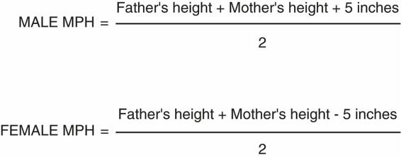
6.Determining the targeted MPH is important in evaluating all patients with short stature (Figure 6-1).
a.Most children, when they have completed their growth, are within ±2 SDs, or 4 inches, of their MPH.
b.A major discrepancy between a child’s present growth percentile and the targeted MPH percentile suggests a pathologic state.
B.History
1.Perinatal history. Assess for prematurity or intrauterine growth retardation (IUGR) or small for gestational age (SGA). A history of neonatal hypoglycemia, prolonged jaundice, and/or microphallus suggests hypopituitarism resulting in growth hormone (GH) deficiency.
2.Chronic diseases such as hypothyroidism, renal failure, central nervous system (CNS) disease, severe asthma with frequent and prolonged steroid use, sickle cell anemia, celiac disease, and inflammatory bowel disease may manifest as short stature.
3.Chronic use of drugs, such as steroids or stimulants for attention deficit/hyperactivity disorder, that result in significant appetite suppression and poor weight gain may lead to short stature.
4.Family history, especially parental growth and pubertal histories, is important. To evaluate for constitutional delay of growth and puberty, or CDGP [see section I.D.1], ask whether the family history is positive for males with “late growth spurts” in high school or college and the age of maternal menarche.
5.Social history is critical because children who live in neglected or hostile environments may exhibit short stature because of psychosocial deprivation.
6.Review of systems should include questions about cold intolerance and constipation (hypothyroidism), abdominal pain, diarrhea or bloody stools (inflammatory bowel disease), and headaches and vomiting (brain tumor).
202
7.Dental history. Delayed dental eruption may suggest CDGP or GH deficiency.
C.Physical examination
1.Accurate height and weight should be plotted on a US National Center for Health Statistics (NCHS) growth chart, along with previous growth points, to assess the child’s growth pattern. The MPH should be noted on the growth chart.
2.Measure the patient’s upper-to-lower (U/L) body segment ratio.
a.Lower segment = pubic symphysis to the heel
b.Upper segment = total height minus lower segment
c.Normal ratios
1.Birth = 1.7
2.3 years of age = 1.3
3.>7 years of age = 1.0
d.Abnormal U/L ratio suggests disproportionate short stature [see section I.D.2.b].
3.Thorough physical examination should include a funduscopic examination, assessment for midline defects (central incisors, cleft palates, etc.), assessment of thyroid size, evaluation for stigmata of genetic syndromes (e.g., web neck, shield chest, and abnormal carrying angles are suggestive of Turner syndrome; see Chapter 5, section IV.C.1), scoliosis screening, and Tanner staging (see Chapter 3, section I.A.2).
D.Categorization of short stature (Figure 6-2)
1.Normal variant short stature refers to children with short stature and normal growth velocity. The two most common categories of normal variant short stature are familial (or genetic) short stature and CDGP.
a.Familial (or genetic) short stature is defined as a height at least 2 SDs below the mean but within 2 SD of MPH with a normal bone age, a normal onset of puberty, and a minimum growth velocity of 2 inches (or 5 cm) per year.
b.Constitutional delay of growth and puberty (CDGP) is defined as short stature with a delayed bone age, late onset of puberty and a minimum growth velocity of 2 inches (or 5 cm) per year.
2.Pathologic short stature. Pathologic short stature, in which height fall more than 3 SDs below the mean with abnormal growth velocity (i.e., growth velocity less than 2 inches [or 5 cm] per year), may be categorized as proportionate or disproportionate.
a.Proportionate short stature is defined as short stature with a normal U/L ratio [see section I.C.2.c]. It is important to distinguish between prenatal onset and postnatal onset.
1.Causes of prenatal-onset proportionate short stature include
a.Environmental exposures (e.g., in utero exposure to tobacco and alcohol)
b.Chromosomal disorders (e.g., Down syndrome, Turner syndrome)
c.Genetic syndromes (e.g., Russell–Silver syndrome, Prader–Willi syndrome; see Chapter 5, sections IV.E.5 and IV.E.2, respectively)
d.Viral infection early in pregnancy (e.g., cytomegalovirus, rubella)
2.Causes of postnatal-onset proportionate short stature
a.Malnutrition
b.Psychosocial causes (e.g., neglect, child abuse)
c.Organ system diseases, including gastrointestinal diseases (inflammatory bowel disease, celiac disease), cardiac diseases (cyanotic congenital heart disease), renal diseases (renal failure, renal tubular acidosis), chronic lung diseases (cystic fibrosis, asthma), and endocrinopathies (hypothyroidism, GH deficiency, and cortisol excess; see also section I.F)
b.Disproportionate short stature is defined as short stature in patients who are short
203
legged with an increased U/L ratio, suggesting rickets or a skeletal dysplasia.
1.Consider rickets in patients with frontal bossing, bowed legs, low serum phosphorus level, and high serum alkaline phosphatase [see section X.C].
2.Consider some form of skeletal dysplasia (e.g., achondroplasia) in patients who are short with short limbs (see Chapter 5, section III.H).
E.Evaluation of pathologic short stature
1.Laboratory studies
a.Complete blood count (CBC), erythrocyte sedimentation rate (ESR), thyroxine (T4), thyroid-stimulating hormone (TSH), tissue transglutaminase IgA (TTG IgA), total serum IgA, serum electrolytes including calcium and phosphorus, and serum creatinine and bicarbonate levels should be obtained.
b.Insulin-like growth factor 1 (IGF-1) and insulin-like growth factor binding protein 3 (IGF-BP3) are indirect measures for GH deficiency. Random GH level should not be measured outside of the neonatal period, as most GH is released in a pulsatile fashion.
c.Chromosome analysis in girls to evaluate for Turner syndrome
d.GH stimulation testing (with agents such as clonidine or arginine) may be needed.
2.Radiographic studies
a.Bone age determination (anterior–posterior [AP] film of the left hand and wrist to assess the characteristics of the epiphyses or growth plates) is helpful to compare with chronologic age (Table 6-1).
b.Neuroimaging (magnetic resonance imaging [MRI] is preferable with thin cuts through the pituitary) is appropriate if GH deficiency or concerns for other CNS pathology exist. It is not otherwise part of the routine evaluation of children with short stature.
3.Key point: Patients with poor growth velocity with normal screening laboratory results, but low IGF-1 and delayed bone age, should have an additional workup for GH deficiency (i.e., GH stimulation testing).
F.Endocrinopathies that cause short stature
1.GH deficiency is uncommon.
a.Clinical features. A neonatal history of prolonged neonatal jaundice, hypoglycemia, microphallus, cryptorchidism, and midline defects (e.g., cleft palate) may be present. Neonates with GH deficiency are typically not SGA. Older children with GH deficiency may have obesity and cherubic facies. The growth curve demonstrates poor growth velocity (less than 2 inches or 5 cm per year).
b.Causes include brain tumors, prior CNS irradiation, CNS vascular malformations, autoimmune diseases, head trauma, and congenital midline defects. (Consider GH deficiency in patients with a single central maxillary incisor or with cleft palate.)
c.Evaluation
1.Imaging studies. See I.E.2.a and I.E.2.b. Key point: All patients with GH deficiency must have neuroimaging, preferably with an MRI with thin cuts through the pituitary.
2.Laboratory studies. Low IGF-1 and IGF-BP3 levels, as well as a poor response on GH stimulation testing (with l-DOPA [L-3,4-dihydroxyphenylalanine], arginine, glucagon, or clonidine)
d.Management. Treatment includes daily subcutaneous injections of recombinant GH until a bone age determination demonstrates that the patient has reached nearly maximal growth potential (by about 13–14 years of age in girls and 15–16 years of age in boys). In cases of CNS tumors, GH should not be used until the tumor has been treated.
204
2.Hypothyroidism. The most common cause of hypothyroidism is Hashimoto thyroiditis [see section IX.B.2.b]. Patients will present with increased TSH, low T4, and positive antithyroid peroxidase antibodies.
3.Hypercortisolism. The most common cause of hypercortisolism is iatrogenic, as a result of prolonged use of steroids [see section IV.E]. Patients present with a history of poor growth and increasing weight gain, purpuric stretch marks and a dorsal neck fat pad on examination, with delayed bone age.
4.Turner syndrome (see also Chapter 5, section IV.C.1). Female patients who are missing a part or all of one of the X chromosomes may present with short stature or lack of puberty. GH treatment has been shown to improve the ultimate height of these patients. Key point: All girls with short stature should have a karyotype, as the physical features of Turner syndrome can be subtle.
FIGURE 6.1 Determination of midparental height (MPH). Most patients, when they have completed their growth, will be within ±2 standard deviations, or 4 inches, of the MPH. For example, if a boy has a father who is 5 feet 9 inches in height and a mother who is 5 feet in height, then the MPH is 5 feet
7 inches ± 4 inches.
205
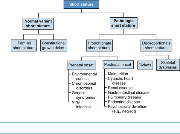
FIGURE 6.2 Differential diagnosis of short stature.
Table 6-1
Using Bone Age in the Differential Diagnosis of Short Stature
Bone Age = Chronologic Age |
Bone Age < Chronologic Age |
Familial short stature |
Constitutional short stature |
Intrauterine growth retardation |
Hypothyroidism |
Turner syndrome |
Hypercortisolism |
Skeletal dysplasia |
Growth hormone deficiency |
|
Chronic diseases |
206
II.Disorders of Puberty
A.Normal puberty (see also Chapter 3, section I.A.2)
1.In the prepubescent state, sex steroids (testosterone and estradiol) are suppressed owing to an inactive hypothalamic–pituitary–gonadal axis (HPGA).
2.Puberty begins when there is a reduction in this hypothalamic inhibition, resulting in activation of the HPGA.
3.The HPGA releases gonadotropin-releasing hormone (GnRH) from the hypothalamus, which binds to receptors in the pituitary gland and causes the release of folliclestimulating hormone (FSH) and luteinizing hormone (LH).
4.Female puberty. The different stages of pubertal development are defined as follows. It is important, however, to be aware that the age of onset and subsequent course of hormonal and physical changes during puberty are variable.
a.Onset is between 8 and 13 years of age.
b.Thelarche is the onset of breast development as a result of the release of estrogen, and adrenarche is the onset of pubic or axillary hair development as a result of the release of adrenal androgens. Breast buds are usually the first sign of puberty, although in 15% of girls, pubic hair develops first.
c.Menarche is the onset of the menstrual cycle. Menstruation begins at 9–15 years of age, with a mean onset of 12.5 years of age.
d.FSH stimulates the ovaries to produce ovarian follicles, which in turn produce estrogen.
e.LH is responsible for the positive feedback in the middle of the menstrual cycle, resulting in the release of an egg.
f.Tanner staging. See Chapter 3, section I.A.2.c.
5.Male puberty
a.Onset is between 9 and 14 years of age.
b.Testicular enlargement is usually the first sign of puberty (≥4 mL as measured with an orchidometer or >2.5 cm in length).
c.Seventy-five percent of the testicular volume is the seminiferous tubules.
d.FSH in boys stimulates the Sertoli cells in the seminiferous tubules of the testes to produce sperm.
e.LH in boys stimulates the testicular Leydig cells to produce androgens, which in turn are responsible for penile enlargement and the growth of axillary, facial, and pubic hair.
f.Tanner staging. See Chapter 3, section I.A.2.c.
6.African American children may develop secondary sexual characteristics earlier than other children.
7.Moderate to severe obesity may be associated with sexual precocity.
B.Precocious puberty
1.Definitions
a.Girls: presence of breast development or pubic hair before 8 years of age
b.Boys: presence of testicular changes, penile enlargement, or pubic or axillary hair before 9 years of age
2.Categories
a.Premature thelarche
1.Definition. There is visible or palpable breast tissue only, with no other secondary sexual characteristics. The growth pattern should be normal, and no pubic hair should be apparent.
207
2.Epidemiology. This is a common and benign condition usually presenting in the first 2 years of life.
3.Etiology. This condition is caused by a transient activation of the HPGA, resulting in transient ovarian follicular stimulation and a release of low levels of estrogen.
4.Generally no workup is necessary as long as no other secondary sexual characteristics (clitoromegaly, pubic hair, enlarged labia minora, pink vaginal mucosa) or growth spurts are noted. Patients should be followed, and if progressive enlargement of the tissue is noted, they should be referred to a pediatric endocrinologist.
b.Premature adrenarche
1.Definition. Early onset of pubic or axillary hair growth without the development of breast tissue or enlarged testes.
2.Epidemiology. This condition is more common in girls than in boys.
3.Classic presentation occurs after 5 years of age, with the onset of pubic hair growth, axillary hair growth, and apocrine odor. No breast tissue or testicular enlargement is noted, and there is no clitoromegaly. Growth is normal without advancement of bone age.
4.Most patients should have a bone age radiograph, as well as laboratory testing for morning levels of 17-hydroxyprogesterone (17-OHP), as some patients with nonclassic congenital adrenal hyperplasia (CAH) may present with premature adrenarche.
5.In most cases, no treatment is indicated.
c.Central (or isosexual) precocious puberty (CPP)
1.Definition. The early onset of gonadotropin-mediated (i.e., mediated by FSH and LH) puberty.
2.Epidemiology. Girls have a higher incidence of CPP than boys.
3.Clinical features
a.In girls, physical examination shows breast development, pubic hair, and rapid growth.
b.In boys, physical examination shows testicular enlargement, pubic hair, and rapid growth.
4.Etiology
a.In girls, most cases are idiopathic.
b.In boys, sexual precocity tends to be pathologic, and all patients need evaluation with an MRI of the head.
c.CNS abnormalities that may cause CPP include hydrocephalus, CNS infections, cerebral palsy, benign hypothalamic hamartomas, malignant tumors such as astrocytomas and gliomas, and severe head trauma.
d.Hypothyroidism may rarely present with CPP, but in this case, there is poor growth and a delayed bone age (unlike all other causes of sexual precocity in which growth is accelerated).
5.Evaluation
a.FSH, LH, and sex steroid levels are elevated. In particular, unstimulated gonadotropin testing should be done with ultrasensitive or pediatricspecific assays, and not routine LH and FSH assays that are designed for use in adults.
b.The GnRH stimulation test can demonstrate premature activation of the hypothalamus. Access to GnRH agonists (GnRHa) is limited in the United States; therefore, short-acting GnRHa such as leuprolide acetate
208
are used in stimulation tests.
1.By injecting synthetic GnRHa into a patient, the LH response, and to a lesser degree the FSH response, can be used as an assessment of the activation of the HPGA. Patients with CPP have a dramatic increase in LH secretion when compared with baseline levels.
2.On the other hand, prepubertal patients whose HPGA has not yet been activated, and patients with peripheral precocious puberty (PPP) in which peripherally produced sex steroids suppress pituitary gonadotropin secretion, would be expected to have a flat response (i.e., no increase in LH secretion) on injection of synthetic GnRHa.
c.An MRI of the head should be performed in all boys, and in girls with any neurologic symptoms (e.g., headaches or seizures) or with rapid pubertal changes.
d.Thyroid tests (TSH and T4) should be performed to rule out hypothyroidism.
d.PPP or heterosexual gonadotropin-independent puberty
1.Definition. Precocious puberty that is independent of the HPGA (i.e., caused by the peripheral production of male or female sex steroids and not FSHor LH-mediated). The hallmark of PPP is suppressed unstimulated gonadotropin levels and/or a flat response on GnRHa stimulation testing, because the HPGA has not been activated.
2.Clinical features
a.Boys present with either feminization (gynecomastia) or with premature onset of pubic hair. Note that there is usually no testicular enlargement because these patients do not have an increase in FSH, which would stimulate seminiferous tubule enlargement [see exceptions in section II.B.2.d.(3).(c)].
b.Girls present with typical secondary sexual characteristics or virilization.
3.Etiology. Causes may differ in boys and girls but, in general, include exposure to exogenous sex steroids (found in some skin lotions or foods), McCune– Albright syndrome, gonadal tumors, adrenal tumors, and nonclassic CAH; see section IV.C.3.a.(3). All of these causes are independent of the HPGA.
a.In boys, consider adrenal tumors, Leydig cell tumors (presenting with asymmetric testicular enlargement), nonclassic CAH, β-human chorionic gonadotropin (β-HCG)–producing tumors, McCune–Albright syndrome, and testotoxicosis.
b.In girls, consider adrenal tumors, virilizing ovarian tumors (arrhenoblastomas), feminizing ovarian tumors (juvenile granulosa tumors), nonclassic CAH, and McCune–Albright syndrome.
c.Specific causes of PPP in males that result in testicular enlargement
1.McCune–Albright syndrome is characterized by fracture-prone bony changes (polyostotic fibrous dysplasia), skin findings (irregularly bordered hyperpigmented macules, or “coast of Maine” jagged-bordered café-au-lait spots), and endocrinopathies (PPP or hyperthyroidism). Patients often have enlarged gonads, but their secretion of sex steroids is independent of the HPGA.
2.Testotoxicosis is a rare disease in which the testes enlarge bilaterally independent of the HPGA.
3.β-HCG—secreting tumors are unique to boys. These tumors are
209
found in the chest, pineal gland, gonad, or liver (hepatoblastoma). Because the β-HCG molecule cross-reacts with LH, it too can bind to LH receptors and enlarge the testes slightly, stimulating Leydig cells and secreting androgens.
4.Evaluation. A GnRH stimulation test may be warranted in addition to the following:
a.In boys, check serum FSH, LH, testosterone, and β-HCG levels.
b.In girls, check serum FSH, LH, and estradiol levels.
c.Perform imaging (testicular, abdominal, CNS, etc.) studies, depending on the suspected etiology.
5.Management. Treatment is based on the underlying cause.
C.Delayed puberty
1.Definitions
a.Boys: No testicular enlargement by 14 years of age.
b.Girls: No breast tissue by 13 years of age, or no menarche by 16 years of age.
2.Classification. Two categories of disorders may result in delayed puberty.
a.Hypogonadotropic hypogonadism. Because of the inactivity of the hypothalamus and pituitary gland, these patients have a low FSH, low LH, and, in turn, low testosterone and low estradiol, with a prepubertal (flat) GnRH stimulation test.
b.Hypergonadotropic hypogonadism. Because of end-organ dysfunction (i.e., gonadal failure), these patients have high FSH and high LH levels with low testosterone or low estradiol levels. There is no abnormality in the hypothalamus or pituitary gland.
3.Etiology of hypogonadotropic hypogonadism
a.CDGP (i.e., immature hypothalamus or “late bloomers”) is much more common in boys than in girls. Often there is a family history in one parent (i.e., mother had late menarche or father had his growth spurt late in high school or in college). Patients with hypogonadotropic hypogonadism frequently have CDGP [see section I.D.1.b for a description of growth pattern].
b.Chronic diseases can cause pubertal delay (e.g., inflammatory bowel disease, anorexia nervosa, renal failure, and heart failure).
c.Hypopituitarism of any cause (e.g., brain tumors)
d.Primary hypothyroidism
e.Prolactinoma
f.Genetic syndromes
1.Kallman syndrome. Isolated gonadotropin deficiency associated with anosmia (inability to smell)
2.Prader–Willi syndrome (see Chapter 5, section IV.E.2)
3.Bardet–Biedl syndrome. Obesity, retinitis pigmentosa, hypogonadism, and polysyndactyly
4.Etiology of hypergonadotropic hypogonadism
a.Chromosomal disorders
1.In boys, consider Klinefelter syndrome (XXY; see Chapter 5, section IV.C.2).
2.In girls, consider Turner syndrome or gonadal dysgenesis (see Chapter 5, section IV.C.1).
b.Autoimmune disorders (e.g., hypogonadism in autoimmune oophoritis, which may also be associated with Hashimoto thyroiditis or Addison disease)
5.Evaluation of delayed puberty. A CBC, ESR, bone age, TSH and T4, testosterone or estradiol, FSH, LH, and prolactin levels are necessary. Neuroimaging may be warranted as well.
210
211
III. Disorders of Sexual Differentiation (DSD or Formerly Known as Ambiguous Genitalia)
A.Normal sexual differentiation (Figure 6-3)
1.During the first 7 weeks of gestation, the gonadal tissue remains undifferentiated. The final appearance of gonadal tissue is dependent on both genetic and hormonal influences.
2.Male sexual differentiation is an active process, whereas female sexual differentiation develops by default when genetic and hormonal influences are absent.
B.Male sexual differentiation is initiated by the SRY gene located on the short arm of the Y chromosome. By 9 weeks’ gestation, the SRY gene differentiates the gonads into fetal testes, which subsequently produce testosterone and anti-Müllerian hormone (AMH or previously called Mullerian inhibiting substance).
1.Internal ducts. In the genetic XY male, testosterone made by fetal Leydig cells stimulates the development of the Wolffian ducts (epididymis, vas deferens, and seminal vesicles), and AMH made by fetal Sertoli cells inhibits the development of the Müllerian structures (fallopian tubes, uterus, and upper one-third of the vagina).
2.External genitalia. The conversion of testosterone to dihydrotestosterone (DHT) by 5αreductase occurs in the skin of the external genitalia. DHT is responsible for penile enlargement, scrotal fusion, and the entire masculinization of the external genitalia. By 12 weeks, this process is complete, except for penile growth, which continues to term.
C.Female sexual differentiation. In the absence of the SRY gene, the gonads become ovaries.
1.Internal ducts. Because there is no testicular tissue, there is no secretion of testosterone or of AMH, resulting in the regression of the Wolffian ducts and the development of the Müllerian structures, respectively.
2.External genitalia. The external genitalia do not virilize because there is a lack of testosterone and of DHT. This results in the development of the labia, the clitoris, and the lower two-thirds of the vagina.
D.Differential diagnosis of the undervirilized male (i.e., a neonate who usually has a 46, XY karyotype with ambiguous genitalia and one or both testes palpable; Figure 6-4).
1.Disorders of testosterone synthesis. Many inherited enzyme deficiencies result in low testosterone levels (i.e., any enzyme deficiency in the pathway of androgen synthesis in
Figure 6-5).
a.Smith–Lemli–Opitz syndrome. This syndrome is due to defective 3β- hydroxysteroid-Δ7 reductase, the last step in cholesterol biosynthesis. Without functional cholesterol biosynthesis, steroid hormone production is defective. Patients can have microcephaly, cleft lip/palate, liver disease, syndactyly of the second and third toes and holoprosencephaly. Genital ambiguity is variable.
b.5α-Reductase deficiency. Patients cannot convert testosterone to DHT, the more potent version of testosterone that binds to androgen receptors and virilizes the external genitalia. Patients may present with ambiguous genitalia, such as varying degrees of hypospadias, or as adolescents with incomplete pubertal progression. Some patients present as adolescent females who do not develop breast tissue (as opposed to patients with androgen insensitivity—see below). A ratio of testosterone:DHT of >20:1 with β-hCG stimulation strongly supports the diagnosis. By comparison, the ratio in normal male infants is typically <10.
2.Disorders of testosterone action are usually due to androgen insensitivity. These patients have X-linked partial or complete peripheral androgen resistance resulting from defective androgen binding to the androgen receptor in the genital tissue. Presentation is
212
highly variable. Complete forms typically present as females with primary amenorrhea and breast development, wheras partial forms may present with some degree of impaired virilization observed in the neonate (such as cryptorchidism or hypospadias). Mullerian structures are usually absent. Patients have normal adrenal metabolites (17OHP levels, etc.), but exaggerated levels of testosterone and DHT are seen after β-hCG stimulation.
3.Disorders of gonad differentiation
a.Gonadal dysgenesis (GD). May be complete, partial, or mixed. Karyotypes are diverse and may include 45, XO/46, XY mosaicism. The clinical presentation is variable, as patients may have completely normal external female genitalia, to some degree of ambiguous genitalia with some internal Mullerian structures (i.e., fallopian tubes, etc.). Typically these patients have low to undetectable levels of AMH, and the gonads do not respond to stimulation with β-hCG.
b.True hermaphroditism (also known as ovotesticular DSD). These patients may have ambiguous genitalia with both ovarian and testicular gonadal tissue and both Mullerian and Wolffian internal ducts. The gonads generally have some function, and levels of AMH are normal. The gonads do respond to β-hCG stimulation. Usually the karyotype is 46, XX, but it can be 46, XY.
E.Differential diagnosis of ambiguous genitalia in the virilized female (i.e., a female who is a genetic XX with ambiguous genitalia and no gonads palpable; Figure 6-6).
1.CAH caused by 21-hydroxylase (21-OH) deficiency is the most common cause of female pseudohermaphroditism [see section IV.C]. 11β-Hydroxylase (11β-OH) deficiency and 3β-hydroxysteroid dehydrogenase deficiency are other causes of CAH.
2.Virilizing drug used by mother during pregnancy
3.Virilizing tumor in mother during pregnancy
F.Evaluation of the patient with ambiguous genitalia
1.Careful history. Maternal history of drugs or virilization during pregnancy, family history of androgen insensitivity, CAH, or consanguinity.
2.Physical examination. Presence or absence of gonads, labioscrotal swelling, bifid scrotum, labial fusion, urogenital sinus, or hypospadias. Palpable gonads suggest that the neonate has an XY karyotype. (Key point: Increased blood pressure suggests CAH with 11β-OH deficiency, and decreased blood pressure suggests adrenal insufficiency; see sections IV.B.1.a and IV.C.3.b.)
3.Chromosome studies (including polymerase chain reaction [PCR] or fluorescent in situ hybridization [FISH] for the SRY gene, and a karyotype)
4.Radiographic studies include pelvic ultrasound and genitogram to define the internal genitourinary anatomy.
5.Laboratory studies
a.Undervirilized males. Besides the abovementioned chromosome studies, electrolytes and 17-OHP levels should be ordered. DHT and testosterone levels (preferably after β-hCG stimulation) with LH, FSH, AMH, and androstenedione levels may be warranted. If serum testosterone is low, further evaluation for an inborn error in androgen synthesis is indicated.
b.Virilized females. Serum electrolytes, testosterone level, and further studies to look for evidence of CAH (17-OHP, etc.) are indicated [see Figure 6-5 and section IV.C.4].
6.Management. Evaluation is complex, and gender assignment should not be rushed. Generally, these patients should be referred to centers with multidisciplinary teams of pediatric endocrinologists, geneticists, and pediatric urologists. The initial focus should be on clear communication with the family regarding the process of evaluation.
213
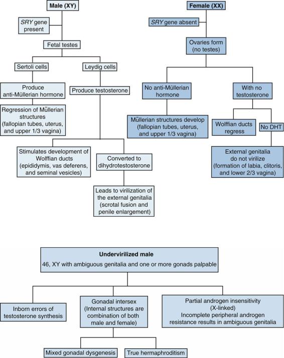
FIGURE 6.3 Normal sexual differentiation in utero. DHT = dihydrotestosterone.
FIGURE 6.4 Differential diagnosis of ambiguous genitalia in an undervirilized male.
214
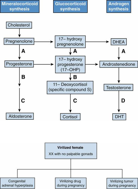
FIGURE 6.5 Steroid pathways in the adrenal cortex. A = 3β-hydroxysteroid dehydrogenase; B = 21hydroxylase (21-OH); C = 11β-hydroxylase (11β-OH); D = 5α-reductase;
DHEA = dehydroepiandrosterone; DHT = dihydrotestosterone.
FIGURE 6.6 Differential diagnosis of ambiguous genitalia in a virilized female.
215
IV. Disorders of the Adrenal Gland
A.General principles of adrenal function
1.The adrenal gland is composed of two parts, the adrenal cortex, which synthesizes a multitude of different steroid compounds, and the adrenal medulla, which produces catecholamines (i.e., epinephrine).
2.Three major pathways in the adrenal cortex result in the production of mineralocorticoids (aldosterone), glucocorticoids (cortisol), and androgens
(dehydroepiandrosterone [DHEA]), as outlined in Figure 6-5.
3.Glucocorticoid and androgen synthesis are regulated by a negative feedback loop by the hypothalamic–pituitary–adrenal axis via adrenocorticotropin hormone (ACTH). Mineralocorticoid synthesis, however, is controlled by the renin–angiotensin system and is independent of the pituitary gland and ACTH.
4.Children may present with disorders of adrenal insufficiency and with disorders of glucocorticoid excess.
B.Classification of adrenal insufficiency. Adrenal insufficiency may be primary or secondary, each with different clinical manifestations.
1.Primary adrenal insufficiency
a.This condition results from destruction of the adrenal cortex or from an enzyme deficiency (i.e., a problem at the level of the adrenal gland).
b.Patients present with signs and symptoms of both cortisol deficiency (anorexia, weakness, hyponatremia, hypotension, and increased pigmentation over recently healed scars) and aldosterone deficiency (failure to thrive, salt craving, hyponatremia, and hyperkalemia).
c.Examples include Addison disease, CAH, and adrenoleukodystrophy (rare, X- linked recessive disorder with neurologic deterioration).
2.Secondary adrenal insufficiency
a.This condition results from any process that interferes with the release of cortisol-- releasing hormone (CRH) from the hypothalamus or ACTH from the pituitary (i.e., a problem at the hypothalamic or pituitary level).
b.In contrast to primary adrenal insufficiency, serum potassium may be normal in secondary adrenal insufficiency because there is no aldosterone deficiency, given an intact renin–angiotensin system. However, these patients are still prone to developing acute adrenal crisis similar to that in primary adrenal insufficiency patients.
c.Examples include pituitary tumors, craniopharyngioma, and Langerhans cell histiocytosis. However, the most common cause is iatrogenic; this occurs when the hypothalamic–pituitary axis has been suppressed by exposure to long-term dosages of glucocorticoids (usually longer than 2 weeks).
C.CAH
1.This autosomal recessive congenital enzyme deficiency in the adrenal cortex is a classic example of primary adrenal insufficiency of childhood. CAH is also the most common cause of DSD in XX infants with no palpable gonads (i.e., virilized females).
2.The enzyme deficiency in patients with CAH may lead to underproduction of cortisol or aldosterone and a buildup of precursors that shunt into androgen pathway leading to an increased production of androgens (see Figure 6-5).
3.Multiple enzyme deficiencies may lead to CAH, and the clinical presentation varies depending on which enzyme is affected. The three main types include:
a.21-OH deficiency (accounts for >90% of cases). Three different subtypes of 21-OH
216
deficiency affect the clinical presentation. There is some degree of correlation between the mutation and clinical phenotype.
1.Classic salt-wasting CAH (i.e., both mineralocorticoid and glucocorticoid pathways are affected, resulting in both cortisol and aldosterone deficiency). Girls present with virilization (mainly varying degrees of clitoromegaly), and at 1–2 weeks of life both boys and girls present with failure to thrive, vomiting, and electrolyte abnormalities. Boys may have no apparent genital abnormality. (Key point: Male infants presenting with saltwasting crisis can be confused for pyloric stenosis. Patients with saltwasting CAH have a metabolic acidosis, whereas patients with pyloric stenosis have a metabolic alkalosis.)
2.Simple virilizing CAH (i.e., only the glucocorticoid pathway is affected, resulting only in cortisol deficiency). Because there is no aldosterone deficiency, there is usually no electrolyte abnormality. Girls present with virilization at birth, and boys present later in life (1–4 years of age) with tall stature and precocious puberty.
3.Nonclassic CAH (i.e., late-onset with mild cortisol deficiency and no mineralocorticoid involvement). These patients usually present at 4–5 years of age. Girls present with premature adrenarche, clitoromegaly, acne, rapid growth, hirsutism, and infertility. Boys present with premature adrenarche, rapid growth, and premature acne.
b.11β-OH deficiency (accounts for 5% of cases). These patients present similarly to patients with the more common 21-OH deficiency, except that they are hypertensive and hypokalemic. Here the enzymatic defect is further down the mineralocorticoid synthetic pathway. The excess precursor metabolite is 11deoxycorticosterone.
c.3β-hydroxysteroid dehydrogenase deficiency (rare). These patients present with salt-wasting crises, glucocorticoid deficiency, and ambiguous genitalia as a result of an early block in all three adrenal cortex steroid pathways.
4.Diagnostic workup varies with the type of CAH (see Figure 6-5):
a.Patients with 21-OH deficiency have increased 17-OHP levels.
b.Patients with 11β-OH deficiency have increased levels of 11-deoxycortisol and 11deoxycorticosterone.
c.Patients with 3β-hydroxysteroid dehydrogenase deficiency have increased levels of DHEA and 17-hydroxypregnenolone.
5.Management
a.Hydrocortisone is administered at a dose that sufficiently suppresses ACTH production so that androgen production decreases, but is not excessive enough to interfere with proper growth. For most patients, this will require doses of 10–
20 mg/m2 per day of hydrocortisone. This is greater than the physiologic dose of hydrocortisone of 6–8 mg/m2 per day. See section IV.D.3 for management of adrenal crisis.
b.If patients are also aldosterone deficient, mineralocorticoid replacement (fluorocortisol) in combination with salt supplements (in young children) may be given at a dosage that normalizes the plasma renin activity (PRA).
c.Frequent follow-up is essential, and growth velocity, physical examination, bone age, and laboratory tests (17-OHP, PRA, androgens, etc.) should be monitored carefully. Parents should be educated and warned about the importance of compliance with medicines and how febrile episodes, vomiting, and surgical operations may require additional steroid therapy to prevent adrenal shock.
217
D.Acquired adrenal insufficiency
1.Etiology. Causes are multiple.
a.Chronic supraphysiologic steroid use (usually greater than 2 weeks)
b.Addison disease is adrenal insufficiency resulting from autoimmune destruction of the adrenal cortex by lymphocytic infiltration. Antibodies to the adrenal gland may be detected (i.e., 21-OH antibodies), and there may be other associated endocrinopathies, including Hashimoto thyroiditis and type 1 diabetes mellitus (DM) (type I polyglandular syndrome), or hypoparathyroidism and chronic mucocutaneous candidiasis (type II polyglandular syndrome).
c.Less common causes of acquired adrenal insufficiency are acute adrenal hemorrhage in the neonate and septicemia (especially associated with meningococcemia, known as Waterhouse–Friderichsen syndrome).
2.Evaluation
a.A high index of suspicion is necessary because the symptoms may be subtle and the conditions can be life-threatening. Patients with Addison disease can present with unusual hyperpigmentation due to simultaneous stimulation of melanocytes from elevated ACTH levels, resulting from lack of cortisol feedback at the level of the pituitary.
b.History of prior steroid use or autoimmune disorders should raise clinical suspicion.
c.Random plasma cortisol levels are usually not helpful (although a cortisol level > 20 µg/dL in the presence of stress excludes adrenal insufficiency).
d.ACTH stimulation test is the test of choice and measures adrenal cortisol reserve by comparing the baseline cortisol level with the cortisol level 1 hour after ACTH injection. Normally, a cortisol value > 18–20 µg/dL after ACTH stimulation is considered an adequate response.
3.Management
a.Some patients may present with adrenal crisis (hypotension, hypoglycemia, severe lethargy), and this is a medical emergency!
b.Prompt treatment requires intravenous fluids with 5% dextrose in normal saline to correct hypotension and hyponatremia, and to prevent hypoglycemia.
c.Parenteral steroids (commonly referred to as “stress doses”) are given in adrenal crisis until the patient is stabilized (50–100 mg/m2 per day of hydrocortisone). Oral mineralocorticoids and dextrose-containing fluids should be given as well.
d.For patients not in adrenal crisis, replacement with oral hydrocortisone ( 8 mg/m2 per day) and mineralocorticoids is sufficient.
E.Glucocorticoid excess
1.Clinical features include poor growth with delayed bone age, central obesity, moon facies, nuchal fat pad, easy bruisability, purplish (hemorrhagic) striae, hypertension, and glucose intolerance.
2.Major causes of hypercortisolism
a.Iatrogenic. The most common cause of glucocorticoid excess is iatrogenic, as seen in patients who have been treated with long-term steroids for chronic diseases, such as asthma, inflammatory bowel disease, and juvenile idiopathic arthritis.
b.Cushing syndrome. This is excessive glucocorticoid production caused by benign or malignant adrenal tumors. Note that most adrenal tumors are virilizing, but on occasion, they may also feminize.
c.Cushing disease. This is excessive glucocorticoid production caused by excessive ACTH production by a pituitary tumor, such as a microadenoma.
3.Laboratory evaluation and diagnosis
218
a.Elevated free cortisol in 24-hour urine collection. Depression, alcohol consumption, and obesity may lead to false positives.
b.Absence of the expected cortisol suppression seen in an overnight dexamethasone suppression test (i.e., dexamethasone given in the evening normally suppresses the following morning’s physiologic rise in cortisol)
4.Key point: Cortisol excess states may be confused with obesity. Hypercortisolism presents with growth impairment and delayed bone age, but obese patients have normal to fast growth and an advanced bone age.
219
V.Diabetes Mellitus
A.Epidemiology. Diabetes melltus (DM) is the second most common chronic disease of childhood, affecting at least 1 of 500 children.
B.Types of diabetes
1.Type 1—insulin deficiency
2.Type 2—insulin resistant
3.Other types of diabetes—Cystic fibrosis–related diabetes, medication induced diabetes, and monogenic diabetes (maturity-onset diabetes of the young [MODY])
220
VI. Type 1 Diabetes Mellitus (Type 1 DM)
A.Etiology. Type 1 DM is an autoimmune disease due to genetic and environmental factors.
1.Genetic factors
a.There are strong genetic influences, but inheritance has not been found to fit into classic Mendelian patterns (autosomal or X-linked). Polymorphisms in human leukocyte antigens (HLAs) (specifically DR3 and DR4 haplotypes) on chromosome 6 are responsible for most of the genetic risk in large genome-wide association studies. HLA is responsible for antigen presentation.
b.Monozygotic twins have a >50% concordance rate, whereas dizygotic twins have only a 30% concordance rate. First-degree relatives of a patient with type 1 DM have 10-fold higher rate of developing type 1 DM.
2.Environmental triggers. Viral infections including enteroviruses (coxsackie) and rubella have been implicated but not definitely proven.
3.Autoimmune factors
a.Autoantibodies against islet antigens (GAD65, ICA512, insulin, and ZnT8) are present in 85% of patients.
b.Autoantibodies may be detected in asymptomatic patients years before the onset of clinical symptoms. Screening of family members of patients with type 1 DM through national screening programs is encouraged.
B.Clinical features
1.The classic presentation includes several weeks of polyuria, polydipsia, nocturia, and occasionally enuresis. As symptoms progress, weight loss, vomiting, and dehydration occur.
2.Diabetic ketoacidosis (DKA) may be the initial presentation in 25% of patients [see section VIII]. The younger the patient, the shorter the course of symptoms before DKA occurs.
3.Girls may present with candidal vulvovaginitis.
C.Diagnosis. Most patients present with hyperglycemia documented by a random blood sugar above 200 mg/dL with polyuria, polydipsia, weight loss, or nocturia.
D.Management
1.Insulin
a.May be administered either via multiple daily injections (MDI) or insulin pumps.
MDI therapy combines long-acting (basal) insulins with short-acting (bolus) insulins. Most newly diagnosed patients are started on MDI and can transition to an insulin pump, if they chose, after several months.
b.Monitoring
1.Daily blood glucose measurements using a glucose meter before all meals and at bedtime.
2.Glycosylated hemoglobin (HbA1c) level, reflecting diabetic control for the past 2–3 months, should be checked every 3 months.
3.Watch for hypoglycemia. All patients should have parenteral glucagon available in case of seizure or coma secondary to low blood sugar.
4.Watch for “honeymoon” period. Within a few weeks after initial diagnosis, many patients exhibit a temporary progressive reduction in their daily insulin requirements. This occurs because of a transient recovery of residual β-cell function, resulting in endogenous release of insulin in response to carbohydrate exposure. This honeymoon period may last from months to 1– 2 years.
221
5.Watch for Somogyi phenomenon. This occurs when the evening dose of insulin is too high, causing hypoglycemia in the early morning hours, resulting in the release of counter-regulatory hormones (epinephrine and glucagon) to counteract this insulin-induced hypoglycemia. The patient then has high blood glucose in the morning. The treatment is to actually lower the bedtime insulin dose and not to raise it.
2.Diet. As with any child, overall carbohydrate intake should be moderate and excess simple sugars should be avoided. All patients should meet with a registered dietician.
3.Education and close follow-up every 3 months.
E.Long-term complications
1.Microvascular complications include diabetic retinopathy, nephropathy, and neuropathy.
2.Macrovascular complications are usually seen in adulthood and include atherosclerotic disease, hypertension, heart disease, and stroke.
3.DKA when ill or noncompliant
4.Autoimmune. Type 1 DM patients are at increased risk for autoimmune thyroid conditions and celiac disease. Annual screening with TSH/T4 and TTG IgA is recommended.
222
VII. Type 2 Diabetes Mellitus (Type 2 DM)
A.Epidemiology. Increasingly common in the pediatric age group, especially after the age of 10, based on epidemiologic studies.
B.Etiology
1.Strong hereditary component (stronger for type 2 than for type 1)
2.The cause is likely a combination of peripheral tissue resistance to insulin and progressive decline in insulin secretion, both of which result in a hyperglycemic state.
C.Clinical features. The clinical presentation is variable.
1.Asymptomatic to mild DKA. Serious DKA is uncommon because children with type 2 DM retain some residual insulin secretion.
2.Obesity and obstructive sleep apnea
3.Acanthosis nigricans (velvety and hyperpigmented skin of the neck and axillary folds) is common.
4.Associated comorbidities may accompany type 2 DM, including hypertension, polycystic ovarian syndrome, and hyperlipidemia. Type 2 DM, along with these comorbidities, are collectively known as the metabolic syndrome.
D.Management
1.Oral agents. Biguanides (i.e., metformin) are the preferred drugs in children and are typically used as monotherapy if HbA1c is <9%.
2.Insulin therapy may be required for those patients who fail oral agents.
223
VIII. Diabetic Ketoacidosis (DKA)
A.Definition. Hyperglycemia that is usually greater than 200 mg/dL with ketonuria/ketonemia, and a serum bicarbonate level <15 mmol/L or a serum pH < 7.30.
B.Pathophysiology
1.Insulin deficiency creates a state of diminished glucose substrate at the cellular level, despite the high serum levels of glucose. The body’s need for substrate to make energy therefore results in gluconeogenesis.
2.Hyperglycemia resulting from this insulin deficiency leads to an osmotic diuresis with polyuria and eventual dehydration.
3.In the face of insulin deficiency, the counter-regulatory stress hormones (glucagon, cortisol, GH, etc.), contribute to fat breakdown (lipolysis), gluconeogenesis and ketone formation, and eventually DKA.
C.Clinical features
1.Patients with mild DKA may present with vomiting, polyuria, polydipsia, and mild to moderate dehydration.
2.Patients with severe DKA may present with severe dehydration, severe abdominal pain that may mimic appendicitis, and deep (Kussmaul) respirations, or coma.
3.It is the presence of ketones that gives the patient with DKA “fruity breath.”
D.Laboratory findings
1.Anion gap metabolic acidosis
2.Hyperglycemia and glucosuria
3.Ketonemia and ketonuria
4.Hyperkalemia caused by metabolic acidosis (potassium moves out of the cells in the face of acidosis) or normokalemia
E.Management
1.Fluid and electrolyte therapy and replacement of the depleted intravascular volume using isotonic saline should begin immediately. Avoid excess fluid administration in patients with DKA, and do not administer bicarbonate to patients with DKA. Excess fluid and bicarbonate administration increases the risk of cerebral edema (see below).
2.A gradual decline in serum glucose (i.e., osmolality) is critical to minimize the risks of cerebral edema, which is a significant cause of morbidity and mortality in the treatment of DKA.
3.Potassium repletion (once urine output has been established) using potassium acetate and potassium phosphate is important, because all patients are potassium depleted, even with a normal serum potassium.
4.Regular insulin (usually a continuous infusion of 0.1 U/kg per hour, although younger children may need less) with careful monitoring of serum glucose levels to ensure a gradual drop in the serum glucose levels. Insulin should not be given in a bolus form to children.
5.The combination of intravenous fluids and insulin should reverse the ketogenesis, stop the hepatic production of glucose, shut down the release of counter-regulatory hormones, and enhance peripheral glucose uptake.
F.Complications
1.Cerebral edema
a.Usually occurs 6–12 hours into therapy and rarely after 24 hours
b.Risk factors include patients younger than 5 years, initial drops in serum glucose levels faster than 100 mg/dL per hour, bicarbonate administration, and fluid administration greater than 4 L/m2 per 24 hours.
224
c.Patients present with worsening mental status and abnormal neurologic examinations in severe cases.
2.Severe hypokalemia
3.Hypocalcemia
225
IX. Thyroid Disorders
A.Thyroid physiology
1.Hypothalamic–pituitary–thyroid axis is regulated by a feedback loop between T4, triiodothyronine (T3), thyrotropin-releasing hormone (TRH), and thyroid-stimulating hormone (TSH).
2.Both T4 and T3 circulate bound to thyroid-binding proteins, including thyroid-binding globulin (TBG) and thyroid-binding prealbumin (TBPA).
3.The free (unbound) forms of T4 and of T3 are the biologically active forms of each hormone.
B.Hypothyroidism
1.Clinical presentation in children and adolescents
a.Suboptimal growth velocity (less than 5 cm per year or 2 inches per year) with a delayed bone age
b.Goiter may sometimes be detected on palpation of the thyroid.
c.Myxedema, or “puffy skin,” dry skin, or occasionally, orange-tinged skin
d.Amenorrhea or oligomenorrhea in adolescent girls
e.Fatigue or decreased energy levels. Sleep may be altered.
f.Constipation
2.Causes are extensive.
a.Congenital hypothyroidism
1.Epidemiology. This condition is one of the most common disorders for which newborns are screened. It is evaluated on newborn screening and has an incidence of 1 in 4000 births.
2.Etiology
a.Thyroid dysgenesis. This is the most common cause (85%) of congenital hypothyroidism. Ectopic thyroid gland (failure of the gland
to properly descend from the base of the tongue during development) is the most common cause ( 80% of cases), followed by agenesis.
b.Thyroid dyshormonogenesis. This refers to multiple inborn errors of thyroid hormone synthesis, which account for about 10–15% of all cases of congenital hypothyroidism. These conditions are autosomal recessive and usually present with a goiter. Pendred syndrome, an organification defect, is the most common of these defects and is associated with sensorineural hearing loss.
c.Use of propylthiouracil (PTU) during pregnancy for maternal Graves disease may result in transient hypothyroidism in the newborn, because PTU crosses the placenta and may temporarily block fetal thyroid hormone synthesis.
d.Maternal autoimmune thyroid disease may also result in transient hypothyroidism, as maternal thyroid-blocking antibodies may cross the placenta and block TSH receptors on the newborn thyroid gland.
3.Clinical features. Most newborns are asymptomatic at birth and have an unremarkable physical examination. However, thyroid hormone is essential for normal brain growth during the first 3 years of life, and with time, the following clinical features become more apparent if the patient goes untreated:
a.Classic historical features include a history of prolonged jaundice and poor feeding.
b.Classic symptoms include lethargy and constipation.
226
c.Classic physical examination findings include large anterior and posterior fontanelles, protruding tongue, umbilical hernia, myxedema, mottled skin, hypothermia, delayed neurodevelopment, and poor growth.
4.Management
a.Repeat lab testing in the context of an abnormal newborn screen should happen immediately. Thyroid hormone replacement should begin immediately with l-T4 (levothyroxine) after confirmation of hypothyroidism (elevated TSH and low-normal to low T4 levels). Thyroid imaging (ultrasound or nuclear imaging) may be useful in distinguishing various forms of congenital hypothyroidism.
b.If treatment is delayed until after the signs and symptoms of hypothyroidism appear, most patients will have suffered permanent neurologic sequelae.
b.Hashimoto disease (chronic lymphocytic thyroiditis [CLT]). This autoimmune disorder is characterized by lymphocytic infiltration of the thyroid gland, resulting in varying degrees of follicular fibrosis and atrophy and follicular hyperplasia.
1.Epidemiology
a.Most common cause of acquired hypothyroidism with or without a goiter
b.More common in girls
2.Etiology. Thyroid autoantibodies develop because of a disturbance in immunoregulation, resulting in a state of thyroid cell cytotoxicity or stimulation. There is often a genetic predisposition.
3.Clinical features. Presentation is variable.
a.Asymptomatic
b.Goiter, which is classically firm and pebbly in nature
c.Short stature
d.Transient hyperthyroidism (“Hashitoxicosis”) may occur in some patients.
4.Management. Thyroid hormone replacement with L-T4 to normalize the TSH level.
3.Diagnosis of hypothyroidism
a.Neonatal screening tests for congenital hypothyroidism (TSH is measured)
b.Increased TSH, which is usually the first sign of thyroid failure
c.Low T4 level
d.Antithyroid antibodies (especially thyroid antiperoxidase antibodies) as a marker for autoimmune thyroid disease
C.Hyperthyroidism
1.Clinical features
a.Eye examination may demonstrate lid lag and exophthalmos.
b.Thyroid gland is enlarged and usually smooth in texture. A thyroid bruit may be appreciated in untreated patients.
c.Cardiac examination demonstrates tachycardia, and patients may complain of palpitations.
d.Skin is warm and flushed. (Key point: The presence of vitiligo or alopecia suggests the possible coexistence of other autoimmune polyendocrinopathies, including Addison disease and DM.)
e.CNS evaluation may be remarkable for nervousness and fine tremors with a history of fatigue and difficulty concentrating in school.
227
f.Pubertal evaluation may be notable for delayed menarche and gynecomastia in boys.
2.Graves disease (diffuse toxic goiter). This autoimmune disorder is characterized by autonomous production of excessive thyroid hormone by the thyroid gland, mediated by a TSH receptor–stimulating antibody.
a.Epidemiology. Graves disease is the most common cause of hyperthyroidism in childhood. Females predominate (male:female = 1:3).
b.Etiology
1.Strong genetic factors
2.Thyroid-stimulating immunoglobulin (TSI), an IgG antibody, cross-reacts with the TSH receptors in the thyroid gland and stimulates T4 production.
c.Laboratory findings. Increased T3 and T4 levels with suppressed TSH level in the presence of TSI.
d.Management
1.Antithyroid medications. The two most commonly used antithyroid medications are PTU and methimazole. Methimazole is the preferred first-line agent given the risks of hepatotoxicity with PTU therapy.
2.Subtotal thyroidectomy may be considered if antithyroid medication fails or if there is a particularly large goiter.
3.Radioactive iodine can be used in older children and adolescents.
228
X.Bone Mineral Disorders
A.Physiology of calcium and vitamin D metabolism
1.Bone. Both vitamin D and parathyroid hormone (PTH) release calcium and phosphorus from bone.
2.Parathyroid gland
a.PTH helps maintain a normal serum calcium level by releasing calcium from the bone and reabsorbing calcium from the kidneys.
b.PTH also releases phosphorus from the bone and excretes phosphorus from the kidneys.
3.Kidney
a.PTH is responsible for calcium reabsorption and phosphorus excretion.
b.The enzyme 1α-hydroxylase vitamin D made in the kidney converts 25-(OH) vitamin D (made by the liver) into the active vitamin D metabolite 1,25-(OH) vitamin D (stimulated by PTH).
4.Gastrointestinal (GI) tract. The main source of calcium absorption is through the intestine due to 1,25-(OH) vitamin D, which is the most potent form of vitamin D.
B.Hypocalcemia
1.Definitions
a.Hypocalcemia. Serum calcium less than 8.0 mg/dL or ionized calcium less than
2.5mg/dL.
b.Pseudohypocalcemia. The lowering of total calcium levels as a result of low serum albumin levels, as seen in nephrotic syndrome. However, the active form of calcium is freely circulating. In patients with low albumin levels, the ionized calcium levels are normal. Therefore, all low total calcium levels should have an accompanying albumin level or an ionized calcium level measured to verify true hypocalcemia.
2.Clinical features
a.Tetany (neuromuscular hyperexcitability)
1.Carpopedal spasm. Hypocalcemia causes hyperexcitability of peripheral motor nerves, resulting in painful spasms of the muscles of the wrists and ankles.
2.Laryngospasm. Spasm of the laryngeal muscles
3.Paresthesias
b.Seizures. Younger patients with hypocalcemia tend to present with seizures or coma, whereas older patients exhibit more signs of neuromuscular hyperexcitability.
3.Etiology
a.Early neonatal hypocalcemia (younger than 4 days) is usually transient and may be associated with prematurity, fetal growth restriction, asphyxia, or infants of diabetic mothers. Hypomagnesemia may also result in hypocalcemia.
b.Late neonatal hypocalcemia (older than 4 days)
1.Hypoparathyroidism. Patients have low calcium and elevated phosphorus levels, usually caused by asymptomatic maternal hyperparathyroidism, in which the mother’s high serum calcium crosses the placenta and suppresses the fetus’s PTH. After delivery this creates a temporary state of hypocalcemic hypoparathyroidism.
2.DiGeorge syndrome (see Chapter 5, section IV.D.1)
3.Hyperphosphatemia leads to hypocalcemia by binding to calcium. It may result from excessive phosphate intake (found in some infant formulas) or
229
from uremia.
c.Childhood hypocalcemia
1.Hypoparathyroidism (parathyroid failure). This condition may be autoimmune (autoimmune polyglandular syndrome) or related to DiGeorge syndrome (see Chapter 5, section IV.D.1) as above.
2.Pseudohypoparathyroidism (parathyroid resistance). This rare autosomal dominant disorder results in PTH resistance. Patients present with short stature, short metacarpals, developmental delay, and low calcium despite elevated PTH levels.
3.Hypomagnesemia. A low magnesium level, as seen in some renal and malabsorptive diseases, may cause hypocalcemia because it interferes with PTH release.
4.Vitamin D deficiency can cause hypocalcemia with low phosphorus levels [see section X.C]. Rickets can present with hypocalcemic symptoms.
4.Laboratory evaluation
a.Serum ionized calcium and phosphorus
b.Serum magnesium
c.Electrocardiogram demonstrating a prolonged QT interval may be found with hypocalcemia.
d.PTH level to distinguish between hypoparathyroidism (low PTH) and pseudohypoparathyroidism (increased PTH)
e.Vitamin D level (in an older child) if both calcium and phosphorus levels are low
f.Radiograph of the wrists or knees to evaluate for rickets [see section X.C]
5.Management
a.Mild asymptomatic hypocalcemia may not require treatment but a search for the etiology must be done.
b.In newborns with serum calcium levels <7.5 mg/dL (or ionized calcium <2.5 mg/dL), or in older children with serum calcium levels < 8.0 mg/dL, calcium should be corrected to prevent CNS hyperexcitability.
c.Calcium supplementation
1.Oral therapy is acceptable if there are no seizures or only moderate tetany.
2.Intravenous calcium gluconate or calcium chloride should be given if patients are more symptomatic.
d.1,25 Vitamin D analogue (calcitriol) should be given to patients with chronic hypoparathyroidism.
C.Rickets
1.Definition. Rickets is a disorder of the growing skeleton (i.e., growth plates) that results in deficient mineralization of growing bones with a normal bone matrix.
2.Predisposing factors
a.Exclusively breastfed infants with minimal sunshine exposure
b.Use of anticonvulsant medications (phenytoin, phenobarbital), which interfere with liver metabolism
c.Renal or hepatic failure
3.Etiology
a.Vitamin D deficiency
b.GI disorders associated with fat malabsorption resulting in vitamin D deficiency (e.g., cystic fibrosis, celiac disease)
c.Nutritional causes (rare in the United States because of vitamin D supplementation)
d.Defective vitamin D metabolism from renal and hepatic failure may cause a
230
deficiency of the important enzymes that synthesize 1,25-(OH) vitamin D, resulting in renal osteodystrophy (see Chapter 11, section XI.). Anticonvulsants may also interfere with vitamin D metabolism through their effects on liver metabolism.
e.Vitamin D-dependent rickets
1.This autosomal recessive condition is rare.
2.Enzyme deficiency in the kidneys of 1α-hydroxylase vitamin D results in the lack of 1,25-(OH) vitamin D.
3.Patients present with increased PTH, low vitamin D levels, low calcium, low phosphorus, and increased alkaline phosphatase.
f.Vitamin D-resistant rickets (X-linked hypophosphatemic rickets)
1.X-linked dominant disorder
2.Caused by a renal tubular phosphorus leak, resulting in a low serum phosphorus level
3.Patients present with rickets in the face of normal calcium and low phosphorus.
4.Patients develop typical bowing of the legs
5.Treatment includes phosphate supplements and 1,25 vitamin D analogues.
g.Oncogenous rickets is a phosphate-deficient form of rickets caused by a bone or soft tissue tumor. It should be considered in patients who present with bone pain or a myopathy.
4.Clinical features
a.Rickets usually occurs during the first 2 years of life and in adolescence, when bone growth is most rapid.
b.Rickets usually involves the wrists, knees, and ribs (at sites of growth plates), presenting with a knobby appearance.
c.Weight-bearing bones become bowed once the patient begins ambulating.
d.Short stature
e.“Rachitic rosary” or prominent costochondral junctions
f.Craniotabes or thinning of the outer skull creates a “Ping-Pong ball sensation” on palpation.
g.Frontal bossing and delayed suture closure
5.Radiographic findings. Wrist radiographs show the earliest changes of rickets with the distal end of the metaphysis appearing widened, frayed, and cupped, instead of showing a welldemarcated zone. There is also widening of the space between the epiphysis and the end of the metaphysis.
6.Laboratory findings. Can vary depending on the etiology of rickets. In vitamin D– deficient rickets, a low serum phosphorus, low to normal serum calcium, elevated alkaline phosphatase, and elevated PTH levels are present. The calcium level can be normal secondary to elevated PTH, which will cause calcium release from the bone.
7.Management. Treatment depends on etiology.
231
XI. Diabetes Insipidus (DI)
A.Definition. Inability to maximally concentrate urine because of either low levels of antidiuretic hormone (ADH) or renal unresponsiveness to ADH.
B.Physiology. ADH is an octapeptide synthesized in the hypothalamic nuclei and transported via axons to the posterior pituitary. The action of ADH is to increase permeability of the renal collecting ducts to water, leading to increased water reabsorption. It is regulated by changes in volume, serum osmolality, and posture.
C.Classification
1.Central DI = ADH deficient
2.Nephrogenic DI = ADH resistant (kidney does not respond to ADH)
D.Etiology of central DI
1.Autoimmune
2.Trauma and hypoxic–ischemic brain injury
3.Hypothalamic tumors (e.g., craniopharyngioma, glioma, germinoma)
4.Langerhans cell histiocytosis
5.Granulomatous disease (e.g., sarcoidosis, tuberculosis)
6.Vascular (e.g., aneurysms)
E.Etiology of nephrogenic DI. Inherited as an X-linked recessive disorder
F.Clinical features. Children present with nocturia, enuresis, poor weight gain, polydipsia, and polyuria.
G.Evaluation and diagnosis
1.If thirst mechanisms are intact and child has access to water, then serum electrolytes can be normal. Otherwise, patients present with hypernatremic dehydration with inappropriately dilute urine in the face of increased serum sodium and increased serum osmolality.
2.Early morning serum and urine specimens for osmolality (collected without recent fluid ingestion) are useful screening tests. A urine osmolality >600 mOsm/kg of water makes DI unlikely. A serum osmolality of >300 mOsm/kg with a urine osmolality of <300 mOsm/kg is diagnostic of DI.
3.Water deprivation test in the hospital may be used to diagnose DI. A rising serum osmolality in the presence of persistent urine output and an inappropriately low urine osmolality is diagnostic. If, at the end of the test, the patient does not respond to administered ADH, then the patient has nephrogenic DI.
4.Neuroimaging is mandatory for all cases of central DI. On an MRI of the brain hyperintense signal normally found in the posterior pituitary is missing.
5.A bone scan may be indicated to rule out Langerhans cell histiocytosis.
H.Management. The drug of choice for central DI is DDAVP (synthetic ADH). Nephrogenic
DI, particuarly in the neonate, can be treated with low-solute formula and thiazide diuretics.
232
XII. Hypoglycemia
A.General principles
1.Definition. This is a controversial area, particularly in the neonate. However, at plasma glucose levels <60 mg/dL counter-regulatory responses (cortisol, GH, and glucagon secretion) designed to raise glucose levels are activated.
2.It is important to recognize hypoglycemia early, especially in newborns and young infants, when the brain is dependent on glucose for proper neurodevelopment.
3.The symptoms of hypoglycemia are age dependent and vary in each patient.
a.Newborns or infants may have varied symptoms that include lethargy, myoclonic jerks, cyanosis, apnea, or seizures.
b.Older children may have symptoms similar to adults, including tachycardia, diaphoresis, tremors, headaches, or seizures.
B.Neonatal hypoglycemia. This condition may be transient (most common) or persistent (less common). (See also Chapter 4, section XIII.)
1.Transient neonatal hypoglycemia is usually detected by screening protocols established for high-risk infants (including prematurity, a history of perinatal asphyxia or fetal distress, sepsis, small for gestational age and large for gestational age infants, and infants of diabetic mothers).
2.Persistent neonatal hypoglycemia is defined as hypoglycemia that persists for longer than 3 days. Typically, the diagnosis is based on differentiating ketotic from nonketotic hypoglycemia. Ketones (specifically β-hydroxybutarate) that are detectable at the time of an episode of hypoglycemia indicate ketotic hypoglycemia. The differential diagnosis of persistent neonatal hypoglycemia includes the following:
a.Nonketotic hypoglycemia
1.Hyperinsulinism due to perinatal stress, genetic defects leading to islet cell hyperplasia, or Beckwith–Wiedemann syndrome: Patients with Beckwith– Wiedemann syndrome can be LGA and present with visceromegaly, hemihypertrophy, macroglossia, umbilical hernias, and distinctive ear creases. Treatment for hyperinsulinism may involve diazoxide, octreotide, or surgical resection of the pancreas.
2.Fatty acid oxidation defects
b.Ketotic hypoglycemia
1.Hormone deficiencies, including GH deficiency and cortisol deficiency. (Key point: Congenital hypopituitarism should be suspected in the neonate who presents with hypoglycemia, microphallus, and midline defects such as a cleft palate.)
2.Hereditary defects in carbohydrate metabolism (e.g., glycogen storage disease type I and galactosemia) or amino acid metabolism (e.g., maple syrup urine disease, methylmalonic acidemia and tyrosinemia) See also Chapter 5, sections VI and VII, and Table 5-5).
C.Hypoglycemia in infancy and childhood. Hypoglycemia is relatively uncommon in older infants and children, but the differential diagnosis is extensive and includes the following:
1.Ketotic hypoglycemia is the most common cause of hypoglycemia in children 1–6 years of age. This form of hypoglycemia typically occurs in the morning in the presence of ketonuria and a low insulin level. This appears to be an inability to adapt to a fasting state. Typically these children are thin and become hypoglycemic after intercurrent infection or prolonged fast. Treatment includes avoiding prolonged fasts and giving foods with long-acting carbohydrates at bedtime (i.e., corn starch).
233
2.Ingestions must always be considered in the differential diagnosis of hypoglycemia in the older child, especially in adolescents.
a.Alcohol metabolism in the liver can deplete essential cofactors needed for adequate gluconeogenesis, resulting in hypoglycemia (especially when the child is in the fasting state).
b.Oral hypoglycemic agents
3.Inborn errors of metabolism
4.Hyperinsulinism, as described in section XII.B.2.a
234